Association of ACE Gene I/D Polymorphisms with Metabolic Syndrome and Type 2 Diabetes in a Jordanian Cohort.
 , Rami Dwairi2, Yousef Al-saraireh3, Maysa Eyalsalman4, Sara Alkhamaiseh5, Batool Gharaibeh6, Sadeel Abuhalimeh7, Hala Abdullah7, Mahmoud Kaswal8and Ashraf Zaghloul9
 1Department of Biochemistry, Molecular Biology and Physiology, Faculty of Medicine, Mutah University, Al-Karak, Jordan
2Department of General Medicine, Faculty of Medicine, Mutah University, Jordan
3Department of Pharmacology, Faculty of Medicine, Mutah University, Jordan
4Jordan University Hospital, Amman, Jordan
5Ministry of health, Amman, Jordan.
6Ministry of health, Ajloun, Jordan.
7Jordan University Hospital, Amman, Jordan.
8Hematology-oncology department, St Bartholomew Hospital, Barts Health NHS Trust, London, UK
9Department of Health Administration and Behavioral Sciences, High Institute of Public Health, Alexandria University, Alexandria, Egypt.
Corresponding Author E-mail:samir_mhgb@yahoo.com
DOI : http://dx.doi.org/10.13005/bpj/3514
Download this article as:

Metabolic syndrome (MetS) is consisted of central obesity, raised blood pressure, insulin resistance, and dyslipidemia, it significantly increases the risk of cardiovascular disease and Type 2 diabetes mellitus (T2DM). MetS prevalence globally is estimated to be between 14% to 32% making MetS a significant a critical public health. The aim of this study was to check the association of angiotensin converting enzyme (ACE) insertion/deletion (I/D) polymorphisms with Type 2 diabetes mellitus (T2DM) and MetS a Jordanian population by a case-control study design. The total participants included 516, 148 and 127 patients with T2DMand MetS, respectively, and 241 healthy control. The I/D polymorphisms of ACE were determined by polymerase chain reaction (PCR) technique. In biochemical analysis, levels of lipid panel, fasting plasma glucose, glycated hemoglobin (HbA1c), and activity of plasma ACE were measured. Enzyme-linked immunosorbent assay (ELISA) was used to quantify levels of apolipoprotein A1 and plasma insulin, urinary microalbuminuria was assessed via semi-quantitative test strips. Overall the distribution of ACE genotypes was not different or the allele frequencies between the study groups. Analysis of clinical outcome, on the other hand revealed that ACE ID polymorphism was significantly associated with increased odds of hypertension, while II polymorphism and I allele were associated with increased odds of microalbuminuria. Conversely, DD genotype showed a significant negative association with both hypertension and microalbuminuria, which might be a protective effect. These data suggest that ACE I/D polymorphisms were not significantly linked to the susceptibility to T2DM or MetS in this population, but may be associated with certain clinical end points of these diseases. Additional research should be made in larger, ethnically diverse groups of participants to confirm these associations.
KEYWORDS:Angiotensin-converting enzyme; Central obesity; Hyperlipidemia; Metabolic syndrome; Polymorphisms; Type 2 Diabetes mellitus
Introduction
MetS is a syndrome of interconnected metabolic disorders involving a central obesity, hypertension, atherogenic dyslipidemia, insulin resistance and impaired glucose metabolism. This syndrome serves as a critical clinical precursor and primary driver for both cardiovascular disease (CVD) and type 2 diabetes mellitus (T2DM).1 Globally, the prevalence of MetS has been increasing from 14% and 32%,which is projected to continue in rising due to population ageing across both sexes, poor dietary habits and inactivity.2 Ultimately, worldwide variations in MetS prevalence remainsconsiderably reliant on discrete demographic issues, including age, ethnicity sex and geographical site.
MetS occurs as a multifaceted condition, motivated by a complex interplay of hereditary susceptibilities and environmental impacts.4 Because of this complicated etiology, recognizing particular genetic indicators is essential for screening and early detection of individuals in high-risk. In spite of the attempts to map the genetic foundation of MetS through genome-wide association studies, conclusive and widely validated results have not yet been achieved.5 Nonetheless, genetic differences assist as a main underlying cause of raised cardiovascular risk, as diverse genes rule the separate metabolic pathways involved. More precisely, specific alleles modulate key mechanisms, including pathways of systemic inflammation and the renin-angiotensin-aldosterone system (RAAS).6 Recently published GWAS analyses of genetic variants mapping in different cohorts have successfully identified multiple such genetic regions, offering deep insight on how they biologically linked to MetS and its features.7
A present evidence suggests that MetS significantly rises the clinical probability of developing different health consequences including T2DM and cardiovascular disorders,8 emphasizing why early screening is necessary for preventive care and diminishing future health hazards.9 However, recent statistics detailing the true rate of MetS in Jordan is inseparably depending heavily on the diagnostic approach, extending from approximately 27% under WHO guidelines (with little difference between sexes) to over 50% using alternative standards, exhibiting a noticeable variation between sexes, with significantly higher rates in females (55.3%) compared to males (46.4%) underscoring the significance of investigating and looking into genetic and environmental determinants of MetS among Jordanians.10
As a central component of RAAS, ACE produces angiotensin II from angiotensin I, a effective vasoconstrictive agent.11 Chromosomal position of the gene of human ACE mapped to 17q23.3 and has within intron 16 a well characterized I/D polymorphism (insertion/deletion) due to a 287-bp Alu polymorphism a common redundant DNA sequence in primates being either present or absent.11 This polymorphism directly influences circulating ACE activity; individuals with DD, ID and II genotypes exhibit the highest, the intermediate and the least ACE activity, respectively.12
Therapeutic inhibition of RAAS pathway lessens insulin resistance, countering angiotensin II-mediated systemic chronic inflammation, vasoconstriction, and apoptosis of pancreatic β-cells. The blockage of RAAS conserve effective beta-cell performance and insulin synthesis by suppressing these pathways, yet direct proof that higher ACE activity arrests or delays insulin resistance is still limited.13 Parallel to these findings, extensive meta-analyses have established ACE I/D polymorphisms as pivotal genetic determinants for essential hypertension and MetS, demonstrating a notably prominent association within Chinese populations. Accordingly, ACE I/D genetic variations appear to exert a significant modulating effect on the susceptibility to and progression of MetS, T2DM, and hypertension.14
The current case-control study thus investigated the correlation of ACE I/D polymorphisms with MetS, T2DM, and with some relevant clinical parameters such as hypertension and microalbuminuria in the Jordanian population
Materials and Methods
Study Population
The present case control study comprised 516 Jordanian subjects who were categorized into three separate groups. The first group included 148 T2DM patients by American Diabetes Association (ADA) diagnostic criteria,15 and Group II, 127 patients diagnosed with MetS by WHO criteria,¹⁶ and Group III, 241 apparently healthy individuals as control group. The following people were not included in the study: participants with a history of hepatic disease, renal disease, or those taking ACE inhibitor drugs. The healthy control group was thoroughly evaluated for endocrine and genetic diseases, and confirmed to be normal. Patients were recruited at the Governmental Hospital, Al-Karak, Jordan, in the Department of General Medicine for routine outpatient visits. Participants who were diagnosed with T2DM and MetS attended the Department of General Medicine of the government hospital in Al-Karak, Jordan for routine consultations.
Sampling
Sample Collection
After an overnight fast of 12–14 hours, all participants were asked to provide the peripheral venous blood and first morning midstream urine samples were taken. HbA1c was measured in blood drawn in EDTA tubes and the extracted genomic DNA from the blood was used for analyzing the polymorphism of ACE gene. Fasting plasma glucose (FPG) and ACE activity were analyzed in blood collected in sodium fluoride tubes, the remaining blood was used for apolipoprotein A1 (ApoA-1) analysis, and serum collected in plain tubes was used for determining serum ACE activity, total cholesterol (TC), triglycerides (TG), high-density lipoprotein-cholesterol (HDL-C) and low-density lipoprotein-cholesterol (LDL-C). Urine samples were first morning midstream and was collected for microalbuminuria assessment. Plasma and serum were separated and stored at −20°C as soon as collected and the urine was sent immediately for biochemical analysis.
Biochemical Analyses
Serum total cholesterol, triglycerides and HDL-C levels were measured by standard enzymatic methods,¹⁸–²⁰ and LDL-C was calculated from Friedewald equation.²¹ ApoA-1 was quantified by ELISA.²² Microalbuminuria was quantified by semi-quantitative reagent test strips.²³ FBG, HbA1c, fasting plasma insulin and ACE activity were measured as described previously.24-27 Fasting insulin was used to estimate insulin resistance via the Homeostatic Model Assessment for Insulin Resistance (HOMA-IR) calculated as follows:HOMA-IR = [FBG (mmol/L) × fasting insulin (μIU/mL)] / 22.5. An HOMA-IR >1.46 was interpreted as being positive for insulin resistance.28
ACE I/D Genotyping
The detection of ACE I/D polymorphism was done using PCR of genomic DNA on an In Situ PCR System 1000 thermal cycler (PerkinElmer, N8040001). The primers used were 5′-CTG GAG ACC ACT CCC ATC CTT TCT-3′ (forward) and 5′-GAT GTG GCC ATC ACA TTC GTC AGAT-3′ (reverse).29 PCR amplification was carried out in a 50 microliter total volume containing template DNA up to 1,000 nanograms, 5 microliters 10X Taq buffer, 0.25 microliters Taq polymerase, 1 microliter of each 10 micromolar primer, 3 microliters MgCl2, 1 microliter of 10 micromolar dNTPs mix, and nuclease-free water. All reaction set up was done on ice before moving to a thermal cycler preheated to 95°C. Thermal cycling conditions comprise an initial step at 95°C for 5 min; 30 cycles of 95°C for 1 min, 60°C for 45 s, and 72°C for 1 min; and a final extension at 72°C for 5 min. PCR products were then separated by agarose gel electrophoresis and visualized by staining. The genotypes were determined by size of fragments: homozygous DD genotype was identified based on a 190 bp fragment; homozygous II genotype was identified based on a 490 bp fragment; heterozygous ID genotype was found if both a 190 bp and a 490 bp fragments were present.
Statistical analysis
The IBM SPSS Statistics version 25.0 programme (IBM Corp., Armonk, NY, USA) was used for statistical analysis. Shaprio-Wilk test of normality for mediumsized groups was used of the continuous variables which were reported as mean ± standard deviation (SD), while categorical variables reported in terms of frequency & percentage. One- and two-tailed statistical tests were used as appropriate to compare the various study groups; and P-values < 0.05 were considered significant. The chi-square test (χ²) was used to determine the association of the ACE I/D genotypes/alleles with clinical outcomes such as hypertension, obesity, microalbuminuria. Due to the case control design presentation of associations was given in odds ratios with 95% confidence intervals.
Results
The characteristics of the subjects of the study at baseline are shown in Table 1. HbA1c level was the highest in the T2DM group (11.1±1.4mmol/L). The fasting blood glucose, fasting insulin, ACE activity, LDL-C, triglycerides, total cholesterol and HOMA-IR were found to be higher in MetS group in comparison with the control group. The control group had the best lipid profile, with the highest HDL-C and ApoA-1 levels.
Table 1: Baseline Biochemical Parameters across the Studied groups (Mean ± SD).
|
|
T2DM | MetS | Control |
|
Number of participants |
148 | 127 | 241 |
| Insulin mmol/l | 12.9±1.2 | 13.2±1.1 |
3.5±0.4 |
|
HbA1c mmol/l |
11.1±1.4 | 11.01±1.3 | 5.9±0.6 |
| Fasting plasma glucose mg/dl | 196.4±16.8 | 198.7±16.0 |
87.7±6.8 |
|
HOMA index |
6.3±0.7 | 6.64±0.6 | 0.973±0.1 |
| Total C mg/dl | 204.6±10.3 | 238.9±12.5 |
173.6±4.6 |
|
TGs mg/dl |
165.5±6.7 | 200.8±11.8 | 94.8±5.4 |
| HDLC mg/dl | 40.86±3.1 | 39.4±3.8 |
53.2±2.5 |
|
LDLC mg/dl |
131.55±4.8 | 159.7±9.7 | 92.3±6.1 |
| APOA-1 mg/dl | 151.6±6.3 | 136.1±9.4 |
160.3±6.3 |
|
ACE activity U/l |
31.7±3.7 | 41.2±6.8 |
14.9±1.4 |
The second table (2) summarizes biochemical parameters with their results of the pairwise comparisons. Patients with MetS showed significantly elevated ACE activity, LDL-C and TG levels compared to T2DM patients, meanwhile ApoA-1 levels were significantly higher in T2DM. In both T2DM and MetS groups significant difference with control group was observed in most of biochemical parameters. ApoA-1 and LDL-C levels did not differ significantly when MetS patients compared to the controls.
Table 2: t-Test Assessment of Biochemical Parameters Across the Studied groups
|
T2DM Versus MetS |
T2DM versus Control |
MetS versus Control |
|
|
Insulin mmol/l |
-1.8 | 109.6* | 118.2* |
| HbA1c mmol/l | 0.3 | 48.2* |
48.8* |
|
Fasting plasma glucose mg/dl |
-1.1 | 88.7* | 92.5* |
| HOMA index | -3.5 | 101.1* |
122.8* |
|
Total C mg/dl |
-30.6* | 112.6* | 116.8* |
| TGs mg/dl | 3.3 | -42.8* |
-41.4* |
|
HDLC mg/dl |
3.3 | -42.8* | -41.4* |
| LDLC mg/dl | -31.1* | 65.6* |
80.9 |
|
APOA-1 mg/dl |
16.1* | -10.9* | -25.2 |
| ACE activity U/l | -14.4* | 62.1* |
57.2* |
| *p< 0.05 | |||
The results of the intergroup comparisons of genotype distribution and allele frequencies of ACE are presented in Table 3. No significant differences were found between the genotype and allele frequency distribution of the T2DM, MetS and control populations. The present results suggest that the distribution of the ACE I/D polymorphisms was similar in all study groups.
Table 3: ACE Polymorphism and Allele Frequencies within the Study Population
|
Groups |
Polymorphism | Allele | |||
| II % | ID % | DD % | I % |
D % |
|
|
T2DM group |
65 (43.9%) | 56 (37.8%) | 27 (18.3%) | 186 (62.8%) | 110 (37.2%) |
| MetS group | 44 (34.6%) | 54 (42.5%) | 29 (22.9%) | 142 (55.9%) |
112 (44.1%) |
|
Controls |
96 (39.8%) | 97 (40.2%) | 48 (20%) | 289 (60%) |
193 (40%) |
Table 4 detailed the risk profiles and odds ratios for ACE variants on hypertension, microalbuminuria, and obesity. Most significantly, ID genotype heterozygosity significantly raised susceptibility of developing hypertension. whereas the DD genotype conferred a protective effect against hypertension. II genotype and I allele carriers demonstrated a statistically significant increase in the risk of microalbuminuria development. In contrast, DD genotype associated with decreased microalbuminuria risk.
Table 4: Evaluating the Clinical Impact of ACE I/D Polymorphisms and Allele Distributions on Hypertension, Microalbuminuria and Obesity
| Hypertension | Microalbuminuria | Obesity | |||||||
| χ² | Odds
Ratio |
95% confidence interval | χ² | Odds
Ratio |
95% confidence interval | χ² | Odds
Ratio |
95% confidence interval | |
| Polymorphisms | |||||||||
| II | – | – | – | 3.2* | 1.4 | 1.02 – 1.92 | 3.2* | – | – |
| ID | 4.3* | 1.4 | 1.01 – 1.88 | – | – | – | – | – | – |
| DD | 4.9* | 0.7 | 0.51 – 0.95 | 3.5* | 0.7 | 0.51 – 0.95 | 3.5* | – | – |
| Alleles | |||||||||
| I | – | – | – | 3.6* | 1.4 | 1.29 – 1.97 | 3.6* | – | – |
| D | – | – | – | – | – | – | – | – | – |
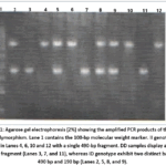 |
Figure 1: Agarose gel electrophoresis (2%) showing the amplified PCR products of the ACE I/D polymorphism. Lane 1 contains the 100-bp molecular weight marker. II genotype is shown in Lanes 4, 6, 10 and 12 with a single 490-bp fragment.
|
Discussion
The main effector molecule of the RAAS, angiotensin II is synthesized via ACE catalysis. In addition to inducing potent vasoconstriction and hypertension, Angiotensin II stimulates aldosterone release. This downstream axis is strongly linked to insulin resistance and hyperinsulinemia, linking RAAS activation to T2DM and MetS development.³⁰ Thus, the ACE insertion/deletion (I/D) polymorphism has been established as the most widely researched genetic variant in this pathway.³¹
The present study revealed non-significant, distinct distribution trends for ACE variants across the studied cohorts. The II polymorphism dominated in T2DM group (DD being least frequent), whereas, the MetS cohort showed a reverse pattern, with the DD and II polymorphisms showing the highest and lowest frequencies, respectively. The assessment of allelic distribution indicated that I allele was most prominent and least frequent in T2DM and MetS groups, respectively, so, it emerged as the primary variant in T2DM group, whereas the D allele predominated in MetS group. Nevertheless, because overall genotype and allele frequencies remained statistically comparable across groups, these findings propose that the ACE I/D polymorphism alone is not significantly associated with susceptibility of T2DM or MetS in the studied groups.
Literature on ACE variants in metabolic diseases shows noticeable geographic and ethnic heterogeneity. Studies consistently showed the II genotype is underrepresented comparable to DD and ID variants in T2DM populations, while DD genotype and D allele predominated.32–34 Regionally, Egyptians Jordanian, and Syrian cohorts exhibit elevated D allele frequencies (67%, 66% and 60%, respectively) aligning with African, Caucasian, and other Arab populations, but conflicting with East Asian cohorts.35–37
Conversely, other studies report that the heterozygous ID genotype predominated in T2DM without significant allele or genotype variance across groups.38–40 Thus, while no universal link exists between ACE I/D variants and T2DM, the DD genotype remains a potential localized risk factor.41Similar inconsistencies extend to MetS. In North Indian, Hungarian, and Mexican cohorts, the D allele and DD genotype were significantly elevated in MetS patients relative to controls, a link further supported by data from a Chinese T2DM cohort.29,42–44 Conversely, adult Moroccan and Chilean studies revealed an unexpected protective effect against MetS, while other reports found no significant association between ACE I/D variants and MetS criteria despite high DD prevalence.45–48 Collectively, these conflicting findings highlight the profound impact of ethnic and geographical variation on T2DM and MetS susceptibility.
Assessment of clinical risk profiles explores that the polymorphisms of ACE gene associate with hypertension and microalbuminuria, independent of the status of obesity. Specifically, ID polymorphism independently predisposes to hypertension, whereas a protective effect is exerted by DD polymorphism. With respect to renal end points, I allele and II polymorphism strongly increase microalbuminuria risk, whereas the DD polymorphism confers a protective association. These findings contrast with data from a Malay cohort, which demonstrated no significant association of ACE variants with obesity or hypertension, showing a borderline interaction between D allele and dyslipidemia.49,50 While an overrepresented I allele in certain populations suggests limited MetS involvement without promoting hypertension, other reports note higher D allele and DD frequencies in MetS that do not achieve statistical significance over controls.51,52
Conversely, Nikzamir et al. found that ACE I/D variants and the D allele correlate with T2DM risk but not MetS.53 Regarding renal sequelae, T2DM patients having DD polymorphism showed markedly higher rates of urinary albumin excretion than the carriers of other variants.54 While earlier data indicated that ACE I/D variants do not tie to obesity with the I allele offering protection against hypertension relative to the D allele findings in an Emirati cohort revealed that the DD genotype lowered hypertension odds exclusively within obese individuals, despite showing no baseline association with joint hypertension and T2DM.55,56 These findings directly challenge studies characterizing the DD genotype as an independent risk factor for hypertension.57
Genotype distribution was not in Hardy–Weinberg equilibrium (HWE) either in control group or T2DM group.
A deviation from HWE happens when a population’s observed genetic frequencies do not match the expected mathematical baseline, meaning the population is actively evolving. HWEPrinciple states that in a stable, idealized population, allele and genotype frequencies will remain constant across generations. This baseline requires five strict conditions to be met: No natural selection: All traits have equal survival and reproductive success, No mutation: Novel alleles are not introduced into the gene pool. No gene flow: The population is closed, with no individual migration in or out. Infinite population size: The population is large enough to prevent genetic drift from causing random shifts in the frequency of alleles. Random mating: Individuals mate by chance, not by specific trait selection. Because natural environments are dynamic, virtually all natural populations deviate from this equilibrium. These deviations are primarily driven by violations of the five conditions: Natural Selection: Certain alleles provide an advantage, causing those genotypes to increase in frequency, Genetic Drift: Random chance alters allele frequencies, which heavily impacts small populations (e.g., following a natural disaster or bottleneck event), Gene Flow: Migration introduces new alleles or alters the ratios of existing ones, Non-Random Mating: (relatively high consanguineous marriage rate)Behaviors like inbreeding increase homozygosity (individuals with identical alleles), while assortative mating skews genotype distributions, Mutation: The spontaneous change of one allele into another.58This finding should be further assessed by larger studies in the future and with independent genotyping of the data.
Some caveats are in order. Firstly, the case-control design makes it impossible to determine cause-effect relationshipsovertime Second, the sample size was relatively small, which might have attenuated the ability to reveal subtle genetic associations. Thirdly, blood levels of ACE were not assessed, which precludes a direct assessment of genotype ACE expression correlations. Finally, and most importantly, if different diagnostic criteria were used, care should be taken when comparing to other studies using different definitions of MetS. Still, larger prospective multi-center studies in a variety of populations are needed to confirm and gain further insight into the relationship between ACE I/D polymorphisms and metabolic disorders.
Conclusion
In this Jordanian cohort, overall ACE I/D genotype distributions and allele frequencies did not differ significantly between controls and patients with T2DM or MetS in the Jordanian population. In investigative subgroup analyses, it was demonstrated that ID genotype was associated with higher odds of hypertension, while the I allele and II genotype were linked to a higher risk of microalbuminuria. Conversely, the DD genotype happened less frequently among patients with renal or vascular complications. These findings suggest that while ACE I/D variants do not seem to be primary markers of susceptibility for T2DM or MetS in this population, certain genotypes may correlate with particular vascular complication within specific patient subgroups. Prospective, multicenter studies with larger sample sizes and statistical corrections for multiple testing are required to validate these exploratory associations.
Acknowledgment
The authors sincerely thank the staff of Internal Medicine Department, Medical School at the University of Mutah, for their indispensable support during this study. Special thanks are also extended to the patients and volunteers for their generous participation in this research.
Funding Sources
The author(s) received no financial support for the research, authorship, and/or publication of this article.
Conflict of Interest
The authors do not have any conflict of interest.
Data Availability Statement
This statement does not apply to this article.
Ethics Statement
Ethical approval for the study was obtained from the Committee of Medical Ethics, of the Medical School at University of Mutah, Jordan (Approval No. 201413)..
Informed Consent Statement
Prior to their involvement in the study, all participants provided written informed consent for experimentation. All participant privacy rights were firmly secured during the study.
Clinical Trial Registration
This research does not involve any clinical trials.
Permission to reproduce material from other sources
Not Applicable
Author contributions
- Samir Mahgoub: proposal writing and administration, experimental work, original draft writing, final manuscript revision and editing.
- Rami Dwairi: selection of participants, final manuscript revision and editing.
- Yousef Al-saraireh: supervision, proposal writing, editing.
- Maysa Eyalsalman: collection of data, supervision.
- Sara Alkhamaiseh: collection of data, supervision.
- Batool Gharaibeh: collection of data, supervision.
- Sadeel Abuhalimeh: collection of data, supervision.
- Hala Abdullah: collection of data, supervision.
- Mahmoud Kaswal: proposal writing, conceptualization, visualization.
- Ashraf Zaghloul: data collection, statistical analysis.
References
- Obeidat AA, Ahmad NM, Ghabashi AM, et al. Developmental Trends of Metabolic Syndrome in the Past Two Decades: A Narrative Review. J Clin Med.2025;14(7):2402. doi: 10.3390/jcm14072402.
CrossRef - Moore JX, Chaudhary N, Akinyemiju T. Metabolic Syndrome Prevalence by Race/Ethnicity and Sex in the United States, National Health and Nutrition Examination Survey, 1988–2012. Prev Chronic Dis. 2017; 16:14: E24. DOI: http://dx.doi.org/10.5888/pcd14. 160287
CrossRef - Ervin RB. Prevalence of metabolic syndrome among adults 20 years of age and over, by sex, age, race and ethnicity, and body mass index: United States, 2003-2006. Natl Health Stat Report. 2009; (13):1-7. PMID: 19634296.https://pubmed.ncbi.nlm.nih. gov/19634296/
- Povel C, Boer JM, Reiling E, et al. Genetic variants and the metabolic syndrome: a systematic review. Obesity Reviews. 2011;12(11):952-67. DOI: 10.1111/j.1467-789X.2011. 00907.x
CrossRef - Chen QR, Jiang SQ, XU XP. Polymorphism of genes encoding homocysteine metabolism-related enzymes and risk for cardio-cerebrovascular disease. Chinese Journal of Cardiology. 2012; 40(9):801-3. PMID: 23141099.https://pubmed.ncbi.nlm.nih. gov/23141099/
- Rywik TM, Szperl M, Ploski R, et al. Is evaluation of complex polymorphism helpful in the assessment of prognosis after percutaneous coronary intervention. A prospective study. Kardiol Pol. 2011;69(9):881-8. PMID: 21928191.https://pubmed.ncbi. nlm.nih.gov/ 21928191/
- Rana S, Ali S, Wani HA, et al. Metabolic syndrome and underlying genetic determinants-A systematic review. Journal of Diabetes & Metabolic Disorders. 2022; 21(1):1095-104. DOI: 10.1007/s40200-022-01009-z
CrossRef - Wonjin Kim KH, DaeKangRD, JeongWY, et al.Metabolic Syndrome Fact Sheet 2024: Executive Report. CardiometabSyndr J. 2024;4(2):70-80. https://doi.org/10.51789/cmsj.2024.4.e14
CrossRef - Kaur J. A Comprehensive review on metabolic syndrome. Cardiol Res Pract. 2014:943162. doi: 10.1155/2014/943162.DOI: 10.1155/2014/943162
CrossRef - Yasein N, Ahmad M, Matrook F, et al. Metabolic syndrome in patients with hypertension attending a family practice clinic in Jordan. EMHJ-Eastern Mediterranean Health Journal. 2010;16 (4): 375-380.PMID: 20795419. DOI: 10.26719/2010.16.4.375
CrossRef - Alharbi KK, Kashour TS, Al-Hussaini W, et al. Association of angiotensin converting enzyme gene insertion/deletion polymorphism and familial hypercholesterolemia in the Saudi population. Lipids in health and disease. 2013;12(1):177. DOI: 10.1186/1476-511X-12-177
CrossRef - Ali A, Alghasham A, Ismail H, et al. ACE I/D and eNOS E298D gene polymorphisms in Saudi subjects with hypertension. Journal of the Renin-Angiotensin-Aldosterone System. 2013,14(4):348-53. DOI: 10.1177/1470320312459976
CrossRef - Jandeleit-Dahm KA, Tikellis C, Reid CM, et al. Why blockade of the renin–angiotensin system reduces the incidence of new-onset diabetes. Journal of hypertension. 2005;23(3):463-73. DOI: 10.1097/01.hjh.0000160198.05416.72
CrossRef - Qu H, Lu Y, Lin S. Meta-analysis on the association of ACE/ID polymorphism and essential hypertension in Chinese population. Zhonghuayu fang yixuezazhi [Chinese journal of preventive medicine]. 2001;35(6):408-11. PMID: 11840772.https://pubmed.ncbi. nlm.nih.gov/11840772/
- Kerner W, Brückel J. Definition, classification and diagnosis of diabetes mellitus. Experimental and Clinical Endocrinology & Diabetes. 2014;122(07):384-6. DOI: 10.1055/s-0034-1366278
CrossRef - World Health Organization. Report of a WHO consultation: definition of metabolic syndrome in definition, diagnosis and classification of diabetes mellitus and its complications. 1999;Part I: Diagnosis and classification of diabetes mellitus. https://iris.who.int/handle/ 10665/66040
- Hurst PL, Lovell-Smith CJ. Optimized assay for serum angiotensin-converting enzyme activity. Clin Chem. 1981;27:2048–2052.PMID: 6273013
CrossRef - Jacobs, N. J. and Vandemark, P. J. The purification and properties of the alpha-glycerophosphate-oxidizing enzyme of Streptococcus faecalis 10C1. Biochem. Biophys. 1960;88:250-5. DOI: 10.1016/0003-9861(60)90230-7
CrossRef - Richmond W. Preparation and properties of a cholesterol oxidase from Nocardia sp. and its application to the enzymatic assay of total cholesterol in serum. Chem. 1973; 19/12:1350-1356. PMID: 4757363. https://pubmed.ncbi.nlm.nih.gov/6273013/
CrossRef - Gordon T, Castelli WP, Hjortland MC, et al. High density lipoprotein as a protective factor against coronary heart disease. The Framingham Study Am. J. Med. 1977;62(5):707-14. DOI: 10.1016/0002-9343(77)90874-9
CrossRef - Teoh Y, Weerasinghe D. Performance of non-HDL cholesterol (via de Cordova equation) and LDL calculated with Friedwald equation compared to LDL cholesterol measured by ultracentrifugation in a lipid clinic population. Atherosclerosis Journal. 2015,241(1):04.43. DOI: https://doi.org/10.1016/j.atherosclerosis.2015.04.433
CrossRef - von Zychlinski A, Williams M, McCormick S, et al. Absolute quantification of apolipoproteins and associated proteins on human plasma lipoproteins. Journal of proteomics. 2014;106:181-190. DOI: 10.1016/j.jprot.2014.04.030
CrossRef - Barrak MP, Shelly LV, Wayne AJ, et al. Detection of Canine Microalbuminuria Using Semiquantitative Test Strips Designed for Use with Human Urine. Veterinary Clinical Pathology. 2002;31 (2): 56–60.DOI: 10.1111/j.1939-165x.2002.tb00280.x
CrossRef - Mehta YP, Munde BP. Study of blood sugar levels in high-risk neonates using glucometer method and laboratory glucose oxidase peroxidase method. J. Contemp. Pediatr. 2017;4:1185-92. DOI: https://doi.org/10.18203/2349-3291.ijcp20172526
CrossRef - Shrikanth NH, Anupama YJ. A case of variant hemoglobin HbE detected incidentally during estimation of Glycosylated hemoglobin (HbA1C). APIK Journal of Internal Medicine. 2018;26(2):38. DOI: 10.4103/2666-1802.260122
CrossRef - Lamy PJ, Sault C, Renard E. High fasting serum insulin level due to autoantibody interference in insulin immunoassay discloses autoimmune insulin syndrome: A case report. In Annales de biologie Clinique. 2016; 74(4):490-494). DOI: 10.1684/abc.2016.1168
CrossRef - Filipovic N, Borcić N, Igić R A simple colorimetric method for estimating plasma angiotensin I converting enzyme activity. Clin. Chim. Acta. 1983;128:177 – 180.DOI:10.1016/0009-8981(83)90068-2
CrossRef - de Azevedo Salgado ALF, de Carvalho L, et al. Insulin resistance index (HOMA-IR) in the differentiation of patients with non-alcoholic fatty liver disease and healthy individuals. Arq Gastroenterol. 2010;47(2):165-9. https://doi.org/10.1590/S0004-28032010000200009
CrossRef - Lee YJ, Tsai JC. ACE gene insertion/deletion polymorphism associated with 1998 World Health Organization definition of metabolic syndrome in Chinese type 2 diabetic patients. Diabetes care. 2002; 25(6):1002-8. DOI: 10.2337/diacare.25.6.1002
CrossRef - Colussi G, Catena C, Lapenna R, et al. Insulin resistance and hyperinsulinemia are related to plasma aldosterone levels in hypertensive patients. Diabetes Care. 2007;30(9):2349-54. DOI: 10.2337/dc07-0525
CrossRef - Moon JY. Recent update of renin-angiotensin-aldosterone system in the pathogenesis of hypertension. Electrolyte Blood Press. 2013; 11:41–5.doi: 5049/EBP.2013.11.2.41
CrossRef - Oh S, Lee J, Shin E, et al. Genome-wide association study of metabolic syndrome in Korean populations. PLoS One. 2020;15(1): e0227357. DOI: 10.1371/journal.pone.0227357
CrossRef - Al-Saikhan F, Abd-Elaziz AM, Ashour R. Association between risk of type 2 diabetes mellitus and angiotensin-converting enzyme insertion/deletion gene polymorphisms in a Saudi Arabian population. BIOMEDICAL REPORTS. 2017;7:56-60. DOI: 10.3892/br.2017.920
CrossRef - Jones H. Angiotensin Converting Enzyme Gene Insertion/Deletion Polymorphism in Gaza Strip-Palestine and Type 2 Diabetic Nephropathy. Medical Reports & Case Studies. 2002;7(1):001-002. doi: 10.2215/CJN.04140907
CrossRef - Al-Harbi ME, Farid ME, Gumaa AK, et al. Genotypes and allele frequencies of angiotensin-converting enzyme (ACE) insertion/deletion polymorphism among Bahraini population with type 2 diabetes mellitus and related diseases. Mol Cell Biochem. 2012;362(1-2):219-23. DOI: 10.1007/s11010-011-1146-1
CrossRef - Al-Hinai TA, Hassan OM, Simsek M, et al. Genotypes and allele frequencies of angiotensin converting enzyme (ACE) insertion/deletion polymorphism among Omanis. J SCIRESMED SCI. 2002;4(1-2):25–27. PMID: 24019722. https://www.ncbi.nlm.nih.gov/pmc/ articles/PMC3174717/
CrossRef - Salem AH, Batzer MA. High frequency of the D allele of the angiotensin-converting enzyme gene in Arabic populations. BMC research notes. 2009;2(1):1-5. DOI: 10.1186/1756-0500-2-99
CrossRef - Domingos BCA, Bonini-Domingos RC, Iacida CE, et al. Angiotensin converting enzyme polymorphism in type 2 diabetes mellitus. Biomarkers and Genomic Medicine. 2014;6: 99e104. https://doi.org/10.1016/j.bgm.2014.06.001
CrossRef - Yang M, Qiu C, Qun XU, et al. Association of Angiotensin Converting Enzyme Gene I/D Polymorphism with Type 2 Diabetes Mellitus. Biomedical and Environmental Sciences. 2006;19:323-327. PMID: 17044652.https://pubmed.ncbi.nlm.nih.gov/17044652/
- Habibullah M, Akter F, Qin X, et al. Association between Angiotensin-Converting Enzyme-Insertion/Deletion Polymorphism and Diabetes Mellitus-2 in Saudi Population. Asian Pac J Cancer Prev. 2021; 22(1):119-123. DOI: 10.31557/APJCP.2021.22.1.119
CrossRef - Barghash A, Al-Gharabli SI, Jweihan M, et al. Angiotensin Converting Enzyme (ACE) Gene Polymorphism in Jordanian Type-1 and Type-2 Diabetic Patients. JJBS. 2020;13(2). https://jjbs.hu.edu.jo/files/vol13/n2/Paper%20Number%2012.pdf
- Mittal G, Gupta V, Haque FS, et al. Effect of angiotensin converting enzyme gene I/D polymorphism in patients with metabolic syndrome in North Indian population. Chin Med J. (Engl). 2011;124(1):45-8. PMID: 21362306.https://pubmed.ncbi. nlm.nih.gov/ 21362306/
- Fiatal S, Szigethy E, Széles G, et al. Insertion/deletion polymorphism of angiotensin-1 converting enzyme is associated with metabolic syndrome in Hungarian adults. Journal of the Renin-Angiotensin Aldosterone System. 2011;12(4):531–538.https://doi.org/10.1177/ 1470320310394231
CrossRef - Alvarez-Aguilar C, Enríquez-Ramírez LM, Figueroa-Nuñez B, et al. Association between angiotensin-1 converting enzyme gene polymorphism and the metabolic syndrome in a Mexican population. ExpMol Med. 2007;39(3):327-334.https://www.nature.com/ articles/ emm20073
CrossRef - Abouelfath R, Habbal R, Laaraj A, et al. ACE insertion/deletion polymorphism is positively associated with resistant hypertension in Morocco. Gene. 2018; 658:178-83. DOI: 1016/j.gene.2018.03.028.
CrossRef - Herrera LC, Castillo W, Estrada P, et al. Association of polymorphisms within the Renin-Angiotensin System with metabolic syndrome in a cohort of Chilean subjects. Endocrinol. Metab. 2016;60 (3):190-198. https://doi.org/10.1590/2359-3997000000134
CrossRef - Sinorita H, Madiyan M, Pramono BR, et al. ACE Gene Insertion/Deletion Polymorphism Among Patients with Type 2 Diabetes, and Its Relationship with Metabolic Syndrome at Sardjito Hospital Yogyakarta, Indonesia. Acta Med Indones-Indones J Intern Med. 2010;42(1):12-16. PMID: 20305326.https://pubmed.ncbi.nlm.nih.gov/20305326/
- Nadalin S, Pavli´c DS, Peitl V, et al. Association between Insertion-Deletion Polymorphism of the Angiotensin-Converting Enzyme Gene and Treatment Response to Antipsychotic Medications: A Study of Antipsychotic-Naïve First-Episode Psychosis Patients and Non adherent Chronic Psychosis Patients. J. Mol. Sci. 2022; 23:12180, 11 pages.https://doi.org/10.3390/ijms232012180
CrossRef - Suokhrie S, Chaudhary V, Mishra S, et al. Association of angiotensin-converting enzyme I/D polymorphism and apolipoprotein B with cardiometabolic abnormalities among young adults: a pilot study from Delhi. Egyptian Journal of Medical Human Genetics. 2023;24:52. https://doi.org/10.1186/s43042-023-00432-y
CrossRef - Apidi E, Sani IA, Johari ZKM, et al. Association of Angiotensin Converting Enzyme (ACE) Gene insertion/deletion (I/D) Polymorphism with Obesity and Obesity Related Phenotypes in Malay Subjects. JJBS. 2020;13(3):267-273. https://jjbs.hu.edu.jo/files/vol13/n3/ Paper%20Number%202.pdf
- Thomas NG, Tomlinson BP, Chan CJ. JF, et al. Renin-Angiotensin System Gene Polymorphisms, Blood Pressure, Dyslipidemia, and Diabetes in Hong Kong Chinese. Diabetes Care. 2001;24:356–361. DOI: 10.2337/diacare.24.2.356
CrossRef - Simsek S, Tekes S, A Tuzcu KAT, et al. Angiotensin-converting enzyme gene insertion/deletion polymorphism with metabolic syndrome in Turkish patients. J Endocrinol Invest. 2013;36(10):860-3. DOI: 10.3275/8967
- Nikzamir A, Nakhjavani M, Golmohamadi T, et al. Association of Angiotensin-Converting Enzyme Gene Insertion/Deletion Polymorphism with Metabolic Syndrome in Iranians with Type 2 Diabetes Mellitus. Arch Iranian Med. 2008;11 (1): 3 – 9. PMID: 18154415. https://pubmed.ncbi.nlm.nih.gov/18154415/
CrossRef - Dudley RC, Keavney B, Stratton M, et al. U.K. Prospective Diabetes Study. XV: Relationship of renin-angiotensin system gene polymorphisms with microalbuminuria in NIDDM. Kidney Int. 1995;48(6):1907-11. DOI: 10.1038/ki.1995.490
CrossRef - Motawi TK, Shaker OG, Shahin NN, et al. Angiotensin-converting enzyme insertion/deletion polymorphism association with obesity and some related disorders in Egyptian females: a case-control observational study. Nutrition & metabolism. 2016;13:68. doi: 10.1186/s12986-016-0127-5. eCollection 2016
CrossRef - Alsafar H, Hassoun A, Almazrouei S, et al. Association of angiotensin converting enzyme insertion-deletion polymorphism with hypertension in emiratis with type 2 diabetes mellitus and its interaction with obesity status. Dis markers. 2015:536041. doi: 10.1155/ 2015/536041
CrossRef - Zhou YF, Yan H, Hou XP, et al. Association study of angiotensin converting enzyme gene polymorphism with elderly diabetic hypertension and lipids levels. Lipids in health and disease. 2013;12(1):1-4. http://www.lipidworld.com/content/12/1/187
CrossRef - Abramovs N, Brass A, Tassabehji M. Hardy-Weinberg Equilibrium in the Large Scale Genomic Sequencing Era. Front Genet. 2020;11:210. doi: 10.3389/fgene.2020.00210
CrossRef
 PlumX:
 Views: 311
  PDF Downloads:
13
PDF Downloads:
13 
Accepted on: 07-09-2026
Second Review by: Dr. Ramdas Bhat
Final Approval by: Dr. Patorn Piromchai