R.Balakrishnan1 and Vijay Ebenezer2
1Professor, Department of Oral and Maxillofacial Surgery, Sree Balaji Dental College and Hospital, Bharath University, Pallikaranai, Chennai-600100 2Vijay Ebenezer, Head of the Department, Department of Oral and Maxillofacial Surgery, Sree Balaji Dental College and Hospital, Bharath University, Pallikaranai, Chennai-600100
DOI : https://dx.doi.org/10.13005/bpj/706
Abstract
The treatment of pediatric maxillofacial fractures is unique due to the psychological, physiological, developmental, and anatomical characteristics of children. The treatment plan in children has to be modified as compared to adults considering the presence of tooth buds and potential disturbances in growth. Use of acrylic splints has been one of the popular techniques in children because of its relatively easy placement and reduced risk of hindrances to growth of jaw. Reduction and immobilization is done with acrylic splint and circum mandibular wiring.
Keywords
Pediatric mandible fracture; acrylic occlusal splint; circum mandibular wiring
Download this article as:| Copy the following to cite this article: Balakrishnan R, Ebenezer V. Management of Mandibular Body Fractures in Pediatric Patients. Biomed Pharmacol J 2015;8(October Spl Edition) |
| Copy the following to cite this URL: Balakrishnan R, Ebenezer V. Management of Mandibular Body Fractures in Pediatric Patients. Biomed Pharmacol J 2015;8(October Spl Edition). Available from: http://biomedpharmajournal.org/?p=3514> |
Introduction
Pediatric mandible fractures account for 32.7% of all facial fractures, followed by nasal bone fractures (30.2%) and midface/zygoma fractures (28.6%) (1). Mandible fractures are rare in the children younger than 5 years (2-6). Many pediatric mandible fractures can be treated without surgical exploration of the fracture site (7). In children it is difficult to use arch bars, plates, interdental ligature due to the absence of teeth due to primary teeth exfoliation and developing tooth buds and the poor retentive shape of the deciduous crown. Majority of the body and symphysis fractures in children are undisplaced because of elasticity of mandible and embedded tooth buds that holds the fragments together “like glue” (8-9). Splinting the fractured mandible with an acrylic splint, retained by circummandibular wires, remains a perfect option.
The following paper will review the management of facial trauma in children. It highlights the role of acrylic splint with the use of circummandibular wiring technique in the management of bilateral body fracture in a 5-year-old child.
Case report
A 5 year old female child came to our department of oral and maxillofacial surgery with a history of fall from 1st floor of her house premises. Extra oral examination of the patient reveals laceration in the lower chin and mild swelling in the anterior region of the mandible (Fig 1). There was open bite in anterior region with deranged occlusion (Fig 2). All primary teeth were present. On palpation there was bilateral step deformity in parasymphysis region. Developing tooth bud was visible in the fracture site(Fig 3). In AP view of skull reveals bilateral parasymphysis mandible fracture. Impression of both upper and lower dental arches were taken with alginate impression material (Fig 4). An acrylic cap splint is prepared on the patient`s arches model.
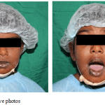 |
Figure 1: Pre operative photos |
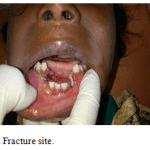 |
Figure 2: Fracture site |
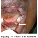 |
Figure 3: Exposed tooth bud in the fracture site |
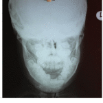 |
Figure 4: Pre operative xray |
Under general anaesthesia nasotracheal intubation done, extraoral skin and intraoral mucosa was prepared with povidione iodine solution. The displaced fractured segments were reduced bimanually (Fig 5). The permanent canine tooth buds were found exposed bilaterally in site of fracture. Reduction of the fracture fragment was difficult due to the entrapment of the permanent canine tooth bud bilaterally (Fig 6). A small stab incision was placed at the inferior border of the mandible in the right side. A bone awl was introduced through the stab incision (Fig 7). The bone awl was guided along the body of the mandible and taken out lingually. Next the wire was tied in and the awl was gently guided along the lower border of the mandible and passed into the buccal sulcus(Fig 8). The wire was held around the mandible and was bound to the cap splint which stabilized the reduced fractured segments (Fig 9). The same procedure was performed on the left side. Then the final occlusion was checked (Fig 10). The external wound on the chin was thoroughly debrided and wound closure was done in layers (Fig 11).
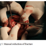 |
Figure 5: Manual reduction of fracture |
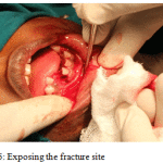 |
Figure 6: Exposing the fracture site |
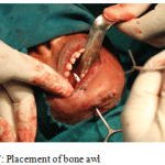 |
Figure 7: Placement of bone awl |
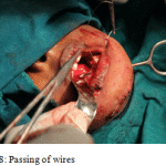 |
Figure 8: Passing of wires |
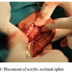 |
Figure 9: Placement of acrylic occlusal splint |
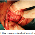 |
Figure 10: Final settlement of occlusal by acrylic splint |
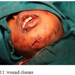 |
Figure 11: wound closure |
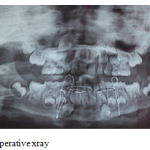 |
Figure 12: Post operative xray |
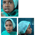 |
Figure 13: Post operative |
Discussion
Facial fractures in children account for the approximately 5% of all fractures. A male predilection is seen in all age groups. The most common fracture in children requiring hospitalization and/or surgery generally involves the mandible and, in particular, the condyle. Fractures in the condylar region are the most common, followed by angle and body fractures. The etiologies of mandibular fractures in children are usually falls and sports injuries. The clinical features of a fractured mandible in a child are the same as in an adult, which includes pain, swelling, trismus, derangement of occlusion, sublingual ecchymosis, step deformity, midline deviation, loss of sensation due to nerve damage, bleeding, TMJ problems, tenderness, movement restriction, open bite and crepitus. Thorough clinical examination, however, may be impossible in uncooperative young trauma patients. Lacerations should be evaluated to reveal injuries to underlying structures.
Problems encountered in management of Pediatric Mandibular Fractures
- Loose anchorage system due to attrition of deciduous teeth and physiologic resorption of roots.[10]
- Difficulties in securing IMF using arch bars and eyelets as primary teeth are not sufficiently stable and may be avulsed due to the pressure exerted. In addition, the partially erupted secondary teeth are not sufficiently stable in the pediatric soft bone.[11]
- Shape of the primary teeth: Conical shape with wide cervical margins and tapered occlusal surface makes placement of wires technically challenging.[12]
- Restricted normal dietary intake in children on IMF was reported to result in significant weight and protein loss and reduced tidal volume.[10]
- Children on IMF are at an increased risk of aspirating gastric contents should they vomit.[10]
- The wires cause discomfort and damage periodontal tissues.[10].
Conclusion
The use of occlusal splint in stabilising the fractured segment of the pediatric dental arches is the most common treatment modality. As the anatomical occlusal relation can be achieved without injuring any developing tooth bud nor injuring to the mucoperiosteum (Fig 12).
Reference
- Imahara SD, Hopper RA, Wang J, et al. Patterns and outcomes of pediatric facial fractures in the United States: a survey of the National Trauma Data Bank. J Am Coll Surg 2008;207(5):710–6
- Ferreira PC, Amarante JM, Silva PN, et al. Retrospective study of 1251 maxillofacial fractures in children and adolescents. Plast Reconstr Surg 2005;115:1500–8.
- Davison SP, Clifton MS, Davison MN, et al. Pediatric mandibular fractures: a free hand technique. Arch Facial Plast Surg 2001;3:185–9.
- Demianczuk AN, Verchere C, Phillips JH. The effect on facial growth of pediatric mandibular fractures. J Craniofac Surg 1999;10:323–8
- Schweinfurth JM, Koltai PJ. Pediatric mandibular fractures. Facial Plast Surg 1998;14:31–44.
- Lindahl L, Hollender L. Condylar fractures of the mandible. II. A radiographic study of remodeling processes in the temporomandibular joint. Int J Oral Surg 1977;6:153–65.
- Tanaka N, Uchide N, Suzuki K, Tashiro T, Tomitsuka K, Kimijima Y, et al. Maxillofacial fractures in children. J Craniofac Surg. 1993;21:289–93.
- B. Mulliken, L. B. Kaban, and J. E. Murray, “Management of facial fractures in children,”Clinics in Plastic Surgery, vol. 4, no. 4, pp. 491–502, 1977.
- A. Fortunato, A. F. Fielding, and L. H. Guernsey, “Facial bone fractures in children,”Oral Surgery Oral Medicine and Oral Pathology, vol. 53, no. 3, pp. 225–230, 1982
- Kaban LB. Diagnosis and treatment of fractures of the facial bones in children 1943-1993.J Oral Maxillofacial Surg. 1993;51:722–9
- Aizenbud D, Hazan-Molina H, Emodi O, Rachmiel A. The management of mandibular body fractures in young children.Dental Traumatol. 2009;25:565–70
- Zimmermann CE, Troulis MJ, Kaban LB. Pediatric facial fractures, recent advances in prevention, diagnosis and management. Int J.Oral Maxillofacial Surg. 2006;35:2–13


