
Manuscript accepted on :January 14, 2016
Published online on: 22-02-2016
Plagiarism Check: Yes
M. Jafarpour1, A. Amniat Talab2*, and A. Nekuie Fard3
1Department of Microbiology، Urmia Branch، Islamic Azad University ، Urmia، Iran 2*Department of Pathobiology، Urmia Branch، Islamic Azad University ، Urmia، Iran 3Iranian Fisheries Research organization, National Artemia Research Center, ARREO, Urmia, Iran
DOI : https://dx.doi.org/10.13005/bpj/940
Abstract
Nowadays by becoming cognizant of the effects of antimicrobial herbs is increasing according to the use of these plants in the world. Mentioned treatment method cause to reduce the side effects and prevention of bacterial resistance to antibiotics. In this regard, the main focus of the paper is centered on Melissa officinalis aqueous Lemon balm extract antibacterial effect of lamiaceae family on Aeromonas hydrophila factor in vitro condition and it cause hemorrhagic septicemia disease in Oncorhychus mykiss. A. hydrophila of rainbow trout with septicemia symptoms were studied after isolation and culturing in different environments in order to definitive diagnosis by using biochemical and molecular (PCR) tests. In order to determine extract antibacterial power; we have used broth micro-dilution ( liquid dilution), minimum bactericide concentration ( MBC) and minimum inhibition concentration (MIC) standard methods. Then, MIC concentration was used in MULLER Hinton agar by creating wells and it was compared zone diameter of bacterial growth inhibition. Results showed that M.Officinalis aqueous lemon balm extract with MIC amount is equal to 2.5 mg/ml and MBC amount is equal to 5 mg/ml. Results domestrated the positive eefect of the extract on bacteria and in order to use it as a medicine, treatment and prevention of fish disease, accomplishment of further clinical research is required.
Keywords
Aeromonas hydrophila – minimum bacterial concentration (MBC) – Inhibitory concentration- bacteria- Melissa officinalis
Download this article as:| Copy the following to cite this article: Jafarpour M, Talab A. A, Fard A. N. In Vitro Study of the Effect of Melissa Officinalis Aqueous Lemon Balm Extract on Aeromonas Hydrophila Causative Hemorrhagic Septicemia Disease in Oncorhychus Mykiss. Biomed Pharmacol J 2016;9(1) |
| Copy the following to cite this URL: Jafarpour M, Talab A. A, Fard A. N. In Vitro Study of the Effect of Melissa Officinalis Aqueous Lemon Balm Extract on Aeromonas Hydrophila Causative Hemorrhagic Septicemia Disease in Oncorhychus Mykiss. Biomed Pharmacol J 2015;9(1). Available from: http://biomedpharmajournal.org/?p=6476 |
Introduction
Herbs are as strong reservoirs of secondary metabolites and indeed they are effective resources of many pharmaceutical materials which contain active ingredient in one or some of their organs. It is less than 1 percent of dry weight of plant and it has organisms. It isn’t known exactly when people commenced to use herbs as medicine as well. Of course, information about medicinal and pharmaceutical properties of plants were available since ancient time at last they were available till the contemporary generations. To date this effort continues and now it is progressing in very fast pace. (Burt, 2004; Tajkarimet.al, 2010). According to the number of chemical compounds in plant extracts, it is impossible to a consider single mechanism for their antibacterial effects; but they will have multiple targets in cells. These Mechanisms aren’t acted separately, but some of them are affected by others.(Burt, 2004). One of the most important property of essential oils and their components is hydrophobic property which leads to penetration of the materials to bacterial cell membrane lipids and mitochondrial which is the main reason that causes of creating disorder in themselves structures and further permeability. This led to exit and leakage of ions and other cell contents.Although the withdrawal of limited quantities of this material is tolerable for bacteria; but it has an impact on on viability and withdrawal of bulky amounts of cell contents or withdrawal of vital ions molecules cause cell death.(Burt, 2004).Generally, when phenolic compounds level is higher in essence; their antibacterial properties against food pathogens will be more. These compounds include carvacrol, eugenol and thymole. Probably, the impact mechanisim of these compounds as the other phenilic compunds are ass follows: disorder in cytoplasmic membrane, disruption the proton motive force and electric current, coagulation of cell contents. (Burt, 2004; Tajkarimet.al, 2010). Also the chemical structure of an essential oil has impact on it’s mechanism. The importance of hydroxyl groups presence in the phenolic compounds such as carvacrol and thymole was approved. The relative position of the phenolic hydroxyl group in phenolic ring doesn’t have much impact in the antibacterial effect. (Burt, 2004).Melissa officinalis is one of the oldest and most vogue herbs. It belongs to lamiaceae family and it is also called lemon balm. Numerous studies have been done on this plant and its various propetires were proved. The most common therapeutic properties of lemon balm are its (Melissa officinalis) Sedative property, antioxidant, antispasmodic, carminative; antibacterial, anti viral and anti-inflammatory cases. (Koch. Heitzman et.al, 1984; lamaison et.al , 1991). Moreover, The effect of this herb studied on known on the nervous system revealed that Melissa officinalis reduces symptoms of neurological disorders. Such as stress, anxiety and irritability (Bisset et. Al 2001).This herb protects nervous cells and eradicates free radicals. (lopeze et al 2009) and because of increasing of increasing valerinan quality effect on sleep. (lopez, et.l 2009).Aqueous and methanol extracts of mentioned herb are mono –amine- oxidase enzyme (MAO) inhibitors and in this case, the effect of methanol is stronger , so it is deduced that the mentioned herb has antidepressant effects. (lopez et al, 2009). aeromonas hydrophia is a factor that cause bacterial septicemia disease. The bacteria usually exists in the fishes digestive system and it is considered as an opportunistic pathogen. Outbreaks of disease is related to changes in environmental conditions such as stress, congestion , sudden temperature changes, fish transportation , poor water quality and high level of nitrite and carbon dioxide. In turkey aeromonas hydrophila infection has been repored for eel and crocodiles and grass carp. (Nielsen et al 2001, Janda et al , 1996, Agniswar et al, 2012). This bacteria is in gram-negative , motile and opportunistic forms which can cause to bacterial hemorrhagic septicemia (BHS) in fishes . (chu et al, 2005). The release of protease, hemolysin and sytolitic toxins are linked to the Pathogenesis of these bacteria. (hu. et al, 2012).Animated aeromonase are mainly found in fresh waters and they are compatible with different environmental conditions such as temperature, salinity and turbidity.( Hazen, et al, 1987).According to results, to fill the knowledge gap related to the effect of Aqueous extract of herb on fish bacterial disease, the main aim of research is to investigate the lemon balm aqueous extract anti- bacterial effect of lemon balm on pathogenic aeromonas hydrophila bacteria in rainbow trouts which is conducted in vitro condition for oncorhychus mykiss.
Materials and Methods
Bacteriological examination
For investigation of the patient fishes, the common diagnostic methods were sued and the seprated bacterias were identified using biochemical and molecular methods. In autopsy, most of the findings for naked-eye , were ascites and visceral congestion. In bacterial culture for whole of patient fishes, we seen severe contamination with aeromonas hydrophila bacteria. So, bacterial species was confirmed with PCR method and it was investigated by biochemical methods. To study the anti- bacterial effects, a new 24 culture was prepared. Before 24 hours of examination ; reserve stock culture medium was inoculated to nutrient agar culture; and incubated at 37 for 24 hours. Then, the colonies on the surface level of the culture environment were washed with normal saline solution and bacterial suspension was diluted with 5 ml of normal saline and uniformity suspension of the bacterias was obtained. Tubes were placed in incubator for 30minutes at 37 in order to create turbidity similar to half Mac Farland standard tubes. So tha, in this case, the concentration of bacteria was about 1 ×10³ cfu/ml. (soltaninejad et al, 2010).After preparation of suspensions and using of swap sterile and dipping it into the bacterial suspension, all levels of Mueller Hilton agar culture in parallel lines and vertical, horizontal and diagonal directions and the entire surface of the plate was covered with a uniform microbial layer. Sterile blank discs cultured via dispenser and they was placed at the proper intervals with 3 iteration. 10 ml of pure extract was poured on sterile discs; then we returned plate and incubated bacterial cultures at 37 for 24 hours in aerobic conditions. After the elapse of the necessary time, we put plates on a dark surface which doesn’t reflect light. In the presence of light, results of calipers diameter were measured by using Koulis and also they were recorded in millimeter scale. (khosravani st al, 2004) In order to determining anti-bacterial activity; erythromycin and doxycycline antibiotics have been used on bacteria culture medium and the rest of process was similar to the extract. In order to conduct quantity experiments and determine minimum inhibition concentration (MIC) and MBC in lemon balm extract (7.5 mg/l concentration) was prepared in Mueller Hilton broth medium. So, for each of concentration per ml of liquid medium 1 CFU/ml active bacteria was added. Furthermore, we have used positive control (culture medium including erythromycin, doxcycline and bacteria without extract) and negative control (culture medium without bacteria). At last the tubes were incubated for 24 hours at 37 and then the result was read. In the last case, we didn’t observe any turbidity for dilute form (inhibition) and it was considered as MIC. All of tubs without turbidity were culture on Mueller Hinton agar. After 24 hours of incubation at 37 , the last extract concentration was considered as MBC which it was able to annihilate of 99 percent initial live bacterias (khosrovani et al , 2004).For data analysis, analysis of variance (ANOVA) was used by considering treatment effect as a fixed effect. The test method was completely random with 3 repeat for each treatment. avermge compare in this reaserch included difference between inhibition diameter for various concentrations of the extract which they were calculated with toukay method. Also, mean diffrence of inhibition diameter for various concentration of the extract were done by using t- test . (Motlagh,2014). For bacterial study, 30 samples were collected from of rainbow trouts liver, spleen, blood, kidney and heart in sterile condition with mentioned signs and they were cultured in nutrient agar. Prepared culture mediums were kept at 25 inside incubator. To identify the bacterial colonies grown and they were harvested in culture medium and also they were stained. Another biochemical diagnostic experiments such as oxidase, catalaze, motile experiment and sugar consumption were done which showed in the table 1.
Table 1: A. hydrophila biochemical diagnostic test for investigated samples
| A.hydrophila | biochemical diagnostic test |
| Gram negative bacillus | Stain |
| + | Motility |
| – | Endol |
| – | Citrate |
| – | H2S |
| – | Lactose |
| – | Urea |
| + | Catalase |
| – | Vp(Voges-Proskauer) |
| + | Oxidase |
| – | Lysine decaboxilase |
| + | Glocuse |
| – | Arabinose |
| – | Sacarose |
| – | Manitol |
| + | β hemolysis |
| + | Growth in Blood Agar |
| + | Growth in MacConkey Agar |
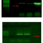 |
Figure 1: PCR molecular diagnostic test |
Molecular diagnostic test (PCR) was used for recognition of pathogenic factor in bacteria species. For separation and extraction of bacteria DNA, we have used boil and centrifuge method. Molecular diagnostic operation was done by using of aeromonas sp. 16s Rrna gene primer. All of PCR stages was accomplished in 0.2 ml sterile microtubes without DNAse in EPFNDORF thermocycle device. In this case, in every microtube of 12.5 microliters master mix include Tag, Dntp,Mgcl2, anzyms ; 8.5microliters DESP water, 1 microliters of each primer and 2 microliters of extracted DNA and in order to reach final volume; 25 microliters have been used. At last 8 microliters of PCR was driven in agar gel at 100 volt for 1 hour which include 1.5 percent safe sat in bands in document gel device. (Nilsen et al , 2001).For DNA electrophoresis, we used gene ladder with 100bp weight. If the band is visible with 599 bp moleculer weight, it is aeromonas species and the band with 685bp moleculor; weight is hydrophila species.
Results
Bacterial culture result after 24hours inside incubator at 25 showed the existence of many small and yellow colonies with +4 intensity. Bacterias are negative –gram in painting stage and according to table 1 biochemical test results were consistent with aeromonas. Hydrophila features and molecular method proved this finding (figures 1 and 2). The result of research were 14mm for diameter without corona creation which it showed meaningful difference in comparison of another groups. (p< 0.05). the most and lowest effects showed by doxycycline and erythromycin respectively. Comparison of MIC and MBC showed that lemon balm extract with 2.5 and 5 mg/ml had more impan than the other groups after doxycycline.
Table 2: Comparison of development of inhabition zone diameter (mm) in lemon balm extract and control groups
| Disk diffusion | herb extract | positive control | Negative control | |
| lemon balm mm | erythromycine mm | doxycycline mm | Sterile distilled water
mm |
|
| Aeromonas Hydrophila | 14 | 10 | 16 | 0 |
Table 3: Comparison lemon balm antibacterial effect and controlling groups against A.hydrophila .
| Test group
|
herb extract | positive control | Negative control | |||||
| lemon balm | erythromycine | doxycycline | Sterile distilled water
|
|||||
| Aeromonas Hydrophila | MIC
mg/ml |
MBC
mg/ml |
MIC
mg/ml |
MBC
mg/ml |
MIC
mg/ml |
MBC
mg/ml |
MIC
mg/ml |
MBC
mg/ml |
| 2.5 | 5 | 5 | 10 | 1.25 | 2.5 | – | – | |
Discussion
In 2004, Mimica duki et al. investigated antioxidant and bacterial activity of lemon balm essential oil against 13 bacterias and 6 mold species.Suitable effective results of this essence were supposed by dosage. Most antimicrobial and antifungal activity was observed against T.shigella and Tricophyton fungal specise. In 2005, Depasqua and et al. showed that mentioned essential is effective against lacto coccus, anthrococcus, pseudomonase and staphylococcus aureus at concentrations which they are more than one percent and also antobacterial effect relate to dosage.Melissa officinalis (lemon balm extract) investigated on streptococcus sanguis salmonella enteritidis, streptococcus mutans, staphylococcus aureus, vibrio anguillarum, aeromonas hyrophila, yersinia ruckeri, pseudomonas aeruginosa, escherichia coli, corynebacterium parvum, listeria spp, lacto coccus garvieae and flavobacterium psychrophilum. Also interaction of this extract studed on anaerobic bacteria and periodontal aerobic flavobacterium such as ; porphyromonas, veillonella parvul, capnocytophaga gingivalis, shigella sonnei, s.enterica, actinomyces odontolyticus, peptostrep tococcus micros, eilenella corrodens. ( mimica- Dukic et al, 2003, frideman et al , 2004; Di Pasqua et al ; 2005).Many researchers studied herb extracts on aeromonas hydrophila and this research is the first case for determinig lemon balm antibacterial effect and it’s aqueous extract on aeromonas hydrophila causative hemorrhagic septicemia disease in oncorhychus mykiss which showed its suitable effect on bacteria controlling in comparison with another antibiotics. We need to do more researches for investigation of pharmaceutical and clinical effects of extract against aeromonas hydrophila.
References
- Agniswar, S., Mousumi, S. and Pranab, R. (2012) Identification and typing of Aeromonas hydrophila through 16S rDNA-PCR Finger printing, Journal of Aquaculture Research Development. 3:6, 1-4.
- Bisset NG, Wichtl M. Herbal drugs and phytopharmaceuticals. 2th ed. Stuttgart: Medpharm GmbH Scientific Publishers; 2001.
- Burt, S. 2004. Essential oils: Their antibacterial properties and potential applications in foods – Areview. International Journal of Food Microbiology, 94, 223-253.
- Chu, W.H. and C.P. Lu. ( 2005) Multiplex PCR assay for the detection of pathogenic Aeromonas hydrophila. Journal of Fish Diseases, 28: 437‒441
- Drebing H, Riemann D, Low H, Schredl M, Reh C, Laux P, et al. Insomnia: are valerian/balm combinations of equal value to enzodiazepine? Therapiewoche. 1992; 42: 726-36.
- Hazen, T. C., Filiermans, C. B., Hirsch, R. P. and Esch, G. W. (1987) Prevalence and distribution of Aeromonas hydrophila in the USA. Journal of. Applied and Environmental Microbiology, 36: 731-738.
- Hu, M., N. Wang, Z.H. Pan, C.P. Lu and Y.J. Lui. ( 2012) Identity and virulence properties of Aeromonas isolates from diseased fish, healthy controls and water environment in China. Letter of Appled Microbiology, 55: 224‒233.
- Janda, J. M., Abboh, S. L., Khashe, S., Kellogg, G. H. and Shimada, T. (1996) Biochemical characteristics and serological properties of the genus Aeromonas. Journal of Clinical Microbiology, 34:1930-1983.
- Khosravi A, Malekan MA. Determination of Alcoholic and aqueous extract of Lavender Astvkas on Staphylococcus aureus and other Gram-negative bacteria. The Journal of Qazvin University of Medical Science. 2004; 29: 3-9.
- Koch-Heitzmann I, Schultze W. Melissa officinalis L. an old medicinal plant with new therapeutic action. Dtsc Apothek Ztg. 1984; 124: 2137-45.
- Lamaison JL, Petitjean-Freytet C, Duband F, Carnat AP. Rosemarinic acid content and antioxidant activity of French lamiaceae. Fitoterapia. 1991; 62: 166-71.
- Lopez V, Martin S, Gomez-Serranillos MP, Carretero ME, Jager AK, Calvo MI. Neuroprotective and neurological properties of Melissa officinalis. Neurochem Res. 2009; 34(11): 1955-61.
- Motlagh, M. K., Kazemi, M., Ghasemi, H. A., & Khaltabadi, A. H. (2014). Antibacterial effect of medicinal plant essence (Thymus vulgaris) on major bacterial mastitis pathogen in vitro. International journal of Advanced Biological and Biomedical Research, 2, 286-294.
- Nielsen, M.E., Hoi, L., Schmidt, A.S., Qian, D., Shimada, T., Shen, J.Y., and Larsen, J.L. (2001) Is Aeromonas hydrophila the dominant motile Aeromonas species that causes disease outbreaks in aquaculture production in the hejiang province of china. Diseases of Aquatic Organisms, 46(22) : 23–29.
- Soltani nejad SH, Satani mokhtari T, Rahbarian P. The study of antibacterial effect of the essential oil and methanol extracts of ziziphora cliniopodiodes on some pathogenic bacteria. Journal of Microbial Biotechnology. 2010; 2: 1-6.
- Tajkarim MM, Ibrahim SA, Cliver DO. Antimicrobial herb and spice compounds in food. Food Control 2010; 21, 1199-18.
- Di Pasqua, R., De Feo, V., Villani, F., & Mauriello, G. (2005). In vitro antimicrobial activity of essential oils from Mediterranean Apiaceae, Verbenaceae and Lamiaceae against foodborne pathogens and spoilage bacteria. Annals of microbiology, 55(2), 139-143.
- Mimica-Dukić, N., Bozin, B., Soković, M., Mihajlović, B., and Matavulj, M. (2003). Antimicrobial and antioxidant activities of three Mentha species essential oils. Planta medica, 69(5), 413-419.
- Friedman, M., Henika, P. R., Levin, C. E., & Mandrell, R. E. (2004). Antibacterial activities of plant essential oils and their components against Escherichia coli O157: H7 and Salmonella enterica in apple juice. Journal of agricultural and food chemistry, 52(19), 6042-6048.

