
Manuscript accepted on :
Published online on: 24-11-2015
Plagiarism Check: Yes
Sadeghi Limanjoob Reza1*, Tarahomi Mohammad1, Farzam Mohsen2, Zare Afshin2, Mohammadi Sahar3, Karimi Ehsan4 and Farzam Mohammad2
1Department of Aquatic Animal Health, Veterinary School, Kazerun Branch, Islamic Azad University, Kazerun, Iran. 2Department of Physiology, Veterinary School, Kazerun Branch, Islamic Azad University, Kazerun, Iran. 3Department of clinical Biochemistry,Kermanshah University of Medical Sciences,Kermanshah,Iran 4Department of Physiology, Medical School, Kazerun Branch, Islamic Azad University, Kazerun, Iran.
DOI : https://dx.doi.org/10.13005/bpj/606
Abstract
Black gill syndrome is caused due to Fusarium Solano in shrimp. Associated diseases such as Vaybr Reeves, as well as Mitosis, the presence of heavy metals such as copper and cadmium can cause the incidence of the disease. The immune system is weakened by stress and the effect of these factors, so the disease happens. The disease symptoms are the Macroscopic and microscopic melanization of shrimp gills. The other pathological signs of the syndrome are hypertrophy and hyperplasia of gill and accumulation of hemisects in the gills. Due to nutritional importance of shrimps and also importance of shrimps in exports, the aim of this study was histopathological study of black gill disease in marine shrimps of coastal city of Bushehr, which is one of the most important areas. In this study, 100 pieces of shrimp were caught off shore in the coastal city of Bushehr. After fixing with the Davidson’s fixing solution, were transferred to Laboratory of Veterinary Medicine in Kazeroon Islamic Azad University and then Histopathological sections were prepared and carefully studied, and the results in the form of photographs and a table were reported. The results show that the risk of this syndrome is38% in the population of marine shrimps in coastal city of Bushehr. The histological results showed that there is necrosis, melanomas and hypertrophy in the gill cells. With regard to the above contents environmental conditions, special bacteria and fungi attack and nutritional deficiencies can weaken the immune system of shrimp, and this make a ground for the black Gill disease to appear.
Keywords
Pathological categorized; Black Gill disease; marine shrimps; Bushehr
Download this article as:| Copy the following to cite this article: Reza S. L, Mohammad T, Mohsen F, Afshin Z, Sahar M, Ehsan K, Mohammad F. Histological Gills Changes of Marine Shrimp in Black Gill Disease in the Bushehr Seacoast. Biomed Pharmacol J 2015;8(1) |
| Copy the following to cite this URL: Reza S. L, Mohammad T, Mohsen F, Afshin Z, Sahar M, Ehsan K, Mohammad F. Histological Gills Changes of Marine Shrimp in Black Gill Disease in the Bushehr Seacoast. Biomed Pharmacol J 2015;8(1). Available from: http://biomedpharmajournal.org/?p=1464 |
Introduction
The need for human food sources of protein, increasing motivation to explore new resources and effort in achieving that is in a certain position among the aquatic and have the attention of managers in various dimensions. Reach, harmless and delicious seafood supplies, in addition to providing a portion of the protein needs of producing countries, it provides an important source of export. And shrimp has a special position among sea foods. Growing demand for shrimp in the world, has led to the emergence of multiple centers of aquaculture-producing countries. Fortunately, the most recent years our country also has joined the shrimp grower countries and tries to produce ever-increasing in it.
One of the most common diseases is the fungal black Gill disease. The disease agent is Fusarium, and mortality caused by these diseases, especially Penaeus Japonicus shrimps is very high and currently there is no way to treat it. spindle shaped macroconidia that causes the disease has grown from hyphae. It rarely infects Panus Mandone shrimp builds and perhaps it is for this reason that it does not tend to hide itself among the sand (1).
In Japan, the black gill disease pathogen is a Saprolignia-like imperfect fungus. Saprolignia can be found in shrimp pools. The scientific name of that is Fusarium Solano that was reported and introduced in more details by the professor Aucussa in 1971. Infected shrimps gill has many black spots. When the yeast infection is severe, Gill blade are fully destroyed by fungi. Conidia were injected into the body of shrimp farming pool environment, and some were wasted over the course of two weeks. Experiment on dead shrimps proved that Conidia and strings has penetrated within the gill blades and were surrounded by dark brown spots. (1)
Tainan marine laboratory studies showed that the cause of the disease, black Gill is remains of food that causes water quality to alter. Daily water replacement therapy for one week can be the solution. Because the replacement of water can stimulate molting in the the shrimp and this can prevent the disease and as a result improve disease (1).
It is shown in the studies that fungus Fusarium Solano and probably other Fusarium species is disease agent. As well as some species of Faicomist fungi, such as Adchensilatwebia and aliphtrus species have recently been separated from gills of cuticles lesions.
The whole family penaid are infected to this mushroom. But some like penuse Janenicuse and penuse californisis are sensitive especially in the early stages. And some like penuse wanomi and penuse stili rusteris have less sensitivity. On the other hand some like penuse monudon show high resistant. Some species of Fusarium was unable to create the black Gill disease in penusedoraon shrimps. But cause the pollution of gills and antenna stages. Shrimps in the ages of young and maturity are sensitive to this disease. (2,3).With regard to the contents mentioned and mortality in shrimp catching effect of the disease, the objective of the present study is histopathologic examination of black gill disease in marine shrimp.
Method
On the research, from several casual fishing shores of Bushehr, marine shrimps were fished by the ship equipped with by bottom trawl nets. And quickly insulin Davidson fixation solution injected ventricular to shrimp, And after doing a few transverse incision in the whole body of the shrimp, they dissolved into Davidson solution that was at least twice the volume of the shrimps and then sent, to categorize according to histopathology laboratories for preparation of microscopic sections. Then, for doing the histological work remove Gill tissue from shrimp body then cut the waste around them with scalpel razor, and then put into tissue processor for other stages, for examination of black Gill disease prevalence rate and the results were reported in the table and figure.
Results
A study on a hundred pieces of marine shrimp in Bushehr port were showed that these shrimps were marked with black Gill disease. So that after histopathology examination of shrimps’ gills, 38% actually experiencing melanization, i.e. so that they were infected with black Gill syndrome. And 45% of them have had inflammation and hyperplasia.
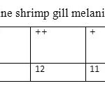 |
Table 1:The amount of marine shrimp gill melanization in the city of Bushehr |
 |
Table 2: The amount of marine shrimp gill hyperplasia in the city of Bushehr. |
In the marine shrimp fishing from the shores of Bushehr city rate of 25 per cent of them had the apparent symptoms like the black Gill. Clinical signs were observed in the shrimp include a black Gill, , blur of whole body, blur of hepatic pancreas, sometimes melanized corrosion on the cuticle surface, and darkening of the dorsal muscles was seen, some results in the table (3) has shown.
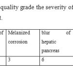 |
Table 3: The amount of quality grade the severity of the marine shrimp body color blur of Bushehr port. |
Gills in shrimp, as the only respiratory organ, and one of the main organs involved in homolangha by osmotic adjustment process of shrimp. And due to the delicate nature of this organ and exposure to substances, pathogens and toxins, gill is one of the most important organs in the shrimp disease detection. (Figure 1) shows the healthy Gill. In samples of infected tissues damages which includes the abundant influence of hemocyte on the lacuna space were observed and in some cases hemocyte nodes with melanized center(Figure 2), increase in the epithelium Lamela layer following, swelling of Gill, hypertrophy (Figure 3), the adhesion of lamela branches and ultimately necrosis of the lamella (Figure 4).
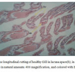 |
Figure 1: the longitudinal cutting of healthy Gill in lacuna space(b), in (a) we have hemocytes in natural amounts. 400 magnification, and colored with hematoxylin eosin. |
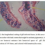 |
Figure 2: the longitudinal cutting of gill infected tissue, in this area we can see hemocytes nodes with dark centers that might be melanin pigments(a), in this area we can see hemocyt entrance into hemocoel space of gill filaments(b), magnification of 100 times, and colored with hematoxylin eosin. |
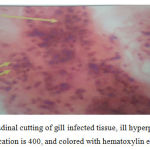 |
Figure 3: longitudinal cutting of gill infected tissue, ill hyperplasia seen in this picture, magnification is 400, and colored with hematoxylin eosin. |
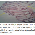 |
Figure 4: the longitudinal cutting of the gill infected tissue we can see Gill filaments adhesion together (a). In this part we see necrosis of first part of Gill filaments, (b) gill cell hypertrophy and melanization, magnification is 400, and colored with hematoxylin eosin. |
Macroscopic lesions observed in this study include darkening of the cuticle, cuticle localized corrosion, darkening of hepatopancreas, sometimes opaque of muscles and existence of melanin pigments in gills. (Figure5).
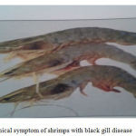 |
Figure 5: Clinical symptom of shrimps with black gill disease |
Discussion and Conclusions
Black Gill disease is a syndrome that is detectable by the accumulation of melanocytes in the gills. Black or brown pigments that founds in the gill are melanin, which is gathered in, inflammation of necrotic tissue. The black gills is an symptomatic event that usually followed by contact with toxic substances and
chemicals such as cadmium, copper, potassium permanganate, ozone, oil, acids, ammonia and nitric. Other caused factors are diseases like Vibrios, Mikoz, infection, Mycobacterium and ascorbic acid deficiency. We found that about 38% of shrimps are affected by black gill disease by studying 100 pieces of
Bushehr shrimps. Ghaednia and associates on a study on on 578 green tiger shrimp at Bushehr, recognize totally 719 fungal Koloni from external surfaces, gills, Hitopankras and nurturing pool water and in this study Fusarium species were included 78.7 percent (4).
Karonasagar and associates in India have been isolated close to 500 yeast species from the sea environment that a few of these fungi are pathogenic for shrimps (5).Ichikawa from Japan in 1968 has been reported an epidemic with 100% mortality in shrimps (6).
Kadusbatiin 1988 has been reported the epidemic in the Philippines with 100% mortality in shrimps (7).Laboratory infections in gills, while the Fusarium Solani has been injected to the fresh skin wounds, and has been lead to 100% mortality during the two weeks (8).Leitner states that Fusarium causes mortal in shrimp that have been weak and have stress due to the pool circumstances and adult shrimps are more sensitive(3).
Brooke et al. in 1994 states that the environment can have an important influence on health, growth and production of shrimps. And environmental change in a parameter can affect the other variable. And a variety of environmental variables related to the disease, including high temperature, low dissolved oxygen, sudden changes sudden changes in salinity, season of the year, and toxins such as nitrite, ammonia, heavy metals, crude oil, the death toll after inadvertently occur during sample preparation and also attacks bacteria and certain fungi in environmental conditions (9).
According to the mentioned research we can say that the black gills outbreaks of shrimp fished from the shores of Bushehr city participated in the unfavorable effect of environmental conditions caused by oil pollution, heavy metals, tumbling of shrimp ecosystem, air heat, high levels of suspended solids in the water and imposition that is resulted after death objectively during sample preparation is effected on the strengthening of this disease. In fact, we can say that a series of dropping population factors of marine shrimp at Bushehr like mentioned factors can be ground black gill disease.
Macroscopic lesions observed in this research include cuticle blur, localized corrosion of cuticle, blur hepatopancreas, sometimes darkening of the muscle and existence of melanin pigments in the Gill.
Brooke et al. in 1990 say that the harm that coused by Fusarium solani, is usually black or Melanized and are limited to the head, tail, and the muscles attached to the gills of the cuticle. Which it’s prevalence has not been reported in the internal organs (10). Synderman stated that after outbreak of Vibriozis Bacterial, black Gill disease happens, which causes darkening of cuticle, appendages and Gill (11).According to the above research, observations in the present study, can say are the same as mentioned researcher’s work.
The gills in fish and shellfish is as the only organ that receive oxygen from water and with the antnal gland considered the main organs of the shrimp that actively interfere in the process of osmotic adjustment of hemolangh and transfer of ions. Also according to the delicate nature of the structure of gill and being directly exposed to environmental pathogens and toxins,gill is one of the most important organs in detection of the shrimp diseases (12).
Microscopic lesions observed in this study consisted of Hemosits ample penetration at Lakuna space, creation of nodal Hemosits with melanin center, the long lamella epithelial layer and inflammation, adhesion coverslip to each other,
Nekrozfilament gill, gill hyperplasia and melanin gill.
Bian et.al.T 1981 at histopathology black gill disease examination reported melanin Kotikoli lesions, granulomatous nodules, hemosits accumulation and melanin gill and also gill hyperplasia that observed made in this study is like
researcher’s work(13). Inflammation changes at shrimp’s gills like Hemosits accumulation at Lakuna space, inflation, Felament spout and Felamini pitelium atop may be a simple defensive mechanism that cause surface reduction and area
expanse of gills that osmotic adjustment applied to perform well (12). In seems that inflammation changes at infected shrimp in this study are not a specialist response to stimulus environmental factors.
Conclusion
According to mentioned matters, it can be stated that the bad environmental conditions, certain bacteria and fungi attack and nutritional deficiencies can provide context to black gill disease by weaken the immune system of shrimp. Also, in the present study, lesions that resulted from this disease include darkening of shrimp’s body, darkening Hetopankras, melanincorrosion at Kotikol and black gill and from microscopic perspective, consisted of Hemosits nodal structure with
melanin center at gill, ample penetration at Lakuna space, gill inflation and Hyperplazi, adhesion and Lamella necrosis.
References
- Shakibazadeh SH, Brine shrimp nurture, second volume, first edition, Assistance of aquaculture nurture, Directorate General Education and Extension, 1379, 243.
- MajidiNasab A., Farmed shrimp disease, first edition Nourbakhsh publication, Tehran,1377, pp. 16-134
- Lighter, D.V. Epizootiology of two mycotic disease in the culture of penaeid shrimp. Proc.Int. colloq. Invertebr. Pathol 1, P:179-183.
- Ghaednia B., Fungal Flora green tiger shrimp farmed in Bushehr. Fisheries Research Institute of IranTehran, 1382
- Karunagar I, Umesha RK. Microbial diseases in shrimp aquaculture in marin microbiology, facets and apportunities, Edithed by Ramaiah N (National institute of oceanography, Goa, India). 2004; 165- 186.
- Ishikawa Y. Preliminary report on black gill disease of the kuruma prawn, penaeus japanicus. Fish pathol, 1998; 3: 34-38.
- Baticados MCL. Diseases of prawns in the Philippines. Asian aquacultures. 1988; 10 (1): 1-8.
- Bagrinao TU, Solis NB, Villaver WR and et al. Important fish and shrimp fry in Philippine coastal water: Identification, collection and handling. Aquaculture excepsion manual, 1986; 10: 41-52.
- Brock JA, Main KL. A guide to the cpmmon problems and disease of cultured penaeus vannamei. World aquaculture society, Baton rouge, Louisiana USA. 1994; 10-19.
- Brock JA, Lightner DV. Chapter3: Diseases of crustacea. Diseases of marine animals, 1990; 4: 245- 424.
- Sindermann, C.J. Principal Disease of marin Fish and shellfish, vol.2,2nd Academic press, New york.
- Bahavan PS, Graldine P. Hiastopathology of hepatopancreas and gills of the prawn macrobrachium malcolmsonii exposed to endosulfan. Aquatic toxicology, 2000; 50: 331-339.
- Bian BZ, Equsa S. Histopathology of black gill disease causes by fusarium solani (Martius) infection in the kuruma prawn, Pnaeus japonicus bate. J. fish disease. 1981; 4: 195-201.

