
Manuscript accepted on :March 10, 2015
Published online on: 08-12-2015
Plagiarism Check: Yes
Nader Saki1*, Soheila Nikakhlagh1, Rayehe Rasekhi2
1Associated Prof of otolaryngology, Hearing and Speech Research Center Ahvaz Jundishahpur University of Medical Sciences, Ahvaz, Iran 2Radiologist, Hearing and Speech Research Center Ahvaz Jundishahpur University of Medical Sciences, Ahvaz, Iran *Correspondence author E-mail: ahvaz.ent@gmail.com
Abstract
Giant mucocele of the frontal sinus is a rare pathology lesion presenting with massive cosmetically unacceptable bony swelling in the frontal region. Most of the patients complain primarily of the ophthalmic symptoms. However, the present case reminds us that frontal mucocele is one of the differential diagnoses for a subcutaneous mass on the forehead. Authors report an unusual case of frontal mucocele in a 64 years old Iranian male who presented with painless slowly progressive subcutaneous swelling of ten-year duration on the forehead. Endoscopic marsupialization, decompression of the mucocele, removal of the sinus mucosa and widening of ostium was performed. The case is discussed and the pertinent literature is reviewed.
Keywords
Giant Mucocele; Frontal Sinus; Subcutaneous Mass; Fess
Download this article as:| Copy the following to cite this article: Saki N, Nikakhlagh S, Rasekhi R. Frontal Mucocele Presenting as a Subcutaneous Giant Mass. Biomed Pharmacol J 2015;8(March Spl Edition) |
| Copy the following to cite this URL: Saki N, Nikakhlagh S, Rasekhi R. Frontal Mucocele Presenting as a Subcutaneous Giant Mass. Biomed Pharmacol J 2015;8(March Spl Edition). Available from: http://biomedpharmajournal.org/?p=2370> |
Introduction
A mucocele is an epithelial-lined, mucus-containing sac completely filling the sinus and capable of expansion. This is in contradistinction to a blocked sinus cavity which simply contains mucus. Mucocele mostly involve the frontal and ethmoid sinuses. It is possible that the frontoethmoidal area is more susceptible to mucocele formation due to the complexity of its drainage, as compare to the sphenoid and maxillary sinuses. It is gradually progressive lesions that grow by expanding, moulding, displacing and destroying the surrounding bony and soft tissues. It can erode through the surrounding bone and spread into intracranial as well as the intraorbital compartment. Frontal sinus mucoceles can also very rarely extend into the subcutaneous region and can present as a forehead mass (2, 3). The authors report an unusual case of frontal mucocele presenting with subcutaneous swelling and displacement of eye-globe.
Case Report
A 64-year-old Iranian male presented with progressive asymptomatic subcutaneous swelling of the left forehead and supraorbital region for ten years. The left eye globe was pushed downward and outward. He also had mild conjunctival chemosis. There was no ulceration or discharging sinus. He did not recall any preceding trauma. He had respiratory problem and cardiomyopathy. He had not undergone any paranasal sinus surgery. Clinical examination of the left eye revealed proptosis, periorbital swelling, ptosis, restricted elevation and abduction, diplopia on left gaze and giant subcutaneous mass. Definite bony defect was palpable around the superior and lateral aspects of the swelling. On neurological examination, her visual acuity was 4|10 in left eye. The rest of the neurological examination was within normal limits. Local examination revealed a massive swelling in left supraorbital and adjoining forehead region that was non-tender, non-pulsatile (Fig 1). Hematological and biochemical parameters were normal. CT scanning and magnetic resonance images revealed a sharply demarcated cystic mass from the subcutaneous area on the forehead expanding into the frontal sinus and orbital cavity. The mass had eroded posterior wall of frontal sinus without extension to the brain. The size of mass was 6.6 ˟ 8.5 cm. The tumor was diagnosed as a frontal mucocele and endoscopic approaches were performed. Local anesthesia and close monitoring of patient by cardiologist and anesthesiologist endoscopic approach to the frontal sinus was used. Endoscopic marsupialization, decompression of the mucocele, removal of the sinus mucosa and widening of ostium was performed. Histopathology showed a fibrous connective tissue cyst wall, partially lined by stratified squamous epithelium, with patchy chronic inflammation. These findings were consistent with the diagnosis of a mucocele. At one year postoperatively, the proptosis had completely resolved, but there was a residual inferior globe displacement.
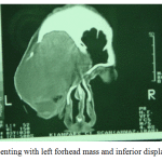 |
Figure 1: Case presenting with left forhead mass and inferior displacement of the globe |
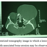 |
Figure 2: Coronal computerized tomography image in which a lesion of the left frontal sinus with associated bone erosion may be observed |
Discussion
The frontal sinus is on average 28 mm by 27 mm by 17 mm and is pyramidal shaped. The sinus is compartmentalized by the intrasinus septa which divides the sinus into halves. The frontal sinus has the most complex and variable drainage of any paranasal sinus. Each frontal sinus narrows down to an inferior margin designated the frontal ostium. The frontal ostium extends between the anterior and posterior walls of the frontal sinus, is demarcated by a variably shaped ridge of bone on the anterior wall of the sinus, and is oriented nearly perpendicular to the posterior wall of the sinus. Frontal sinus mucoceles are mucus-containing cysts caused by obstruction of the sinus orifice. The causes of blockage of paranasal ostia include inflammatory sinusitis, allergy, polyp and trauma and rarely past surgery. Surgery of the frontal sinus requires exquisite knowledge of the anatomy of the frontal sinus, the frontal sinus drainage pathway, and the surrounding structures such as the orbit, the brain, and the other paranasal sinuses. Frontal sinuses are the most common site for mucoceles and these can be frontoethmoidal or frontal only, but bilateral frontal involvement is rare (2, 3). These lesions are usually observed in the fourth to sixth decade of life. No gender preference has been observed. Gradual distension, thinning and erosion of the bony wall of the sinus are caused by progressive accumulation of mucoid material. The mucocele can extend into the orbit or intracranial compartment by eroding the bony limits and producing bony defects. Mucoceles can present with diminution of vision, visual field defect, diplopia, orbital swelling, retroorbital pain, displacement of eye globe, ptosis, and proptosis (4, 6). Very rarely these lesions can present as a forehead swelling. The definitive treatment of mucocele is surgery. Surgical treatment of mucoceles can be accomplished with a minimally invasive endoscopic procedure or craniotomy with craniofacial surgery. Endoscopic surgery has increased the safety and efficacy of intranasal marsupialization for the treatment of mucoceles in all paranasal sinuses (3). Some surgeons prefer the combined endoscopic and craniotomy approach for the treatment of frontal mucoceles (5).
Conclusion
Early recognition and management of mucoceles is of paramount importance. A high index of suspicion and appropriate radiological studies are necessary for the diagnosis of mucocele. Transnasal endoscopic evacuation is a viable surgical option to more invasive procedures.
References
- David Lewis MD, Nicolas Busaba MD, Surgical management; Sinusitis; Taylor and Francis Group, 2006; pp257-264
- Akiyama M, Inamoto N, Hashigucci K: Frontal mucocele presenting as a subcutaneous tumour on the forehead. Dermatology 199(3):263-264, 1999
- Tan CSH, Yong VK, Yip LW, Amrith S: An unusual presentation of a giant frontal mucocele manifesting with a subcutaneous forehead mass. Ann Acad Med Singapore 34:397-398, 2005
- Al›nda Cilt Alt› Kitlesi Olarak Belirti Veren Frontal Mukosel Olgusu:Frontal Mucocele Presenting with Forehead Subcutaneous Mass: An Unusual PresentationNadir bir Olgu Turkish Neurosurgery 2008, Vol: 18, No: 2, 200-203
- Daniels et. Al.; The frontal sinus drainage pathway and related structures, American Society of Neuroradiology 24, August 2003.
- Dale Rice MD; Steven Schaefer MD; Endoscopic Paranasal Sinus Surgery, Third Edition, Lippincott Williams and Wilkins, 2004; pp36-38
- Nikakhlagh S, Saki N*. Functional Endoscopic Sinus Surgery Sinus Mucocele. The Iranian Journal of Otorhinolaryngology. 2004;16(3)37; 36-41.(English abstract)
- Har-El G. Endoscopic management of 108 sinus mucoceles. Laryngoscope 2001;111:2131–4.

