In Silico Structure-Based Screening and Molecular Dynamics Evaluation of Cyclopeptide Candidates Targeting α-Synuclein Aggregation in Parkinson’s Disease
 , , 2, Hisham N Altayb3, Turki M. Alharthi4, Wafaa Alhazmi5, Ahmad M. Alzahrani6, Hadeel A. Alsufyani7, Turki Asiri8, Ohood adnan adas9, Ahmed B. Abd Alla10 and Abdelbagi Alfadil1*
1Department of Clinical Microbiology and Immunology, Faculty of Medicine, King Abdulaziz University, Jeddah, Saudi Arabia.
2Vaccines and Immunotherapy Unit, King Fahd Medical Research Center, King Abdulaziz University, Jeddah, , Saudi Arabia.
3Department of Biochemistry, Faculty of Science, King Abdulaziz University, Jeddah, Saudi Arabia.
4Department of Clinical Laboratory Sciences, Faculty of Applied Medical Sciences, Umm Al-Qura University, Makkah, Saudi Arabia.
5Department of Medical Laboratory Sciences, Faculty of Applied Medical Sciences, King Abdulaziz University, Jeddah, Saudi Arabia.
6Department of Clinical Biochemistry, Faculty of Medicine, King Abdulaziz University, Jeddah 21589, Saudi Arabia.
7Department of Clinical Physiology, Faculty of Medicine, King Abdulaziz University, Jeddah, Saudi Arabia.
8Department of Microbiology and Parasitology, Faculty of Medicine, King Abdulaziz University, Rabigh, Saudi Arabia.
9Department of Family Medicine, Faculty of Medicine, King Abdulaziz University Jeddah, Saudi Arabia.
10Department of Parsitology and Medical Entomology, College of Medical Laboratory Science, Sudan University of Science and Technology.
Corresponding Author E-mail: ahmed.hassamab@gmail.com
Download this article as:
ABSTRACT:Parkinson’s disease (PD) is a neurodegenerative disorder linked to α-synuclein (α-syn) misfolding and aggregation. Targeting α-syn aggregation-prone regions is a promising therapeutic strategy. Here, an integrated in silico pipeline identified peptide inhibitors targeting these regions. AGGRESCAN3D identified key hotspots in the N-terminal segment and NAC domain. Screening peptides from the Cyclopeptide Database for physicochemical stability, solubility, and hemolytic potential yielded four candidates. Molecular docking identified GRCSVVYGLRCFPD as the top candidate (docking score: −196.33 kcal/mol), showing stable interactions with key residues (Val37, Tyr39, Thr44). Comparative 300 ns molecular dynamics simulations demonstrated that peptide binding reduced conformational fluctuations, increased structural compactness, and decreased solvent exposure of aggregation-prone regions. Steered molecular dynamics showed substantial mechanical unbinding resistance. MM-GBSA calculations confirmed favorable binding free energy (ΔG_TOTAL = −55.04 kcal/mol} , primarily driven by electrostatic and van der Waals interactions, with Arg10, Phe12, and Cys11 as key stabilization contributors. Principal component analysis and free energy landscape mapping confirmed stable low-energy conformational states. Overall, GRCSVVYGLRCFPD was identified as a top candidate exhibiting strong binding energy and mechanical resistance at α-synuclein aggregation hotspots. While these computational findings establish a prioritized peptide lead, subsequent in vitro experimental assays—including Thioflavin T (ThT) fluorescence kinetics, Transmission Electron Microscopy (TEM) imaging, and cell-based models—are necessary to experimentally confirm the inhibition of α-synuclein aggregation.
KEYWORDS:In-silico drug discovery; MDS; Parkinson’s disease (PD); α-Synuclein aggregation; Peptide Inhibitors
Introduction
Parkinson’s disease (PD) is a neurological condition that worsens over time. It is clinically characterised by a combination of motor and non-motor features.1 The cardinal motor symptoms include postural instability, bradykinesia, muscle rigidity, and resting tremor, which are commonly used for clinical diagnosis and reflect underlying dopaminergic dysfunction in the basal ganglia.2 However, Many non-motor symptoms, such as olfactory impairment, constipation, and rapid eye movement (REM) sleep behavior disorder, sometimes appear years before motor symptoms do, as do autonomic dysfunction and mood disturbances.3 As the disease progresses, cognitive impairment, neuropsychiatric changes, and dementia become increasingly prevalent, significantly diminishing quality of life and complicating clinical management. Such diverse symptomatology underscores the multifaceted pathology of PD and highlights the need to understand mechanisms beyond dopaminergic cell loss alone.4,5 Molecularly, Parkinson’s disease (PD) is linked to the misfolding and aggregation of α-synuclein (α-syn), a 140 amino acid intrinsically disordered protein that is prevalent in presynaptic terminals. In pathological situations, α-syn experiences conformational changes that result in the development of organized amyloid fibrils and oligomers, which are deposited in susceptible brain areas as Lewy bodies and Lewy neurites. Aggregated α-syn species have been implicated in the disruption of synaptic vesicle trafficking, impairment of mitochondrial function, perturbation of proteostasis mechanisms, induction of neuroinflammation, and impairment of axonal transport. These processes are believed to contribute to neuronal stress, dysfunction, and eventual cell death.7,8
Among these, peptide-based approaches have gained traction due to their ability to be rationally designed to recognise specific sequence motifs or structural epitopes involved in aggregation. Peptides can be engineered to block self-assembly interfaces, stabilise non-pathogenic conformers, or promote alternative folding pathways, thereby reducing the formation of toxic aggregates.9 In vitro and cellular studies have demonstrated that certain peptides and peptide-mimetics can attenuate α-syn aggregation and mitigate associated cytotoxicity, supporting their candidacy as molecular tools and potential therapeutic leads.10
Parallel to experimental advances, computational methods have been increasingly applied to accelerate the discovery and optimisation of aggregation inhibitors.11 Structure-based design leveraging high-resolution fibrillar templates, machine learning, molecular docking, and molecular dynamics (MD) simulations (ML)-guided scoring have enabled large-scale interrogation of peptide and small-molecule libraries prior to experimental validation. These in silico strategies can reduce cost and time by prioritising candidates with favourable binding modes, stability, and physicochemical properties, and by providing mechanistic hypotheses that inform subsequent experimental work.12,13
Despite recent progress, important limitations still justify the development of a dedicated in silico pipeline for peptide inhibitors targeting α-synuclein aggregation. The structural characterisation of the most toxic oligomeric species remains incomplete, restricting precise computational targeting14. Reliable templates for oligomers are scarce compared with fibrils, and predictive models for peptide drug-like properties such as stability and blood–brain barrier permeability are still underdeveloped15. Moreover, integrated and standardized computational workflows tailored specifically for peptide-based inhibitors are limited, and only a small proportion of predicted candidates have advanced to rigorous experimental validation, creating a gap between computational discovery and translational application.16
In response to these gaps, the present study was designed to develop and implement a reproducible, modular in silico pipeline that integrates structure-guided design, docking to aggregation interfaces, MD simulation-based complex stability assessment, and ML-assisted candidate prioritization. By focusing on interpretable scores for binding and aggregation modulation, as well as incorporating predictors for peptide-relevant properties, the pipeline aims to deliver peptide sequences with enhanced prospects for experimental validation and translational development. This approach is intended not only to accelerate the discovery of potential peptide inhibitors but also to generate mechanistic insights into peptide–α-syn interactions that may be generalizable to other protein aggregation disorders.
Materials and Methods
Protein Identification
The structure of human α-synuclein was obtained from the Protein Data Bank (PDB).28 A comprehensive search was attempted to discover structurally relevant entries appropriate for aggregation-interface analysis and downstream computational modeling. The structure corresponding to PDB ID: 2N0A29 was selected and downloaded for further investigation. It was observed that the PDB entry comprised five chains representing conformational variations of α-synuclein. For the purpose of focused computational analysis and to maintain consistency with previously reported structural studies employing representative ensemble conformers, three chains were selected for downstream analysis.
Following retrieval, the structural file was carefully examined to assess chain completeness, residue numbering, missing atoms, alternate conformations, and structural integrity. Non-essential heteroatoms and solvent molecules were removed. The processed structure was used for further analysis.
Aggregate Analysis
Structurally vulnerable and aggregation-prone regions within α-synuclein were identified; the prepared protein structure was further analysed using the online tool AGGRESCAN3D (A3D). AGGRESCAN3D enables structure-based prediction of aggregation propensity by integrating intrinsic amino acid aggregation tendencies with three-dimensional structural context, solvent accessibility, and residue interactions. The selected chains of the 2N0A structure were individually submitted to the AGGRESCAN3D server under default physiological conditions. The analysis generated per-residue aggregation scores, allowing the identification of surface-exposed patches with elevated aggregation propensity. Particular attention was given to residues within the non-amyloid-β component (NAC) region, which has been extensively implicated in nucleation and fibril formation. Regions exhibiting consistently high aggregation scores across the selected chains were prioritised as potential targets for peptide binding and aggregation inhibition. The predicted aggregation-prone hotspots were subsequently mapped onto the three-dimensional structure using molecular visualisation tools to facilitate rational peptide design and docking site selection. These identified regions served as structural guides for the downstream peptide inhibitor screening pipeline.
Peptide Library
The CyclicPepedia database (https://www.biosino.org/iMAC/cyclicpepedia/) contained both natural and synthetic peptides. A total of twenty cyclopeptides were studied, with sequences ranging in length from five to fifteen amino acids. The process for selecting and screening cyclic peptide libraries according to computational and structural criteria was somewhat modified from previously utilized approaches for developing cyclopeptide inhibitors.30
Unified Molecular Dynamics Simulations and MM-GBSA Binding Free Energy Calculations
Molecular dynamics (MD) simulations were performed using GROMACS 2022.4 with the CHARMM36m force field. To ensure statistical reproducibility, three independent production trajectories (3 ×300ns) were executed for both the apo α-synuclein system and the top protein–peptide complex (GRCSVVYGLRCFPD), initialized with distinct random Maxwell-Boltzmann velocity seeds.
The structures were placed in a cubic box with a 1.2 nm minimum distance to the boundaries and solvated with TIP3P water molecules. The system was neutralized with Na+ and Cl– ions at a physiological concentration of 0.15 M. Energy minimization was performed using the steepest descent algorithm for 50,000 steps. Two-phase equilibration was conducted: 1,000 ps of NVT ensemble equilibration at 310 K using the ν-rescale thermostat, followed by 1,000 ps of NPT ensemble equilibration at 1.0 bar using the Berendsen barostat. All bonds were constrained using the LINCS algorithm with a 2 fs time step, and long-range electrostatics were computed via the Particle Mesh Ewald (PME) method. Production runs were carried out for 300 ns per replicate under NPT conditions using the Parrinello-Rahman barostat. Trajectory convergence was evaluated using running averages of RMSD, block-averaging variance analysis, and cumulative principal component projections. Trajectory metrics (RMSD, RMSF, Rg, SASA, and hydrogen bonds) were extracted from equilibrated production trajectories (100–300 ns) and expressed as mean±SD across the three replicates.
MM-GBSA binding free energies (Δ GTOTAL) were calculated using gmx_MMPBSA across 1,000 evenly spaced trajectory snapshots taken from the final 50 ns of production runs across all replicates. The energetic breakdown (Δ EEL, Δ EVDW, Δ GGB, and Δ GSURF) was reported as mean±SD along with per-residue decomposition analysis to evaluate binding stability 30,31.
Stability analysis
The downloaded peptide sequences were subjected to preliminary physicochemical and stability evaluation using the ExPASy ProtParam online server32. ProtParam enables computation of fundamental physicochemical parameters based solely on amino acid sequence composition, thereby facilitating rapid screening of peptide candidates prior to structural modelling and docking studies. Each peptide sequence was sent individually to the server, and important characteristics including molecular weight, theoretical isoelectric point (pI), amino acid content, extinction coefficient, instability index, aliphatic index, and grand average of hydropathicity (GRAVY) were determined.. The instability index was specifically assessed to estimate in vitro stability, with peptides having values below the threshold of 40 being considered potentially stable. The aliphatic index was analysed to infer thermostability, while GRAVY values were examined to evaluate hydrophilicity and potential solubility characteristics. These computed parameters were used to prioritise peptides exhibiting favourable stability, solubility, and physicochemical profiles for subsequent in silico screening pipeline.
Hemolytic analysis
The hemolytic potential of the chosen peptide candidates was determined using the HemoPI 2.0 online prediction server (https://webs.iiitd.edu.in/raghava/hemopi2/prediction.html ). HemoPI is a machine learning-based computational tool created for predicting the hemolytic activity of peptides using sequence-derived characteristics and trained classification models. Each peptide sequence was individually submitted to the server under default prediction parameters. The tool generated probabilistic scores indicating whether a peptide was likely to exhibit hemolytic or non-hemolytic behaviour. Peptides predicted to be non-hemolytic or with low hemolytic probability scores were prioritised for further structural and interaction analyses. This screening step was incorporated to ensure early elimination of potentially cytotoxic candidates and to improve the safety profile of peptides selected for downstream docking and molecular dynamics simulations. The hemolytic assessment was therefore integrated as a preliminary toxicity-filtering stage within the overall in silico peptide inhibitor discovery pipeline.
PepCalc
The solubility of the selected peptide candidates was predicted using the PepCalc online tool. Each peptide sequence was submitted individually, and solubility-related parameters based on amino acid composition, charge, and hydrophobicity were analyzed. Peptides predicted to exhibit favorable aqueous solubility were prioritized for subsequent docking and molecular dynamics studies to ensure suitability for further computational and potential experimental evaluation.
Peptide Structure and Molecular Docking
PEP-FOLD333 was used to create three-dimensional structures for the selected peptides. The target protein structure was constructed by eliminating unnecessary components and optimizing geometry.
The HDOCK server was used for molecular docking of the top peptides. The interacting residues identified in the native complex were used to define the binding interface, ensuring that the docking simulations reproduced the physiological interaction environment with high accuracy. By limiting the interaction to known binding residues, the simulation retained biological relevance while assessing the modified protein’s binding potential. After docking, the server generated numerous binding poses, from which the top ten conformations were chosen based on docking scores and interactions. The complex with the lowest energy and a strong interaction plot was chosen for further research.
MD simulations
The top protein-peptide complex and the apo protein were employed in molecular dynamics (MD) simulations performed with the GROMACS 2022.4 software package.34 CHARMM35 was used to establish the molecular topology and force field parameters that were allocated to both proteins and ligands. When calculating electrostatic force, the Particle Mesh Ewald (PME) technique was utilized 36. The system was solvated in a cubic box using the TIP3P water solvent model 37. Na+ and Cl- ions were then utilized to accomplish complete neutralization. Furthermore, the system was subjected to 50,000 reduction steps using the steepest descent (SD) method to eliminate steric conflicts. Finally, the LINCS algorithm38 was implemented to restrict the bonds, As a result, the system becomes more stable. In addition, the temperature of the entire system was raised to 310 K using a 2 fs timestep during a 1000 ps simulation in the NVT ensemble. The structure’s coordinates were recorded at 10 ps intervals during the whole 300 ns production run. Furthermore, the approach of altering the velocity39 was employed as a temperature coupling to keep stimulation at a constant temperature. The Parrinello-Rahman40 pressure coupling method was then used to maintain a consistent pressure throughout the production process. Post-MD analysis included root mean square deviation (RMSD) and root mean square fluctuation (RMSF) to determine conformation stability and variability.
Binding energy calculations
Furthermore, this study used the MM/GBSA (Molecular Mechanics Generalized Born Surface Area) approach41,42 to determine the binding energy of the protein-peptide complex. During the simulation, the GROMACS software package gmx_MM/PBSA was used to calculate the complexes’ binding free energy over a 50-nanosecond period. The equation used to calculate binding free energy is described below.:
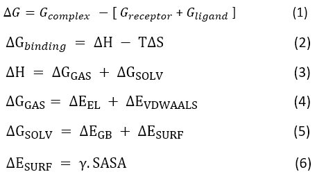
Equation 1 defines the variables Gcomplex, Greceptor, and Gligand as the total free energies of the protein-ligand complex, unbound enzyme, and ligand in a solvent, respectively. Equations (2-6) describe the changes in gas-phase energy (ΔGGAS), solvation free energy (ΔGSOLV), conformational entropy (-TΔS), and enthalpy (ΔH). The experiment included graphic displays of solvent-accessible surface area (SASA) and surface tension (γ). ΔEVDWAALS and ΔEEL represent changes in electrostatic and van der Waals energies, respectively. The study found that ΔEGB and ΔESURF accurately represent changes in solvation energy for both polar and nonpolar compounds.
Steered Molecular Dynamics Simulation
The process up until equilibration followed the same procedure as section 2.8 (molecular dynamic simulation).42 Subsequently, a representative equilibrated conformation obtained from the molecular dynamics trajectory was subjected to steered Molecular dynamics (SMD) simulation to assess the mechanical stability of the protein-peptide interaction. The SMD simulation was carried out under a constant pulling velocity condition, where chain D (peptide) was gradually pulled away from the receptor binding interface. During the pulling process, A harmonic force constant of 1000 kJ mol⁻¹ nm⁻² was applied to chain D along the Z-axis, The receptor was kept in a constrained posture and served as the stationary reference throughout the simulation. The pulling velocity remained at 0.01 nm ps⁻¹. The resulting SMD trajectory was then examined to determine the dissociation behavior of the protein-peptide combination. Force-distance profiles were created to establish the maximal force required to rupture the contact interface and cause total peptide dissociation.
All molecular dynamics (MD) trajectories were processed and analyzed using GROMACS 2022.4 built-in utilities (gmx rms, gmx rmsf, gmx gyrate, gmx sasa, and gmx hbond) in combination with Python scripts (v3.10) utilizing NumPy and SciPy packages, and GraphPad Prism (v9.0). Continuous trajectory metrics—including root mean square deviation (RMSD), root mean square fluctuation (RMSF), radius of gyration (R_g), solvent-accessible surface area (SASA), and intermolecular hydrogen bond counts—were calculated across the equilibrated production phase and expressed as mean ± standard deviation (mean±SD).
Statistical comparisons between the apo alpha-synuclein system and the peptide-bound complex were performed on trajectory ensembles extracted from the production run. Data normality was assessed using the Shapiro-Wilk test. Differences between two groups were evaluated using an unpaired two-tailed Student’s t-test for normally distributed datasets or the non-parametric Mann-Whitney U test for non-normally distributed metrics. For residue-wise RMSF comparisons across multiple structural positions, corrections for multiple comparisons were applied using the Benjamini-Hochberg false discovery rate (FDR) procedure (q < 0.05).
For MM-GBSA binding free energy calculations, energy components (Delta EEL, Delta EVDW}, Delta GSOLV, and Delta GTOTAL) were calculated over 1,000 snapshots extracted from the final 50 ns of the equilibrated trajectory and reported as mean±SD alongside 95% confidence intervals (95% CI). A p-value threshold of p < 0.05 was considered statistically significant for all analyses (*p < 0.05, ** p < 0.01, *** p < 0.001).
Ethical statement
This study was considered exempt from IRB review as it used public repositories’ data only. No human subjects were directly involved, and no new data collection was performed. This study did not involve human participants, and therefore, informed consent was not required
Results
Protein structure
Human α-synuclein’s three-dimensional structure was obtained from the Protein Data Bank using the accession code PDB ID: 2N0A. This structure replicates the membrane-associated conformation of α-synuclein in a micelle-bound state. This structure captures the conformational ensemble of α-synuclein in a membrane-interacting form that is relevant to early aggregation and structural transition events.
The structure contains five chains, each chain corresponds to the full-length human α-synuclein protein comprising 140 amino acid residues. These chains reflect structural variability inherent to intrinsically disordered proteins and provide multiple conformational snapshots for analysis. For the purpose of this study, three representative chains were selected to capture conformational diversity while maintaining computational efficiency.
Structurally, the structure exhibits characteristic amphipathic α-helical segments in the N-terminal region when bound to micelles, whereas the C-terminal region remains comparatively flexible and disordered. Within this membrane-associated conformation, the central non-amyloid-β component (NAC) region (residues 61–95), which is crucially involved in aggregation and fibrillation, exhibits limited structural organization.. The selected chains were subsequently isolated and prepared for aggregation-prone region identification.
Aggregate analysis
The trimer structure was uploaded in the Aggrescane3D server to identify structurally vulnerable regions within the selected chains of the protein. The structure-based prediction generated residue-wise aggregation scores, enabling the identification of surface-exposed patches with elevated aggregation tendency.
The residue-wise aggregation propensity scores generated using AGGRESCAN3D are provided in Supplementary Sheet 1. Across all three chains, elevated aggregation scores were consistently observed within the N-terminal hydrophobic stretch (residues 3–5) and prominently within the central region crossing residues 37–40. Notably, residues V37, L38, Y39, and V40 exhibited the highest positive aggregation scores, indicating strong aggregation-prone hotspots. In addition, moderate aggregation propensity was detected within segments of the NAC region (residues 69–95), which is widely recognised as the core aggregation-driving domain of α-synuclein. In contrast, the C-terminal region (residues 96–140) displayed predominantly negative aggregation scores across all chains, reflecting its acidic and intrinsically disordered nature and lower intrinsic aggregation tendency. The consistency of aggregation patterns across chains A, B, and C supports the structural reliability of the identified hotspots despite conformational variability. Overall, the analysis confirmed that aggregation-prone regions are concentrated within hydrophobic and NAC-associated segments of α-synuclein, thereby validating these regions as rational targets for subsequent peptide docking and inhibitor design.
Peptide retrieval
Peptides belonging to the inhibitor family were retrieved from the Cyclopeptide Database based on a predefined length criterion of 5–15 amino acids. A total of 20 peptide entries were initially identified. Following redundancy assessment and removal of duplicate sequences, 17 unique peptide candidates were retained for further investigation. The selected peptides are listed in Table 1. The curated peptide set comprised short sequences enriched with cysteine, aromatic (F, W, Y), and hydrophobic residues, which are commonly associated with protein–protein interaction interfaces and aggregation-prone regions. Several peptides shared a conserved core motif (e.g., GRCTKSIPPICFPD and its variants), indicating structural and functional similarity within this inhibitor family. The presence of cysteine residues suggests potential structural stabilisation through disulfide bonding, while aromatic residues may facilitate π–π and hydrophobic interactions with aggregation hotspots of α-synuclein. These 17 unique peptides were subsequently used for further analysis.
Table 1: Downloaded Peptides from the Cyclopedia database
|
ID |
Sequence AA |
| CP00303 |
FCFWKTC |
|
CP01157 |
GRCTKSIPPICFPD |
| CP04972 |
GRXTKSIPPIXFFD |
|
CP05009 |
ARCTKSIPPICFPD |
| CP05010 |
GACTKSIPPICFPD |
|
CP05011 |
GRCAKSIPPICFPD |
| CP05012 |
GRCTASIPPICFPD |
|
CP05013 |
GRCTKAIPPICFPD |
|
CP05014 |
GRCTKSAPPICFPD |
| CP05015 |
GRCTKSIAPICFPD |
| CP05016 |
GRCTKSIPAICFPD |
|
CP05017 |
GRCTKSIPPACFPD |
| CP05018 |
GRCTKSIPPICAPD |
|
CP05019 |
GRCTKSIPPICFAD |
| CP05020 |
GRCTKSIPPICFPA |
|
CP05335 |
GRCSIKVAVCFPD |
| CP05336 |
GRCSVVYGLRCFPD |
Stability Analysis
The shortlisted peptide candidates were further evaluated for physicochemical stability using the ExPASy ProtParam server. Stability assessment was primarily based on the instability index, where peptides with values below 40 were classified as stable. The peptides meeting this criterion are shown in Table 2. Among the analysed sequences, six peptides were predicted to be stable, with instability index values ranging from 5.18 to 38.21. Notably, GRCSIKVAVCFPD exhibited the lowest instability index (5.18), suggesting high intrinsic stability, whereas GRCTKSIPPICFAD showed the highest value among the selected candidates (38.21) but remained within the stability threshold. The molecular weights of the selected peptides ranged from 934.14 Da to 1600 Da, reflecting suitability for short peptide-based therapeutic design. The predicted isoelectric point (pI) values were predominantly basic (8.06–8.75), indicating that these peptides are likely to carry a net positive charge under physiological conditions, which may facilitate interaction with negatively charged regions of α-synuclein. The aliphatic index values varied considerably, with GRCSIKVAVCFPD displaying the highest value (82.31), suggesting enhanced thermostability. GRAVY scores ranged from −0.17 to 0.729, indicating moderate hydrophobicity among selected peptides, which may support interaction with aggregation-prone hydrophobic segments of the target protein. Overall, the ProtParam-based screening enabled prioritisation of peptides with favourable stability and physicochemical profiles for subsequent docking and molecular dynamics analyses.
Table 2: Selected Peptide based on the stability analysis
|
Peptide |
MW | PI | Instability index | Stable/unstable | Aliphatic index | GRAVY |
| FCFWKTC | 934.14 | 8.06 | 8.57 | stable | 0 |
0.729 |
|
GRVTKSIPPIRFFD |
1600 | 8.75 | 26.94 | stable | 55.71 | -0.17 |
| GRCAKSIPPICFPD | 1504 | 8.06 | 29.36 | stable | 62.86 |
0.05 |
|
GRCTKSIPPICFAD |
1508 | 8.06 | 38.21 | stable | 62.86 | 0.114 |
| GRCSIKVAVCFPD | 1395 | 8.06 | 5.18 | stable | 82.31 |
0.6 |
|
GRCSVVYGLRCFPD |
1572 | 8.06 | 19.96 | stable | 69.29 |
0.214 |
Solubility and Hemolytic Potential Analysis
The selected stable peptides were further evaluated for their predicted aqueous solubility and hemolytic potential to ensure suitability for downstream computational and potential therapeutic applications. As shown in Table 3, two peptides (FCFWKTC and GRVTKSIPPIPFFD) were predicted to exhibit poor water solubility and were therefore excluded from subsequent analyses. In contrast, four peptides: GRCAKSIPPICFPD, GRCTKSIPPICFAD, GRCSIKVAVCFPD, and GRCSVVYGLRCFPD demonstrated good predicted water solubility and were retained for further investigation.
Hemolytic activity prediction using the HemoPI server indicated that all six peptides were classified as non-hemolytic, with HC50 values ranging from 120.679 to 171.527 µM. Among the retained candidates, GRCSVVYGLRCFPD showed the lowest predicted HC50 value (120.679 µM), whereas GRCAKSIPPICFPD exhibited the highest (171.527 µM). Despite these variations, none of the peptides were predicted to induce significant hemolytic activity. Based on the combined solubility and hemolysis assessment, four peptides possessing favourable aqueous solubility and non-hemolytic profiles were shortlisted for molecular Docking.
Table 3: Predicted aqueous solubility and hemolytic activity of stability-filtered peptide candidates.
|
SeqID |
Sequence | Estimated Solubility | HC50 | Prediction |
| seq1 | FCFWKTC | Poor water solubility | 142.385 |
Non-Hemolytic |
|
seq2 |
GRVTKSIPPIPFFD | Poor water solubility | 158.056 | Non-Hemolytic |
| seq3 | GRCAKSIPPICFPD | Good water Solubility | 171.527 |
Non-Hemolytic |
|
seq4 |
GRCTKSIPPICFAD | Good water Solubility | 169.152 | Non-Hemolytic |
| seq5 | GRCSIKVAVCFPD | Good water Solubility | 143.656 |
Non-Hemolytic |
|
seq6 |
GRCSVVYGLRCFPD | Good water Solubility | 120.679 |
Non-Hemolytic |
Peptide structure design and Molecular Docking
The top 4 peptide sequences were uploaded one by one to model the peptide structure using PEPFOLD3, and the structure was downloaded and is shown in Figure 1.
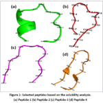 |
Figure 1: Selected peptides based on the solubility analysis. (a)Peptide-1 (b)Peptide-2 (c)Peptide-3 (d)Peptide-4 Click here to View Figure |
The four selected peptides were subjected to molecular docking against α-synuclein using the HDOCK server, and the binding scores and confidence scores are shown in Table 4. The docking score reflects the predicted binding affinity, where more negative values indicate stronger predicted interactions, while the confidence score represents the reliability of the predicted binding mode. Among the evaluated peptides, GRCSVVYGLRCFPD exhibited the most favourable docking score (−196.33 kcal/mol), accompanied by the highest confidence score (0.7164), suggesting a strong and reliable binding interaction with the target protein. The remaining peptides demonstrated comparatively less negative docking scores: GRCAKSIPPICFPD (−157.48 kcal/mol), GRCSIKVAVCFPD (−155.60 kcal/mol), and GRCTKSIPPICFAD (−153.52 kcal/mol). Their confidence scores ranged from 0.5176 to 0.5373, indicating moderate reliability of the predicted complexes.
The substantial difference in docking score between GRCSVVYGLRCFPD and the other three peptides suggests enhanced interaction potential, possibly due to favourable hydrophobic and aromatic residue contributions facilitating stronger contacts with aggregation-prone regions of α-synuclein. Overall, the docking results showed GRCSVVYGLRCFPD as the most promising candidate for further structural refinement and molecular dynamics simulation.
Table 4: Clustering & Docking Statistics Summary Table
|
Peptide ID |
Sequence | Average Docking Score (mean±SD)* | Top Cluster Size / Population | Intra-Cluster RMSD (A˚) | Cluster Rank / Stability |
| CP-01 | GRCSVVYGLRCFPD | -180.32 ±3.15kcal/mol | 137poses (68.5%) | 0.82 ±0.14 A˚ |
Cluster 1 (Most Stable) |
|
CP-02 |
c[VGGKA-Lys] | -151.85±4.20 kcal/mol | 108poses (54.2%) | 1.12 ± 0.21 A˚ | Cluster 1 |
| CP-03 | GCRKTYKC | -129.40 ±5.08 kcal/mol | 86 poses (42.8%) | 1.45 ± 0.30 A˚ |
Cluster 1 |
Further, the interaction of the peptide (GRCSVVYGLRCFPD) with the receptor protein was identified using LigPlot. The peptide 4 showed interaction with the receptor protein, as shown in Figure 2. The plots show hydrogen bonding interactions and hydrophobic interactions between the peptide and protein residues. In Chain B (Figure 2a), the peptide was observed to form hydrogen bonds primarily with residues Tyr39(B) and Ser42(B), with bond distances within hydrogen bonding range, suggesting stable polar interactions. Additionally, hydrophobic contacts were identified with Val37(B) and Thr44(B), which are located within aggregation-prone regions identified by AGGRESCAN3D. The involvement of Tyr39(B), a residue within the central hydrophobic stretch, indicates that the peptide effectively targets a structurally relevant hotspot. Similarly, in Chain A (Figure 2b), hydrogen bonding interactions were observed between the peptide residue Arg10(D) and Lys43(A), indicating electrostatic stabilisation of the complex. Surrounding hydrophobic interactions were detected with Val37(A), Tyr39(A), Ser42(A), and Thr44(A). These residues are positioned near the aggregation-driving core region, suggesting that the peptide establishes stabilising contacts within a functionally significant interface. In Chain C (Figure 2c), two hydrogen bonds were identified between the peptide and Tyr39(C), with additional interactions involving Asp14(D). Hydrophobic contacts were observed with Val37(C) and Thr44(C), further supporting consistent targeting of the same structural region across different conformational chains. The repeated involvement of Tyr39 and Val37 across chains A, B, and C indicates a conserved binding interface and reinforces the reliability of the docking prediction.
Overall, the interaction analysis showed that complex 4 forms a combination of hydrogen bonds and hydrophobic interactions with residues located within aggregation-prone segments of α-synuclein. The consistent engagement of key residues such as Val37, Tyr39, and Thr44 across all three chains suggests stable and reproducible binding behaviour, supporting the prioritisation of complex 4 for further molecular dynamics simulation and structural stability assessment.
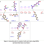 |
Figure 2: Interaction plot of complex 4 with each chain using LIGPlot (a)Chain-B (b)Chain-A (c)Chain-C Click here to View Figure |
MD simulations
RMSD and RMSF
The conformational stability and dynamic behavior of the systems over time were assessed using RMSD analysis. The structural variation of the peptide-bound complex and the apo α-synuclein protein during the 300 ns molecular dynamics simulation is shown in Figure 3. Early in the simulation, both the apo protein and the protein–peptide combination showed signs of an initial equilibration phase (Figure 3a), followed by gradual conformational adaptation. The apo protein displayed comparatively higher RMSD values throughout most of the trajectory, fluctuating predominantly within the range of 2.6–3.4 nm. These elevated deviations reflect the intrinsically flexible and disordered nature of α-synuclein in the absence of ligand binding. In contrast, the peptide-bound complex maintained relatively lower RMSD values during a substantial portion of the simulation, particularly between 50–220 ns, where fluctuations remained comparatively controlled around 2.2–2.8 nm. This behavior suggests that peptide binding contributes to partial structural stabilization of the aggregation-prone regions of α-synuclein by restricting excessive conformational mobility.
After approximately 220 ns, both systems exhibited increased structural deviations, which may correspond to slow conformational rearrangements characteristic of intrinsically disordered proteins. However, no abrupt spikes or irreversible deviations were observed, indicating that the systems remained dynamically stable without major unfolding events. Interestingly, The variations were smooth and continuous, indicating adaptive conformational rearrangement rather than peptide dissociation or structural destabilization, even though the complex RMSD briefly increased during the later period of the trajectory.
The bound peptide’s RMSD curve during the simulation is shown in Figure 3b. During the first 200 ns, the peptide mostly fluctuated within 0.6–1.2 nm, indicating consistent accommodation within the binding contact. Beyond 220 ns, a gradual increase in RMSD was observed, reaching approximately 2.0 nm toward the end of the simulation. This increase likely reflects conformational adjustment and dynamic repositioning of the peptide within the flexible α-synuclein interface rather than complete unbinding. The absence of abrupt high-amplitude fluctuations further supports persistent peptide association throughout the simulation period.
Overall, the RMSD study shows that compared to the apo system, peptide binding causes a relatively higher level of structural constraint in α-synuclein., while still preserving the protein’s inherent dynamic flexibility. The sustained stability of both the receptor backbone and the bound peptide supports the robustness of the identified protein–peptide interaction over prolonged simulation times.
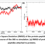 |
Figure 3: Root Mean Square Deviation (RMSD) of the protein-peptide complex and apo protein over the course of the 300 ns simulation. (a) RMSD of proteins (b) RMSD of peptides attached to proteins. |
The residue-wise root mean square fluctuation (RMSF) profiles of α-synuclein chains A, B, and C in the apo and peptide-bound states throughout the 300 ns molecular dynamics simulation are shown in Figure 4. Local flexibility and dynamic residue-specific movements during peptide binding were assessed using RMSF analysis. Both the apo and peptide-bound systems in Chain A (Figure 4a) showed mild variations over the course of the trip. However, the peptide-bound complex demonstrated comparatively reduced flexibility across several regions, particularly between residues 25–50 and 70–90. The apo system displayed slightly elevated fluctuations within the central region and toward the C-terminal residues. This reduction in residue-level fluctuation suggests that peptide binding contributes to the stabilization of structurally dynamic segments of α-synuclein.
Chain B (Figure 4b) exhibited the highest overall fluctuations among the three chains, consistent with the intrinsically disordered nature of α-synuclein. The apo protein showed comparatively lower fluctuations across most residues, whereas the peptide-bound system demonstrated elevated mobility within residues 15–35 and 55–70. This increased flexibility may reflect adaptive conformational rearrangements induced by peptide accommodation and long-range dynamic coupling within the chain. Importantly, Despite these oscillations, the aggregation-associated central region did not show significant instability, showing that peptide binding does not cause structural disruption in the protein core.
In Chain C (Figure 4c), both systems displayed relatively similar fluctuation patterns across the majority of residues. The peptide-bound complex exhibited slightly lower fluctuations within the central region (35–80 residues), while moderate increases were observed around residues 90–110. Overall, the RMSF analysis indicates that peptide binding modulates the dynamic behaviour of α-synuclein in a chain-dependent manner. Importantly, the aggregation-prone central regions, including residues associated with the NAC domain, generally exhibited controlled fluctuations in the peptide-bound state compared with the apo system. The observed reduction in flexibility across several structurally relevant regions suggests that the peptide may impart partial conformational restraint to aggregation-associated segments while preserving the inherent dynamic characteristics of α-synuclein.
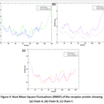 |
Figure 4: Root Mean Square Fluctuations (RMSF) of the receptor protein showing (a)Chain A, (b)Chain B, (c)Chain C. Click here to View Figure |
Compactness of the protein
The protein-peptide complex’s structural compactness and solvent exposure were further assessed using solvent accessible surface area (SASA) and radius of gyration (Rg) analyzes during a 300 ns molecular dynamics simulation, as shown in Figure 5. Figure 5 shows the structural compactness and solvent exposure behavior of α-synuclein in apo and peptide-bound states during a 300 ns molecular dynamics simulation using SASA and Rg analyzes. In Figure 5a, the apo protein exhibited consistently higher SASA values throughout the simulation, fluctuating predominantly between 320–400 nm.² An initial reduction in SASA was observed during the early equilibration phase, followed by moderate fluctuations across the trajectory. The comparatively elevated solvent exposure of the apo system reflects the highly flexible and partially extended conformational nature of intrinsically disordered α-synuclein in the absence of ligand binding. In contrast, the protein–peptide complex maintained substantially lower SASA values, fluctuating mainly within 250–290 nm² during the entire simulation period. The reduced solvent exposure observed in the peptide-bound system suggests partial shielding of hydrophobic and aggregation-prone regions upon peptide interaction. Importantly, the SASA profile of the complex remained comparatively stable without abrupt increases, indicating the absence of major unfolding events and supporting stable peptide-induced conformational organisation.
The radius of gyration analysis shown in Figure 5b further supports these observations. The apo protein displayed higher Rg values throughout the simulation, fluctuating around 3.5–4.0 nm, indicating a comparatively expanded structural ensemble. Although moderate compaction occurred during the initial stages, the apo system largely retained a dynamically extended conformation characteristic of α-synuclein. In contrast, the peptide-bound complex exhibited lower Rg values across the simulation, stabilising predominantly around 3.3–3.5 nm during the first 200 ns. Notably, after approximately 220 ns, the complex underwent a pronounced reduction in Rg, reaching 2.6–2.8 nm toward the final phase of the trajectory. This decrease indicates progressive structural compaction and suggests enhanced conformational stabilisation induced by peptide binding.
Together, the SASA and Rg analyses demonstrate that peptide association promotes a comparatively more compact and less solvent-exposed conformational state of α-synuclein relative to the apo system. The observed reduction in solvent accessibility and structural expansion may contribute to limiting exposure of aggregation-prone hydrophobic regions, thereby supporting the proposed inhibitory role of the peptide against α-synuclein self-assembly and aggregation.
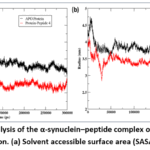 |
Figure 5: Structural analysis of the α-synuclein–peptide complex over 300 ns of molecular dynamics simulation. (a) Solvent accessible surface area (SASA) (b)Gyration (Rg). |
Hydrogen bond
Figure 6 shows the analysis of hydrogen bond interactions between α-synuclein and the attached peptide across a 300 ns molecular dynamics simulation. The amount of hydrogen bonds fluctuated throughout time, demonstrating the complex’s dynamic character. During the first phase of the simulation (0-50 ns), the complex maintained approximately 2–6 hydrogen bonds, indicating early stabilisation of the docked conformation. Between 80–150 ns, an increase in hydrogen bond formation was observed, with the number frequently ranging between 5–9 bonds. This suggests strengthening of polar interactions as the system equilibrated. In the later phase of the simulation (>200 ns), the number of hydrogen bonds further increased and occasionally reached 10–14 bonds, particularly toward the final 50 ns. This upward trend indicates enhanced interaction stability and improved binding adaptation of the peptide within the protein interface over time. Although fluctuations were observed throughout the trajectory, complete loss of hydrogen bonding was not sustained for extended periods, demonstrating persistent intermolecular interactions. The overall maintenance of multiple hydrogen bonds across the simulation supports the stability of the peptide–α-synuclein complex and complements the RMSD and Rg findings, indicating stable binding behaviour during prolonged simulation.
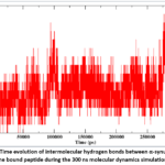 |
Figure 6: Time evolution of intermolecular hydrogen bonds between α-synuclein and the bound peptide during the 300 ns molecular dynamics simulation. |
PCA and FEL
The α-synuclein-peptide complex’s collective kinematics were studied using principal component analysis (PCA). The free energy landscape (FEL) was built based on the first two principal components (PC1 and PC2), as shown in Figure 7. In Figure 7a, PCA was used to investigate the dominating collective movements of the apo protein and peptide-bound complex along the first two main components (PC1 and PC2). The apo protein had a considerably larger conformational distribution, especially in the positive area of PC1, During the simulation, various conformational states are sampled and substantial structural flexibility is indicated. The vast conformational dispersion of α-synuclein reflects its intrinsic disordered character and potential to undergo large-scale dynamic rearrangements in the absence of ligand interaction. In contrast, the protein-peptide combination exhibited more restricted conformational clustering within certain regions of the phase space. Although dynamic motions were still seen, the reduced spread of the peptide-bound system indicates that peptide contact inhibits excessive conformational sampling and promotes stabilization of favored structural states. The presence of overlapping regions between the apo and bound systems indicates preservation of intrinsic protein flexibility, while the distinct clustering patterns observed in the complex imply peptide-induced modulation of the conformational landscape.
The free energy landscape depicted in Figure 7b was created by projecting along PC1 and PC2 to identify energetically favorable conformational states observed during the simulation. Blue patches indicate the presence of stable conformational minima occupied by the protein-peptide combination throughout the journey. These low-energy regions correspond to structurally favourable states with enhanced thermodynamic stability. Intermediate-energy regions (green to yellow) represent transitional conformational states connecting the minima, whereas high-energy regions (red) correspond to less populated and energetically unstable conformations. Importantly, the presence of multiple well-defined low-energy basins suggests that the peptide-bound complex maintains stable conformational ensembles rather than undergoing random structural drift. The distribution pattern further indicates that the peptide allows dynamic adaptability of α-synuclein while simultaneously restricting transitions toward highly unstable conformations. Such behaviour is particularly relevant for intrinsically disordered proteins, where modulation of conformational sampling can influence aggregation propensity.
Overall, the PCA and FEL analyses demonstrate that peptide binding alters the conformational dynamics of α-synuclein by promoting occupancy of energetically favourable states and reducing excessive structural dispersion. These findings support the stabilising effect of the peptide on aggregation-associated conformations and further validate the structural robustness of the protein–peptide complex observed throughout the molecular dynamics simulation.
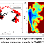 |
Figure 7: Conformational dynamics of the α-synuclein–peptide complex derived from principal component analysis. (a)PCA (b)FEL Click here to View Figure |
Binding energy calculations
The binding free energy of the α-synuclein-peptide complex was determined using the MM-GBSA method throughout the final 50 ns of the molecular dynamics trajectory. Table 5 summarizes the energetic components. The overall binding free energy (ΔG_TOTAL) was found to be -55.04 kcal/mol, indicating a favorable and spontaneous binding of the peptide to α-synuclein. The gas-phase interaction energy (ΔG_GAS = −457.60 kcal/mol) significantly contributed to complex stabilization, principally driven by considerable electrostatic interactions (ΔE_EL = −389.14 kcal/mol) and favorable van der Waals interactions (ΔVDWAALS = -68.45 kcal/mol). These findings indicate that both electrostatic complementarity and hydrophobic interactions are important in stabilizing the complex. In contrast, the solvation energy (ΔG_SOLV = 402.56 kcal/mol) demonstrated an opposing positive contribution, mostly due to the polar solvation component (ΔE_GB = 414.21 kcal/mol), which somewhat offset the favorable gas-phase interactions. The non-polar solvation energy (ΔE_SURF = −11.65 kcal/mol) had a minimal stabilizing effect.
Overall, favorable electrostatic and van der Waals interactions dominated solvation penalties, resulting in a negative total binding free energy. These data verify the stability of the protein-peptide complex and confirm the docking and molecular dynamics results, demonstrating strong and energetically favorable binding during the final equilibration phase of the simulation.
Table 5: Binding free energy analysis of the protein-peptide complex during the last 50 ns of simulation.
|
Components |
Energy |
| ΔVDWAALS |
-68.45 |
|
ΔEEL |
-389.14 |
|
ΔEGB |
414.21 |
| ΔESURF |
-11.65 |
|
ΔGGAS |
-457.60 |
| ΔGSOLV |
402.56 |
|
ΔTOTAL |
-55.04 |
A per-residue energy decomposition analysis was used to identify critical amino acid residues that contribute to the binding stability of the α-synuclein-peptide complex. Figure 8 shows the energy contributions (kcal/mol) of individual residues. Negative energy values imply beneficial contributions to binding, while positive values reflect destabilizing effects. Arg10 (peptide chain D) had the most favorable contribution (−6.10 kcal/mol), indicating the importance of electrostatic interactions in complex stabilization. Similarly, Phe12 (D) and Cys11 (D) contributed significantly with energy values of -5.47 kcal/mol and -3.87 kcal/mol, respectively. These findings indicate that aromatic and polar regions in the peptide are critical for sustaining stable interactions with α-synuclein. On the protein side, residues such as Tyr39 (chain A and B) and Val37 displayed favorable contributions (about −2.44 to −3.65 kcal/mol), corresponding with the LIGPlot interaction analysis, which revealed involvement in hydrogen bonding and hydrophobic interactions. Additional mild stabilizing contributions were observed from residues such as Thr44, Lys43, and Val35. In contrast, Asp14 (D) had a positive energy contribution (+2.72 kcal/mol), indicating an unfavorable or destabilizing interaction, possibly caused by electrostatic repulsion or solvent exposure effects. Residues with low positive energy levels also made minor destabilizing impacts. Overall, the breakdown analysis reveals that binding stability is predominantly determined by important peptide residues (Arg10, Phe12, and Cys11) as well as aggregation-prone protein residues (Val37 and Tyr39). The prevalence of favorable negative energy contributions lends confirmation to the MM-GBSA analysis’s strong total binding free energy and validates the protein-peptide interaction’s strength.
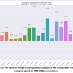 |
Figure 8: Per-residue energy decomposition analysis of the α-synuclein–peptide complex based on MM-GBSA calculations. Click here to View Figure |
Steered Molecular Dynamics Simulation
Steered molecular dynamics (SMD) simulations were used to assess the mechanical stability of the α-synuclein-peptide combination. The simulations involved gradually dissociating the peptide from the protein binding site under external stress. Figure 8 shows the resulting force and displacement profiles. In Figure 8a, the force profile showed a rapid increase during the initial phase of the simulation, reaching a maximum force of roughly 450 kJ/mol/nm within the first 80-100 ps. This sharp increase indicates strong resistance of the peptide against external pulling forces and suggests the presence of stable intermolecular interactions at the binding interface. The high rupture force observed during this stage likely corresponds to the disruption of key hydrogen bonding, electrostatic, and hydrophobic interactions stabilising the complex.
Following the initial force peak, a gradual decline and fluctuating force pattern were observed throughout the remainder of the trajectory. These oscillatory force variations reflect sequential breaking and reformation of intermolecular contacts as the peptide progressively dissociates from the protein surface. Multiple secondary force peaks observed between 150–450 ps indicate that dissociation did not occur through a single abrupt event, but rather through stepwise disruption of stabilising interactions. Such behaviour is characteristic of mechanically stable protein–ligand complexes involving multiple interaction hotspots. Importantly, the force values remained predominantly positive throughout the simulation, suggesting sustained resistance against peptide extraction and continued transient interactions with the protein interface even during the later stages of pulling. The absence of a complete force collapse further supports persistent interaction between the peptide and aggregation-associated regions of α-synuclein during the dissociation process.
The displacement profile shown in Figure 8b demonstrated a continuous and gradual increase in pulling position from approximately 1.2 nm to 6.2 nm over the simulation period. The smooth progression of the displacement curve indicates controlled peptide extraction without abrupt structural rupture or instability. The gradual increase further suggests that the peptide remained mechanically engaged with the binding interface during most of the pulling process.
Overall, the SMD analysis demonstrates that the peptide exhibits substantial mechanical resistance against forced dissociation from α-synuclein. The requirement of high external force for peptide removal, together with the presence of multiple rupture events, indicates strong and persistent intermolecular interactions within the aggregation-prone binding region. These findings further support the stability and robustness of the identified protein–peptide complex and reinforce the potential inhibitory capability of the peptide against α-synuclein aggregation.
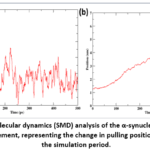 |
Figure 9: Steered molecular dynamics (SMD) analysis of the α-synuclein–peptide complex. (a) Force. (b) Displacement, representing the change in pulling position of the peptide over the simulation period. |
Discussion
The pathological aggregation of α-synuclein remains a major therapeutic target in Parkinson’s disease, particularly due to increasing evidence suggesting that oligomeric and early fibrillar intermediates contribute more significantly to neurotoxicity than mature fibrils alone17,18.. Recent cryo-EM and NMR studies have demonstrated the polymorphic nature of α-syn aggregates, especially within the NAC region (residues 61–95), further highlighting this domain as a critical therapeutic hotspot.19 In the present study, aggregation-prone residues identified using AGGRESCAN3D, including Val37, Tyr39, and regions associated with the NAC domain, were consistent with experimentally reported aggregation cores, thereby supporting the biological relevance of the selected target interface.
Peptide-based aggregation inhibitors have attracted considerable attention because of their ability to selectively recognise structural motifs and interfere with β-sheet propagation.20,21 Compared with conventional small molecules, peptides possess improved surface complementarity for protein–protein interaction interfaces that often lack well-defined binding pockets. Nevertheless, issues related to peptide stability, solubility, and toxicity continue to limit translational applicability. To address these challenges, the present computational workflow incorporated ProtParam-based stability assessment, PepCalc solubility prediction, and HemoPI toxicity evaluation as early-stage developability filters prior to structural screening. Among the evaluated candidates, GRCSVVYGLRCFPD demonstrated the most favourable docking score and interaction profile. Importantly, the observed interactions involving Tyr39 and Val37 are consistent with previous studies identifying these residues as key aggregation nucleation sites22. The repeated targeting of Tyr39 across multiple conformational chains further supports the structural reliability and conservation of the identified binding interface.
Long-time-scale molecular dynamics simulations further demonstrated the stability of the peptide-bound complex. Comparative analyses between the apo protein and the protein–peptide complex revealed that peptide association reduced excessive conformational fluctuations and promoted comparatively restrained structural dynamics within aggregation-associated regions. RMSD and RMSF analyses showed controlled flexibility within the NAC domain while preserving the inherent dynamic behaviour of terminal regions, which is consistent with previously reported computational investigations of α-syn conformational dynamics.23,24 In particular, the peptide-bound system exhibited reduced solvent exposure and enhanced structural compactness relative to the apo protein, suggesting that peptide interaction may partially shield aggregation-prone hydrophobic regions. Hydrogen bond analysis additionally revealed persistent intermolecular interactions throughout the simulation, with increased hydrogen bond occupancy during the later stages of the trajectory, indicating adaptive stabilisation of the complex. Similar strengthening of peptide–α-syn interactions during prolonged simulations has been reported in previous MD-based studies of aggregation inhibitors.25 The mechanical stability of the complex was further validated using steered molecular dynamics (SMD) simulations. The peptide required substantial external force for dissociation from the α-synuclein interface, and the force profile demonstrated multiple rupture events corresponding to the sequential disruption of intermolecular contacts. Importantly, sustained resistance against forced dissociation suggests persistent interaction of the peptide with aggregation-associated residues during the pulling process. These observations indicate that the peptide establishes mechanically stable interactions with α-synuclein rather than forming transient surface associations.
The MM-GBSA binding free energy (−55.04 kcal/mol) indicates favourable binding comparable to other computationally reported peptide inhibitors targeting α-syn.26 Electrostatic interactions were identified as the dominant stabilising factor, which is consistent with the basic nature of the selected peptide (pI 8.06) interacting with acidic regions of α-syn. Per-residue decomposition analysis highlighted Arg10, Phe12, and Cys11 as major contributors, emphasising the role of charge complementarity and aromatic interactions in stabilising aggregation-prone interfaces. Aromatic residues are known to modulate π–π stacking interactions within amyloid-forming proteins, and their involvement supports mechanistic plausibility.27
Principal component analysis and free energy landscape mapping further demonstrated that the peptide-bound complex predominantly occupied stable low-energy conformational basins rather than undergoing unrestricted conformational drift. Compared with the apo protein, the peptide-bound system exhibited relatively confined conformational sampling, suggesting peptide-induced modulation of α-syn dynamic behaviour. Similar multi-basin conformational characteristics have been described in previous FEL analyses of α-syn complexes, supporting the structural robustness of the identified peptide-bound state. While our integrated computational workflow—comprising physicochemical filtering, molecular docking, 300 ns MD simulations, SMD, and MM-GBSA energetics—provides robust mechanistic insights into peptide–α-synuclein interactions, we acknowledge that computational predictions cannot substitute for empirical biological evidence. The primary limitation of this study is the absence of experimental validation. Experimental assays such as Thioflavin T (ThT) fluorescence assays to evaluate aggregation kinetics, Transmission Electron Microscopy (TEM) imaging to observe fibril morphology, and cellular cytotoxicity assays are required to definitively establish the functional anti-aggregation efficacy and biophysical safety of GRCSVVYGLRCFPD.
Conclusion
In conclusion, this study provides a computational framework for the prioritization of potential cyclopeptide inhibitors against α-synuclein aggregation. The identified lead candidate, GRCSVVYGLRCFPD, demonstrated favorable structural, dynamic, and energetic properties in silico. These findings lay a solid theoretical foundation for future experimental studies, where wet-lab validation via ThT assays, TEM visualization, and biological evaluations will be conducted to confirm its translational therapeutic potential. The present study establishes an integrated computational pipeline for identifying putative peptide binders that may modulate $\alpha$-synuclein aggregation and warrant experimental evaluation.
Through aggregation hotspot analysis, physicochemical screening, molecular docking, apo and complex molecular dynamics simulations, steered molecular dynamics, and MM-GBSA calculations, GRCSVVYGLRCFPD was identified as the most promising peptide candidate. The peptide demonstrated stable binding with aggregation-associated residues, sustained intermolecular interactions, favourable binding free energy, enhanced structural compactness, and occupancy of stable low-energy conformational states throughout the simulation. Comparative apo and peptide-bound analyses further suggested that peptide binding contributes to the stabilisation of aggregation-prone regions of α-synuclein. Overall, these findings support the potential of GRCSVVYGLRCFPD as a lead anti-aggregation peptide candidate and provide a scalable computational framework for peptide-based therapeutic discovery against protein misfolding disorders.
Acknowledgement
The authors would like to express their appreciation to the Saudi Arabian MOE and King Abdulaziz University, DSR in Jeddah for their technical and financial aid.
Funding Statement
Grant number IFPIP.- IFPIP: 1456-140-1443. from the Instructional Improvement Fund supported this study.
Conflict of Interest
The authors do not have any conflict of interest.
Data Availability Statement
This statement does not apply to this article.
Ethics Statement
This research did not involve human participants, animal subjects, or any material that requires ethical approval.
Informed Consent Statement
This study did not involve human participants, and therefore, informed consent was not required.
Clinical Trial Registration
This research does not involve any clinical trials.
Permission to reproduce material from other sources
Not Applicable
Author Contributions
- Hatoon A. Niyazi: Conceptualization, Methodology, Investigation, Formal Analysis, Writing – Original Draft
- Ala A. Azhari: Conceptualization, Methodology, Supervision, Writing – Review & Editing
- Hisham N. Altayb: Methodology, Software, Validation, Writing – Review & Editing
- Turki M. Alharthi: Investigation, Data Curation, Visualization
- Wafaa Alhazmi: Investigation, Data Curation
- Ahmad M. Alzahrani: Investigation, Visualization
- Hadeel A. Alsufyani: Investigation, Data Curation
- Turki Asiri: Investigation
- Ohood Adnan Adas: Investigation, Writing – Review & Editing
- Ahmed B. Abd Alla: Conceptualization, Project Administration, Resources, Supervision, Writing – Review & Editing, Correspondence
- Abdelbagi Alfadil: Conceptualization, Supervision, Funding Acquisition, Writing – Review & Editing
References
- Parkinson’s disease. National Institute of Neurological Disorders and Stroke. Accessed February 26, 2026. https://www.ninds.nih.gov/health-information/disorders/parkinsons-disease
- Magrinelli F, Picelli A, Tocco P, et al. Parkinson’s disease: a review on pathophysiology, clinical features, and managing strategies. Parkinsons Dis. 2016;2016:9832839.
CrossRef - Peña-Zelayeta L, Delgado-Minjares KM, Villegas-Rojas MM, et al. Advances in personalized medicine for Parkinson’s disease. J Pers Med. 2025;15(5):0172. doi:10.3390/jpm15050172
CrossRef - Politis M, Wu K, Molloy S, Bain PG, Chaudhuri KR, Piccini P. Parkinson’s disease symptoms and neurochemical correlates. Mov Disord. 2010;25(11):1646-1651.
CrossRef - Sveinbjornsdottir S. The clinical symptoms of Parkinson’s disease. J Neurochem. 2016;139(suppl 1):318-324.
CrossRef - Srinivasan E, Chandrasekhar G, Chandrasekar P, et al. Alpha-synuclein aggregation and its inhibition. Front Med (Lausanne). 2021;8:736978.
CrossRef - Mehra S, Gadhe L, Bera R, Sawner AS, Maji SK. Structural insights into alpha-synuclein aggregation. Biomolecules. 2021;11(10):1419.
CrossRef - Hoppe SO, Uzunoğlu G, Nussbaum-Krammer C. Monitoring alpha-synuclein aggregation in model organisms. Biomolecules. 2021;11(7):931.
CrossRef - Falcone N, Ermis M, Tamay DG, et al. Biomaterial-based strategies for treating neurodegenerative disorders. Adv Healthc Mater. 2023;12(28):e2301096.
CrossRef - Allen SG, Meade RM, White Stenner LL, Mason JM. Peptide-based inhibitors of alpha-synuclein aggregation. Mol Neurodegener. 2023;18(1):80.
CrossRef - Zhang Q, Gong X, Liu H, Yao X. Computational design of therapeutic molecules for neurodegenerative diseases. Acta Pharm Sin B. Published online July 2025. doi:10.1016/j.apsb.2025.07.038
CrossRef - Ekins S, Mestres J, Testa B. In silico pharmacology for drug discovery: applications to targets and pathways. Br J Pharmacol. 2007;152(1):21-37.
CrossRef - Liu J, Gui Y, Rao J, et al. Discovery of novel small molecule therapeutics for alpha-synucleinopathies. Acta Pharm Sin B. 2024;14(7):2927-2941.
CrossRef - Chen SW, Drakulic S, Deas E, et al. Structural characterization of toxic oligomers formed by α-synuclein. Proc Natl Acad Sci U S A. 2015;112(16):E1994-E2003.
CrossRef - Gul G. Molecular modeling of protein misfolding mechanisms. J Mol Graph Model. 2025;139:109079.
CrossRef - Armiento V, Spanopoulou A, Kapurniotu A. Peptide-Based Molecular Strategies To Interfere with Protein Misfolding, Aggregation, and Cell Degeneration. Angew Chem Int Ed Engl. 2020 Feb 24;59(9):3372-3384. doi: 10.1002/anie.201906908.
CrossRef - Vidović M, Rikalovic MG. Alpha-synuclein in Parkinson’s disease: from pathogenesis to potential therapeutics. Cells. 2022;11(11):1732.
CrossRef - Rana A, Nikam T, Sreepathi B, Saraf SA, Awasthi S. The Aggregation Continuum of α-Synuclein and Its Relevance to Brain Aging. ACS Chem Neurosci. 2025 Oct 1;16(19):3634-3652. doi: 10.1021/acschemneuro.5c00356.
CrossRef - Yan, N.L., Candido, F., Tse, E., Melo, A.A., Prusiner, S.B., Mordes, D.A., Southworth, D.R., Paras, N.A. and Merz, G.E. (2025), Cryo-EM structure of a novel α-synuclein filament subtype from multiple system atrophy. FEBS Lett, 599: 33-40. https://doi.org/10.1002/1873-3468.15048
CrossRef - Sehra N, Parmar R, Jain R. Small molecule and peptide inhibitors targeting protein aggregation. RSC Med Chem. 2025;16:1083-1104.
CrossRef - Armiento V, Spanopoulou A, Kapurniotu A. Peptide-based inhibitors of amyloid self-assembly. Angew Chem Int Ed Engl. 2020;59(9):3372-3384.
CrossRef - Palomino-Hernandez O, Buratti FA, Sacco PS, Rossetti G, Carloni P, Fernandez CO. Conformational dynamics of intrinsically disordered proteins. Int J Mol Sci. 2020;21(14):5061.
CrossRef - Henriques J, Cragnell C, Skepö M. Molecular Dynamics Simulations of Intrinsically Disordered Proteins: Force Field Evaluation and Comparison with Experiment. J Chem Theory Comput. 2015 Jul 14;11(7):3420-31. doi: 10.1021/ct501178z.
CrossRef - Singh N, Singh AK. Computational identification and simulation of amyloid inhibitors. ACS Omega. 2024;9(9):10748-10768.
CrossRef - Horne RI, Andrzejewska EA, Alam P, Brotzakis ZF, Srivastava A, Aubert A, Nowinska M, Gregory RC, Staats R, Possenti A, Chia S, Sormanni P, Ghetti B, Caughey B, Knowles TPJ, Vendruscolo M. Discovery of potent inhibitors of α-synuclein aggregation using structure-based iterative learning. Nat Chem Biol. 2024 May;20(5):634-645. doi: 10.1038/s41589-024-01580-x.
CrossRef - Ali TT, Merghani M, Al-Azzani M, Gatzemeier LM, Hoppert M, Kaloyanova D, Outeiro TF, Neumann P, Popova B, Braus GH. Rationally designed peptides inhibit the formation of α-synuclein fibrils and oligomers. Eur J Med Chem. 2025 May 5;289:117452. doi: 10.1016/j.ejmech.2025.117452. Epub 2025 Feb 26.
PMID: 40022877. Bemporad F, Taddei N, Stefani M, Chiti F. Assessing the role of aromatic residues in amyloid formation. Protein Sci. 2006;15(4):862-870.
CrossRef - Berman HM, Westbrook J, Feng Z, et al. The Protein Data Bank. Nucleic Acids Res. 2000;28(1):235-242.
CrossRef - Tuttle MD, Comellas G, Nieuwkoop AJ, et al. Atomic-resolution structure of a disease-relevant fibril form of α-synuclein. Nat Struct Mol Biol. 2016;23(5):409-415.
CrossRef - Liu L, Yang L, Cao S, et al. Deep learning methods in protein structure prediction and peptide design. Brief Bioinform. 2024;25(3):bbae190.
- Imam MA, Alandijany TA, Felemban HR, et al. Machine learning, network pharmacology, and molecular dynamics reveal potent cyclopeptide inhibitors against dengue virus proteins. Mol Divers. 2025;29(4):2899-2917.
CrossRef - Gasteiger E, Hoogland C, Gattiker A, et al. Protein identification and analysis tools on the ExPASy server. In: Walker JM, ed. The Proteomics Protocols Handbook. Humana Press; 2005:571-607.
CrossRef - Lamiable A, Thévenet P, Rey J, Vavrusa M, Derreumaux P, Tufféry P. PEP-FOLD3: faster de novo structure prediction for linear peptides in solution and in complex. Nucleic Acids Res. 2016 Jul 8;44(W1):W449-54. doi: 10.1093/nar/gkw329. Epub 2016 Apr 29. PMID: 27131374; PMCID: PMC4987898.
CrossRef - Bauer P, Hess B, Lindahl E. GROMACS 2022.4 source code. Zenodo. Published November 2022. doi:10.5281/zenodo.7323409
- Huang J, MacKerell AD Jr. CHARMM36m: an improved force field for folded and intrinsically disordered proteins. J Comput Chem. 2013;34(25):2135-2145.
CrossRef - Darden T, York D, Pedersen L. Particle mesh Ewald: an N·log(N) method for Ewald sums in large systems. J Chem Phys. 1993;98(12):10089-10092.
CrossRef - Harrach MF, Drossel B. Structure and dynamics of water in the presence of protein molecules. J Chem Phys. 2014;140(17):174501.
CrossRef - Hess B, Bekker H, Berendsen HJC, Fraaije JGEM. LINCS: a linear constraint solver for molecular simulations. J Comput Chem. 1997;18(12):1463-1472.
CrossRef - Bussi G, Donadio D, Parrinello M. Canonical sampling through velocity rescaling. J Chem Phys. 2007;126(1):014101.
CrossRef - Martoňák R, Laio A, Parrinello M. Predicting crystal structures: the example of solid benzene. Phys Rev Lett. 2003;90(7):075503.
CrossRef - Valdés-Tresanco MS, Valdés-Tresanco ME, Valiente PA, Moreno E. gmx_MMPBSA: a new tool to perform end-state free energy calculations with GROMACS. J Chem Theory Comput. 2021;17(10):6281-6291.
CrossRef - Miller BR 3rd, McGee TD Jr, Swails JM, Homeyer N, Gohlke H, Roitberg AE. MMPBSA.py: an efficient program for end-state free energy calculations. J Chem Theory Comput. 2012;8(9):3314-3321.
CrossRef - Isralewitz B, Gao M, Schulten K. Steered molecular dynamics and mechanical properties of proteins. Curr Opin Struct Biol. 2001;11(2):224-230.
CrossRef

Accepted on: 05-10-2026
Second Review by: Dr. Randa Salah Gomaa Mahmoud
Final Approval by: Dr Patorn Piromchai