Antioxidant and Cytotoxic Constituents of Paramignya trimera and a Multi-marker HPLC Assay for Quality Control
 , Tran Hoang Khanh2, Nguyen Bao Phuc3and Vuong Thi Huyen Trang4*
1Faculty of Traditional Pharmacy, University of Medicine and Pharmacy, Ho Chi Minh City 70000, Vietnam
2British International School Ho Chi Minh City, Ho Chi Minh City, Vietnam
3Foreign Language Specialized School – VNU, Hanoi, Vietnam
4Vietnam Academy of Science and Technology, Hanoi, Vietnam
Corresponding Author. E-mail: Vuongthihuyentrang@gmail.com
Download this article as:
ABSTRACT:Paramignya trimera (Oliv.) Burkill (Rutaceae), known in Vietnam as “Xáo tam phân”, is widely used against liver disorders and cancer, yet its active constituents and quality standards remain poorly defined. Total extracts and solvent-partitioned fractions of roots, stems and leaves were assessed for total phenolic content (Folin–Ciocalteu), antioxidant activity (DPPH and malondialdehyde assays) and cytotoxicity (MTT against MDA-MB-231, HepG2 and HAK1B cells). Constituents of the most active fractions were isolated by column chromatography and identified by UV, IR, ESI-MS and 1D/2D-NMR, and a simultaneous HPLC–PDA assay for three marker coumarins was developed and characterised. Roots were the most cytotoxic (HepG2 IC50 24.9 µg/mL), whereas stems had the highest phenolic content and DPPH activity; the fractions with the highest antioxidant activity were also the most cytotoxic, an association reported here as an observation only. Twelve compounds were obtained; limonin and 6-(2′-hydroxy-3′-methylbut-3′-enyl)-7-hydroxycoumarin are reported for the first time from the species, and a further pyranocoumarin remains tentatively assigned. Ostruthin and 8-methoxyostruthin were moderately cytotoxic (MDA-MB-231 IC50 32.4 and 47.8 µM); in an exploratory, correlative experiment, siRNA knockdown of the histone demethylase KDM2A was associated with reduced cellular sensitivity to 8-methoxyostruthin, without evidence of direct target engagement. The assay quantified 6.55% ostruthin in cultivated roots and showed wide marker variability among ten commercial samples. The study provides certified reference standards and a multi-marker assay to support authentication and quality control of this species.
KEYWORDS:Acridone alkaloid; Cytotoxicity; HPLC quality control; KDM2A; 8-Methoxyostruthin; Ostruthin; Paramignya trimera
Introduction
Paramignya trimera (Oliv.) Burkill (Rutaceae), known in Vietnam as “Xáo tam phân”, is a spiny woody climber of the genus Paramignya, which comprises about thirty species distributed across South and Southeast Asia, including Thailand, Malaysia, India and Indonesia. In Vietnam the species has been recorded around Nui Lap Vo (Binh Duong) and Hon Heo (Khanh Hoa), where the roots and stems are used in traditional medicine to cool the body, nourish and protect the liver, and to treat hepatitis and cirrhosis.1 In 2012 the National Institute of Medicinal Materials reported that root extracts of material from Hon Heo were cytotoxic to five human cancer cell lines—HepG2 (liver), HCT116 (colon), MDA-MB-231 (breast), OVCAR-8 (ovary) and HeLa (cervix)—with the strongest effects on HepG2 and HeLa.2 Following this report, demand for the root increased, harvesting of wild populations intensified, and the species is now cultivated in several provinces; adulteration and substitution of the traded drug have also been documented.
Interest in plant-derived anticancer leads reflects the continuing burden of cancer, which is increasing in many developing countries. Vietnam recorded 182,563 new cases and 122,690 deaths in 2020, with liver (14.5%), lung (14.4%), breast (11.8%), gastric (9.8%) and colorectal (9.0%) cancers the most frequent3; corresponding global estimates for 2022 were approximately 20 million new cases and 9.7 million deaths.4 Synthetic cytotoxic agents are effective but costly and frequently dose-limiting in their toxicity, and plant-derived compounds continue to be examined as additional sources of antiproliferative activity. For P. trimera, however, the constituents responsible for the reported activity, their molecular targets and the composition of the traded material remain incompletely characterised.
Systematic phytochemistry of the species began in 2013. Nguyen et al.5 reported the first constituents of the roots; in the same year Pham et al. isolated and quantified ostruthin—the 6-geranyl-7-hydroxycoumarin now used as the species’ marker.6 Further constituents were reported over the following decade: Nguyen et al. characterised paratrimerins A and B, two new dimeric monoterpene-linked coumarin glycosides, from the roots and stems7; the acridone series was later extended by the cytotoxic paratrimerin I8; and Quan et al. reported an entire family of dimeric coumarins, paratrimerins J–Y, from the stems.9 These studies indicate that simple 7-hydroxy(geranyl/prenyl)coumarins, pyranocoumarins, dimeric coumarin glycosides and acridone alkaloids are characteristic constituents of the species, together with triterpenoids, flavonoids and phenolics.10
Comparable constituent classes have been reported from other species of the genus, consistent with the chemotaxonomic position of Paramignya within the Rutaceae, a family whose phytochemistry has been reviewed recently.11
Pharmacological research has centred on anticancer and hepatoprotective activity. Beyond the National Institute of Medicinal Materials’ initial five-cell-line screen,2 Son summarised a methanol root extract with an IC50 of about 23 µg/mL against MCF-7.10 Nguyen et al. showed that a methanol extract induces apoptosis in HepG2 cells, albeit at a far higher IC50 (≈ 582 µg/mL); the difference between these values illustrates the influence of plant origin and extraction conditions on measured potency.12 Nguyen et al. examined the relationship between extraction conditions and activity, reporting that solvent choice and extraction method markedly affect the root’s phenolic content and antioxidant capacity,13 and that root and leaf extracts and their fractions are both antioxidant and anti-proliferative across a cancer-cell panel (IC50 ≈ 15–32 µg/mL), the activity concentrating in the less-polar, coumarin-rich fractions14,15; more recent work has characterised the peel essential oil16 and developed root-extract–derived gold nanoparticles with anticancer activity.17 Hepatoprotective activity has also been reported: root extracts and the coumarins ostruthin/8-methoxyostruthin protect mouse liver against paracetamol-induced injury.18,19 Several coumarins are anti-inflammatory, suppressing NO, PGE2, iNOS and COX-2 in stimulated macrophages and microglia,20 while the isolated coumarins and acridones also show antioxidant and α-glucosidase-inhibitory activity.21 A concise overview of these studies is given in Table 1.
Table 1: Overview of representative previous studies on P. trimera and related Paramignya species.
|
Study |
Material | Focus |
Key finding |
|
NIMM2 |
Roots | Cytotoxicity |
Active against HepG2, HCT116, MDA-MB-231, OVCAR-8, HeLa; strongest on HepG2/HeLa |
|
Nguyen et al.5 |
Roots | Phytochemistry |
First chemical constituents reported |
|
Pham et al.6 |
Roots | Chemistry / QC |
Isolated and quantified ostruthin (marker) |
|
Nguyen et al.18 |
Roots | Toxicology / activity |
Acute toxicity, hepatoprotection and cytotoxicity |
|
Nguyen et al.7 |
Roots, stems | Phytochemistry | Paratrimerins A, B (new dimeric coumarin glycosides) |
| Le et al.20 | Roots/stems | Anti-inflammatory |
Coumarins suppress NO, iNOS and COX-2 |
|
Nguyen et al.14 |
Roots, leaves | Antioxidant / cytotoxicity | Extracts and fractions active (IC50 ≈ 15–32 µg/mL) |
| Trinh et al.21 | Roots | Chemistry / activity |
Coumarins/acridones; α-glucosidase & antioxidant |
|
Nguyen et al.12 |
Roots | Anticancer | Induces apoptosis in HepG2 |
| Quan et al.9 | Stems | Phytochemistry |
Paratrimerins J–Y (dimeric coumarins) |
Despite these advances, the studies summarised above leave several gaps. First, most bioactivity assessments have relied on crude extracts, and the individual constituents responsible for the observed cytotoxicity—together with their molecular targets—have rarely been characterised. Second, although ostruthin is regarded as the marker compound of P. trimera, it also occurs in other Paramignya and Rutaceae species; quantifying ostruthin alone is therefore insufficient either to authenticate the crude drug or to guarantee its quality, and P. trimera indeed still lacks a monograph in the Vietnamese Pharmacopoeia. Third, the leaves of this plant are widely used in herbal preparations yet have received little chemical or pharmacological attention. The present study addresses these gaps through the extraction, fractionation and chemical characterisation of its constituents and the evaluation of their biological activities.
The specific objectives were as follows: (1) to evaluate the antioxidant activity (DPPH radical scavenging and inhibition of lipid peroxidation) and cytotoxicity of the roots, stems and leaves and their solvent-partitioned fractions; (2) to isolate and elucidate the structures of the constituents from the most active plant parts; (3) to carry out an exploratory assessment of whether the histone demethylase KDM2A contributes to cellular sensitivity to the two most abundant coumarins, ostruthin and 8-methoxyostruthin; and (4) to establish reference standards and develop an HPLC method for the simultaneous quantification of three marker coumarins, for application to the cultivated and commercial root samples analysed in this study.
Materials and Methods
Plant material
Whole plants of P. trimera bearing roots, stems and leaves were collected on Hon Lon island, Van Phong Bay, Van Thanh commune, Van Ninh district, Khanh Hoa province (March 2013) and botanically authenticated by Dr Vo Van Chi; roots (4.5 kg), stems (5.3 kg) and leaves (2 kg) were used for chemical and biological study. For method development, cultivated roots (2.0 kg) were obtained from the Quang Minh medicinal-plant farm (Dong Nai; March 2017). For the market survey, ten commercial root samples (100 g each; M1–M10) were purchased in August 2022 from herbal retail outlets in Dong Nai, Da Lat (Lam Dong), Ho Chi Minh City, Tay Ninh and Binh Phuoc, and were sold as dried, sliced root. None of the vendors was able to state the geographical origin, the harvest date, or whether the material had been wild-collected or cultivated, and no accompanying documentation was issued, so no provenance beyond the place and month of purchase could be established for any sample. Material was chopped, shade-dried, oven-dried, milled and sieved. Voucher specimens are deposited at the Faculty of Pharmacy, University of Medicine and Pharmacy at Ho Chi Minh City (XTP 01-2013 and XTP 02-2017). The study was carried out between August 2014 and August 2022 at the University of Medicine and Pharmacy at Ho Chi Minh City (Departments of Pharmacognosy, Analytical Chemistry–Quality Control and Pharmacology), the University of Medicine and Pharmacy Faculty of Traditional Medicine, the Faculty of Medicine of Vietnam National University Ho Chi Minh City, and the Department of Molecular Pharmacy, Faculty of Pharmacy, Takasaki University of Health and Welfare (Japan).
Chemicals, reference substances and cell lines
Ethanol, methanol, chloroform, ethyl acetate and n-hexane (analytical grade) and HPLC-grade acetonitrile and methanol were from Merck (Germany). Silica gel 60 (40–63 µm) and pre-coated TLC silica gel 60 F254 plates (0.25 mm) were from Merck; DPPH and pyrogallol were from Sigma-Aldrich. Reference antioxidants were ascorbic acid (99.17%), silymarin and quercetin (> 99%). Positive cytotoxic controls were paclitaxel and doxorubicin (> 99%; Ebewe Pharma, Austria) and sorafenib (> 99%; Bayer HealthCare, USA). MDA-MB-231 (triple-negative breast adenocarcinoma) and HepG2 (hepatocellular carcinoma) cells were obtained from ATCC and maintained at the Pasteur Institute, Ho Chi Minh City; MDA-MB-231 and HAK1B (hepatocellular carcinoma) were maintained at Takasaki University. Cells were grown in EMEM or DMEM supplemented with 10% fetal calf serum, 2 mM L-glutamine, 100 IU/mL penicillin and 100 µg/mL streptomycin at 37 °C under 5% CO2.
Phytochemical screening, quality parameters and total phenolic content
Preliminary phytochemical screening of 20 g of each plant part followed the Ciulei procedure (as modified locally), partitioning the drug into diethyl-ether, ethanol and aqueous fractions before characteristic colour/precipitation tests.22 Loss on drying, total ash, acid-insoluble ash and water- and ethanol-extractable matter were determined (n = 3) per the Vietnamese Pharmacopoeia V (appendices 9.6–9.8).23 Polyphenols were confirmed qualitatively (2% FeCl3, 20% NaOH, 1% lead acetate) and quantified in 96% ethanol extracts by the Folin–Ciocalteu method24: 100 g of powder was exhaustively percolated with 96% ethanol, concentrated to constant weight, dissolved in 70% ethanol (50 µg/mL) and reacted with Folin–Ciocalteu reagent and 29% Na2CO3; absorbance was read at 760 nm against a pyrogallol calibration curve (y = 0.0233x − 0.158, R² = 0.9974) on a Powerwave HT plate reader, and content expressed as mg pyrogallol equivalents (PGE)/g of dry drug. The Folin–Ciocalteu reaction measures total reducing capacity rather than any individual phenolic constituent; values are therefore reported as pyrogallol equivalents and interpreted only as a non-specific index of total phenolic content.
Antioxidant assays
DPPH radical scavenging followed Kulisic et al.,25 one of several complementary assays now recommended for phytochemical antioxidant profiling26: 2 mL of 0.2 mM DPPH in methanol was mixed with 2 mL of test solution and, after 30 min in the dark, absorbance was read at 517 nm; total extracts were screened at 1000 µg/mL and then over a concentration range to obtain EC50 values, with ascorbic acid and quercetin as references. Inhibition of membrane lipid peroxidation was measured by the malondialdehyde (MDA)/thiobarbituric-acid method27 using a rat-liver homogenate in 1.15% KCl as substrate: 0.1 mL of sample at various concentrations was reacted with 0.5 mL homogenate in 50 mM phosphate buffer, incubated 15 min at 37 °C, stopped with 10% trichloroacetic acid, and, after adding 0.8% thiobarbituric acid and heating (100 °C, 15 min), the pink chromophore was read at 532 nm; activity was expressed as EC50 with quercetin as reference. All assays were performed in triplicate and EC50 values obtained from regression.
In vitro cytotoxicity
Growth inhibition was measured by the MTT assay,28,29 in which mitochondrial succinate dehydrogenase of viable cells reduces MTT to a formazan read at 570 nm. Cells were seeded in 96-well plates (104 cells/well), allowed to attach for 24 h, then treated for 72 h with test samples, positive controls (paclitaxel 0.5 µM; doxorubicin 10 µg/mL) or vehicle (DMSO, final < 0.05%). Formazan was dissolved in acidified isopropanol and absorbance read on a Powerwave HT reader. Total extracts and fractions were tested at 6.25–100 µg/mL and isolated ostruthin and 8-methoxyostruthin at 2.5–100 µM (each concentration in triplicate). Percentage growth inhibition and IC50 values were derived from concentration–response regression.
Extraction, fractionation and isolation
Root powder (4.5 kg) was extracted by hot maceration with 96% ethanol (35 L × 4), filtered and concentrated to 1.6 L of a viscous extract, which was suspended in water and partitioned in order of increasing polarity with n-hexane, chloroform and ethyl acetate to give, after concentration, an n-hexane–insoluble precipitate (82 g) and n-hexane (86 g), chloroform (200 g), ethyl acetate (40 g) and aqueous (330 g) fractions. Stem powder (5.3 kg) was percolated with 96% ethanol (90 L), concentrated to 4.3 L and partitioned similarly to give n-hexane (36 g), chloroform (19 g), ethyl acetate (39.7 g) and aqueous (92 g) fractions. Guided by antioxidant and cytotoxic screening, the less-polar n-hexane and chloroform fractions were prioritised. Compounds were purified by vacuum-liquid chromatography (VLC) and classical open-column chromatography over silica gel (40–63 µm) with n-hexane–chloroform–ethyl acetate gradients, followed by recrystallisation; purity and homogeneity were monitored by TLC (UV 254/365 nm; vanillin–sulfuric acid and 10% H2SO4 charring). Eight compounds (PT-1–PT-8) were obtained from roots and four (PT-9–PT-12) from stems.
Structure elucidation
Structures were established from UV, IR and ESI-MS spectra together with 1D and 2D NMR (¹H, ¹³C, DEPT, HSQC, HMBC and COSY) recorded on a Bruker Avance-500 spectrometer (Institute of Chemistry, Vietnam Academy of Science and Technology, Hanoi) and ESI-MS on a Micromass Quattro micro API (Waters), and by comparison with published data. Reported structures were checked against the Reaxys and SciFinder databases (30 July 2022). Only low-resolution ESI-MS was available; high-resolution mass measurements, and therefore formal confirmation of molecular formulae, were not obtained, and no optical-rotation or electronic-circular-dichroism data were recorded. Full ¹H and ¹³C assignments, multiplicities, coupling constants and the key HMBC/COSY correlations underpinning each assignment are given in the Supporting Information. Novelty of occurrence within the species was assessed by searching Reaxys, SciFinder, PubMed, Google Scholar and Vietnamese-language journal archives for Paramignya trimera together with each compound name and its synonyms (last searched 30 July 2022 and repeated in September 2026 with the same outcome).
KDM2A knockdown and Western blot
To test whether the cytotoxicity of ostruthin and 8-methoxyostruthin involves the histone lysine demethylase KDM2A, MDA-MB-231 and HAK1B cells were seeded at 2 × 10⁵ cells/mL and transfected with a KDM2A-targeting siRNA (Si97; pre-designed siRNA, Applied Biosystems, USA; sense strand 5′-GAACCCGAAGAAGAAAGGAUUCGUU-3′) or a non-targeting control siRNA (SiM) at 40 nM using Lipofectamine RNAiMAX (Invitrogen, USA) according to the manufacturer’s instructions (3 µL of reagent per 3 mL of culture medium); cells were incubated for 48 h at 37 °C under 5% CO₂ and washed three times with phosphate-buffered saline before compound treatment. Knockdown was verified by Western blot30 with an anti-KDM2A primary antibody and anti-GAPDH (Santa Cruz Biotechnology) as loading control, followed by horseradish-peroxidase-conjugated anti-rabbit or anti-mouse secondary antibody (Amersham) and detection with an ECL Plus kit (GE Healthcare), on a ChemiDoc XRS+ system (Bio-Rad) with Quantity One software; band intensities were quantified by densitometry and normalised to GAPDH; representative membranes, including uncropped images, and their densitometric quantification are shown in the Supporting Information. No formal statistical comparison was performed, consistent with the exploratory status of this experiment. Transfected and control cells were then treated with ostruthin or 8-methoxyostruthin (2.5–100 µM), or with sorafenib (0.39–25 µM), for 72 h, and the viability of KDM2A-silenced cells was compared with that of controls. Because this experiment used a single siRNA sequence without rescue by KDM2A over-expression, it was treated throughout as exploratory.
Reference standards and HPLC method
Ostruthin and 8-methoxyostruthin, isolated in large amounts and of high purity, were fully characterised (appearance, DSC melting point, UV, IR, ESI-MS, ¹H/¹³C NMR, TLC, related substances and assay), evaluated for vial homogeneity (one-way ANOVA) and inter-laboratory transferability at two GLP laboratories of the Ho Chi Minh City Institute of Drug Quality Control, and assigned certified contents by robust statistics (ISO 13528, Algorithm A),31 in line with ASEAN guidance on the establishment of reference substances. A reversed-phase HPLC method for the simultaneous determination of demethylsuberosin, ostruthin and 8-methoxyostruthin was developed on a Shimadzu LC-2030C 3D system with PDA detection using a Cosmosil 5C18-AR-II column (4.6 × 250 mm, 5 µm) at 1.0 mL/min, 10 µL injection, 332 nm detection and a 25-min acetonitrile–water gradient (55:45 for 0–15 min, 80:20 for 15–20 min, 100:0 for 20–25 min). Samples were prepared in methanol with a single 30-min sonication (20 mL). Method performance was assessed for specificity (peak purity), linearity, LOD and LOQ, precision and accuracy with reference to ICH and AOAC guidance32,33. A full ICH validation — system-suitability testing over replicate injections, separate repeatability and intermediate-precision studies, and accuracy at defined spiking levels — was not undertaken within the scope of this study, and the method is therefore described as characterised rather than validated. The method was applied to the cultivated root and the ten market samples.
Statistics
Antioxidant data are means ± SD (n = 3) with EC50 from regression (Microsoft Excel). Cytotoxicity data are means ± SEM; group differences were tested by the Mann–Whitney test in SPSS 20.0, and IC50 values obtained by regression. A p-value < 0.05 was considered significant.
Results
Quality parameters and polyphenol content
Water-extractable matter was highest in roots (17.8%), more than tenfold that of stems (1.74%) and leaves (1.53%); 96% ethanol gave a similar pattern (roots 17.73%; stems 2.03%; leaves 1.42%). Loss on drying (7.25–8.81%), total ash (3.25–4.31%) and acid-insoluble ash (0.027–0.036%) were within acceptable ranges (Table 2). Screening indicated coumarins as the dominant class, with alkaloids, triterpenoids, flavonoids, essential oil and saponins; all polyphenol colour reactions were positive. Total polyphenol content decreased stem (154.3 mg PGE/g) > root (106.7) > leaf (56.0) (Table 2).
Table 2: Quality parameters and total phenolic content of P. trimera by plant part (mean ± SD, n = 3).
|
Plant part |
Loss on drying (%) | Total ash (%) | Acid-insoluble ash (%) | Water-extractable matter (%) | Ethanol-extractable matter (%) |
Total phenolic content (mg PGE/g) |
|
Root |
7.61 ± 0.23 | 3.30 ± 0.14 | 0.036 ± 0.010 | 17.80 ± 0.12 | 17.73 ± 0.14 | 106.7 |
| Stem | 7.25 ± 0.10 | 4.31 ± 0.22 | 0.030 ± 0.020 | 1.74 ± 0.21 | 2.03 ± 0.22 |
154.3 |
|
Leaf |
8.81 ± 0.26 | 3.25 ± 0.12 | 0.027 ± 0.005 | 1.53 ± 0.15 | 1.42 ± 0.18 |
56.0 |
PGE, pyrogallol equivalents.
Antioxidant activity
At 1000 µg/mL total extracts scavenged 90.7% (stem), 88.8% (root) and 63.6% (leaf) of DPPH radicals. EC50 values increased in the order stem (158.1) < root (274.4) < leaf (593.9 µg/mL); because a lower EC50 denotes greater potency, DPPH-scavenging capacity decreased in the order stem > root > leaf, matching the rank order of total phenolic content. Ascorbic acid gave an EC50 of 5.27 µg/mL. On fractionation the less-polar fractions were generally most active; in the MDA assay the n-hexane and chloroform fractions were most protective, lowering MDA formation 26–35-fold relative to the aqueous fraction (root n-hexane EC50 325.9 vs aqueous 11,435 µg/mL) (Table 3).
Table 3: Antioxidant activity (EC50, µg/mL) of total extracts and fractions by DPPH and MDA assays.
|
Sample |
DPPH – Root | DPPH – Stem | MDA – Root | MDA – Stem |
| Total ethanol extract | 274.4 | 158.1 | – |
– |
|
n-Hexane fraction |
377.4 | 329.3 | 325.9 | 383.2 |
| Chloroform fraction | 133.4 | 182.3 | 420.7 |
429.2 |
|
Ethyl acetate fraction |
180.6 | 86.3 | 442.6 | 654.0 |
| Aqueous fraction | 599.2 | 377.7 | 11,435 |
8,530 |
Note: ascorbic acid EC50 5.27 µg/mL (DPPH, total extracts); quercetin 4.94 (DPPH, fractions) and 13.47 (MDA). Lower EC50 values indicate greater antioxidant potency.
Cytotoxicity of extracts and fractions
Root extract was consistently the most cytotoxic (HepG2 IC50 24.9 µg/mL; MDA-MB-231 29.6 µg/mL); leaf extract was moderately active (HepG2 86.9; MDA-MB-231 58.4 µg/mL), while stem extract was weak (IC50 > 100 µg/mL). Fractionation localised HepG2 activity to the n-hexane (36.8 µg/mL) and chloroform (40.7 µg/mL) fractions, the ethyl acetate and aqueous fractions being inactive (Table 4). The n-hexane and chloroform fractions ranked highest in both the MDA and the HepG2 assay; with only four fractions, two of which were inactive in the cytotoxicity assay, no formal correlation analysis was possible, so this co-occurrence is reported as an observation rather than as evidence that the same constituents drive both activities.
Table 4: Cytotoxic IC50 (µg/mL) of P. trimera extracts and root fractions after 72 h (MTT assay).
|
Sample |
MDA-MB-231 | HepG2 |
| Root total extract | 29.6 |
24.9 |
|
Leaf total extract |
58.4 | 86.9 |
| Stem total extract | > 100 |
> 100 |
|
Root n-hexane fraction |
– | 36.8 |
| Root chloroform fraction | – |
40.7 |
|
Root ethyl acetate fraction |
– | inactive |
| Root aqueous fraction | – |
inactive |
Note: Positive controls: paclitaxel (0.5 µM) and doxorubicin (10 µg/mL; 86.8% HepG2 inhibition).
Isolated compounds
Twelve compounds were obtained (Figure 1). From roots: ostruthin (PT-1, 520 mg), 8-methoxyostruthin (PT-2, 300 mg), xanthyletin (PT-3, 55 mg), oriciacridone (PT-4, 10 mg), citrusinin-1 (PT-5, 25 mg), a tentatively assigned pyranocoumarin provisionally formulated as 3′-hydroxy-3′-methyl-2′-(3″-isoprenyl)-pyranocoumarin (PT-6, 4 mg), demethylsuberosin (PT-7, 24 mg) and limonin (PT-8, 24 mg); from stems: 6-(2′-hydroxy-3′-methylbut-3′-enyl)-7-hydroxycoumarin (PT-9, 12 mg), 6-(6′-hydroxy-3′,7′-dimethylocta-2′,7′-dienyl)-7-hydroxycoumarin (PT-10, 10 mg), 6-(7′-hydroxy-3′,7′-dimethylocta-2′,5′-dienyl)-7-hydroxycoumarin (PT-11, 15 mg) and a stigmasterol/β-sitosterol mixture (PT-12, 36 mg). Six are 7-hydroxycoumarins, two pyranocoumarins and two acridone alkaloids (Table 5). Compound PT-6 returned no match in the Reaxys or SciFinder databases, and its spectroscopic data are consistent with a pyranocoumarin differing from paramicoumarin B by an additional hydroxyl and by the position of the pyran methyl; because only 4 mg was isolated and only low-resolution ESI-MS was available, this structure is reported as a tentative assignment and no claim of novelty is made pending HR-ESI-MS, complete 1D/2D-NMR assignment and stereochemical analysis. Xanthyletin (PT-3), oriciacridone (PT-4) and citrusinin-1 (PT-5) were first isolated from P. trimera by our group34,35 and have since been reported by others21; they are re-isolated here and are not claimed as new to the species. Limonin (PT-8) and PT-9 are, to our knowledge, reported for the first time from P. trimera. Of the twelve isolates, only limonin (PT-8) and PT-9 are new records for the species; PT-1, PT-2, PT-7 and PT-10–PT-12 have been reported previously from P. trimera, PT-3–PT-5 were described by us earlier,34,35 and PT-6 is a tentative assignment. The contribution of the present study therefore lies in the bioactivity-guided localisation of activity and in the multi-marker assay rather than in the discovery of new chemistry.
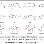 |
Figure 1: Chemical structures of the twelve compounds isolated from P. trimera. Coumarins and pyranocoumarins (PT-1, PT-2, PT-3, PT-6, PT-7, PT-9, PT-10, PT-11), acridone alkaloids (PT-4, PT-5), limonin (PT-8) and phytosterols (PT-12). |
Table 5: Compounds isolated from P. trimera, with class, source, yield and novelty.
|
Code |
Compound | Class | Part | Yield | Note |
| PT-1 | Ostruthin | 7-Hydroxycoumarin | Root | 520 mg |
Major marker |
|
PT-2 |
8-Methoxyostruthin | 7-Hydroxycoumarin | Root | 300 mg | Major marker |
| PT-3 | Xanthyletin | Pyranocoumarin | Root | 55 mg |
Previously reported from species [33] |
|
PT-4 |
Oriciacridone | Acridone alkaloid | Root | 10 mg | Previously reported from species [34] |
| PT-5 | Citrusinin-1 | Acridone alkaloid | Root | 25 mg |
Previously reported from species [34] |
|
PT-6 |
3′-Hydroxy-3′-methyl-2′-(3″-isoprenyl)-pyranocoumarin | Pyranocoumarin | Root | 4 mg | Tentative structure; novelty not claimed |
| PT-7 | Demethylsuberosin | 7-Hydroxycoumarin | Root | 24 mg |
Assay marker |
|
PT-8 |
Limonin | Limonoid | Root | 24 mg |
First from species |
|
PT-9 |
6-(2′-Hydroxy-3′-methylbut-3′-enyl)-7-hydroxycoumarin | 7-Hydroxycoumarin | Stem | 12 mg |
First from species |
|
PT-10 |
6-(6′-Hydroxy-3′,7′-dimethylocta-2′,7′-dienyl)-7-hydroxycoumarin | 7-Hydroxycoumarin | Stem | 10 mg |
– |
|
PT-11 |
6-(7′-Hydroxy-3′,7′-dimethylocta-2′,5′-dienyl)-7-hydroxycoumarin | 7-Hydroxycoumarin | Stem | 15 mg | – |
| PT-12 | Stigmasterol + β-sitosterol | Phytosterol | Stem | 36 mg |
Mixture (~4:1) |
Cytotoxicity of ostruthin and 8-methoxyostruthin and an exploratory KDM2A knockdown experiment
As the most abundant and readily isolated constituents, ostruthin and 8-methoxyostruthin were selected for further characterisation. Both were concentration-dependently cytotoxic, more potent against MDA-MB-231 (IC50 32.4 and 47.8 µM) than HAK1B (40.1 and 60.1 µM); ostruthin was consistently stronger than its 8-methoxy analogue. Sorafenib was markedly more potent (6.10 µM on MDA-MB-231; 8.22 µM on HAK1B) (Table 6; Figure 2).
Table 6: Cytotoxic IC50 (µM) of ostruthin and 8-methoxyostruthin vs sorafenib (72 h, MTT).
|
Compound |
MDA-MB-231 | HAK1B |
| Ostruthin (PT-1) | 32.4 |
40.1 |
|
8-Methoxyostruthin (PT-2) |
47.8 | 60.1 |
| Sorafenib | 6.10 |
8.22 |
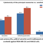 |
Figure 2: Cytotoxic potency (IC50, µM) of ostruthin and 8-methoxyostruthin relative to sorafenib against MDA-MB-231 and HAK1B cells. |
KDM2A (lysine-specific demethylase 2A), an epigenetic regulator over-expressed in liver, breast, lung and gastric cancers and implicated in tumour-cell stemness and angiogenesis, was silenced by siRNA. Western blotting showed a marked reduction of KDM2A protein in transfected (Si97) MDA-MB-231 and HAK1B cells relative to control (SiM); the blots, loading controls and densitometric quantification are provided in the Supporting Information. When challenged with the test compounds, KDM2A-silenced cells were less sensitive than controls to 8-methoxyostruthin at 25 µM, and likewise to sorafenib. These observations derive from a single compound concentration and a single siRNA sequence, and were not accompanied by formal statistical testing, by full dose–response comparison in both conditions, by a second independent siRNA, by rescue through KDM2A over-expression, or by measurement of H3K36me2 or of enzymatic KDM2A activity. They therefore support only the limited conclusion that KDM2A knockdown was associated with reduced cellular sensitivity to 8-methoxyostruthin, and do not establish that the compound inhibits KDM2A or that KDM2A is a direct molecular target.
Reference standards and HPLC assay performance
Ostruthin and 8-methoxyostruthin met primary reference-substance requirements, with certified contents of 99.0% and 97.7% (as-is; n = 12, robust statistics), passing appearance, UV, IR, MS, NMR, TLC, related-substance (≤ 3%) and homogeneity criteria with no significant inter-laboratory difference. The simultaneous assay resolved demethylsuberosin, ostruthin and 8-methoxyostruthin at 7.0, 14.4 and 15.3 min (peak purity > 99.9%) (Figure 3), was linear (R² > 0.99), with LOD 2.84/8.0/3.82 µg/mL and LOQ 8.6/24.2/11.6 µg/mL, RSD < 2% and recovery 95–105%, within the ICH and AOAC acceptance criteria for these characteristics. Cultivated roots (Dong Nai) contained 6.55% ostruthin, 1.04% demethylsuberosin and 0.64% 8-methoxyostruthin (dry basis). Across the ten market samples (M1–M10) composition varied widely—some contained all three markers, several only demethylsuberosin plus ostruthin, others ostruthin alone—all below the cultivated reference (Table 7).
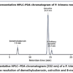 |
Figure 3: Representative HPLC–PDA chromatogram (332 nm) of a P. trimera root extract showing baseline resolution of demethylsuberosin, ostruthin and 8-methoxyostruthin. |
 
Table 7: Performance characteristics of the three-marker HPLC–PDA method determined in this study, and marker content of the cultivated root.
|
Parameter |
Demethylsuberosin | Ostruthin | 8-Methoxyostruthin |
| Retention time (min) | 7.0 | 14.4 |
15.3 |
|
Specificity – peak purity index |
> 0.999 | > 0.999 | > 0.999 |
| Linearity (r2) | > 0.99 | > 0.99 |
> 0.99 |
|
LOD (µg/mL) |
2.84 | 8.0 | 3.82 |
| LOQ (µg/mL) | 8.6 | 24.2 |
11.6 |
|
Precision – RSD (%) |
< 2 | < 2 | < 2 |
| Accuracy – recovery (%) | 95–105 | 95–105 |
95–105 |
|
Content in cultivated root (%, w/w) |
1.04 | 6.55 |
0.64 |
Note: values are those determined in the present study. LOD and LOQ were obtained from the signal-to-noise ratio; precision is the relative standard deviation of replicate determinations and accuracy the recovery of added standard. System-suitability testing, calibration regression equations, separate intermediate-precision data and accuracy at defined spiking levels were not evaluated. Contents are on a dry-weight basis; the marker content of the ten market samples (M1–M10) is described in the text, and their place and month of purchase are given in Materials and Methods.
Discussion
Antioxidant capacity is polyphenol-linked and part-specific. The parallel decline of DPPH activity and polyphenol content across parts (stem > root > leaf) agrees with Nguyen et al., who attributed the antioxidant activity of P. trimera to phenolics, flavonoids and proanthocyanidins acting by hydrogen donation and metal-ion reduction13,14—the same reactive-oxygen-species–buffering chemistry that underpins their cytoprotective role.36 In the present work, however, only total reducing capacity was measured, so no individual phenolic class can be assigned from our data. A distinctive feature here is the parallel use of DPPH and the cell-membrane MDA assay: whereas DPPH captures direct scavenging, the MDA model on liver homogenate reflects the ability of lipophilic constituents to intercalate into membranes and interrupt the peroxidation chain—better aligned with in vivo hepatoprotection.37 That n-hexane and chloroform fractions were most protective explains why polarity-guided isolation converged on lipophilic coumarins, dovetailing with reports that stem and root extracts protect mouse liver against paracetamol toxicity.18,19
Cytotoxicity is concentrated in the roots. Root extracts were the most cytotoxic (HepG2 IC50 24.9 µg/mL), which is consistent both with the screen of the National Institute of Medicinal Materials2 and with the value of about 23 µg/mL reported for a methanol root extract against MCF-7.10 Our HepG2 IC50 is far lower than the 582 µg/mL of Nguyen et al.12; such divergence is expected from differences in plant origin, solvent and composition—variables that, as our market survey shows, strongly affect marker content. Activity across a wider cancer-cell panel has been reported for root and leaf extracts (IC50 ≈ 15–32 µg/mL).14,15 The fractions that were most active in the MDA assay were also the most cytotoxic, which is consistent with, but does not demonstrate, the association between antioxidant chemistry and antiproliferative effect described for polyphenol-rich drugs.37 Antioxidant capacity measured by chemical assays does not establish that antioxidant activity is responsible for the cellular cytotoxicity; differences in extraction efficiency, compound composition, cell-line sensitivity and the presence of other constituents may each contribute independently.
The chemical inventory of the species is further extended. Of the twelve isolates, limonin (PT-8) and PT-9 are reported here for the first time from P. trimera, whereas xanthyletin, oriciacridone and citrusinin-1 confirm the isolations we reported earlier from this species34,35; PT-6 remains a tentative assignment. The identities of the known compounds were established by comparison with published data, including those for xanthyletin44 and for the C-geranyl/C-prenyl 7-hydroxycoumarin series.45 The recurrent 7-hydroxycoumarin scaffold of PT-1, PT-2, PT-7 and PT-9–PT-11 is consistent with the concentration of cytotoxic activity in the less-polar fractions, since coumarins of this series are documented cytotoxic and anti-inflammatory agents.38-40,43 Ostruthin in particular has established antibacterial and antiproliferative activity,41,42 which supports its selection here as both a bioactive constituent and an analytical marker. The two acridones and limonin broaden the structural range of the root extract and may contribute to the multi-target activity of the crude drug,20,46 although their individual contributions were not dissected in this study.
An exploratory observation links 8-methoxyostruthin to KDM2A. Earlier mechanistic accounts of ostruthin-type coumarins centred on redox and NF-κB–dependent anti-inflammatory signalling.43 KDM2A is a JmjC histone H3K36 demethylase that promotes proliferation, stemness, ribosomal-RNA synthesis and tumour angiogenesis and is over-expressed in liver, breast, lung and gastric cancers47-52; in hepatocellular carcinoma it is recruited by ZHX2 to demethylate H3K36 at stemness genes,49 and it drives breast-tumour growth by silencing tumour-suppressor genes via TET2.48 In the present work, siRNA knockdown of KDM2A reduced the sensitivity of MDA-MB-231 and HAK1B cells to 8-methoxyostruthin at 25 µM. Sorafenib, the reference systemic agent for advanced hepatocellular carcinoma,53 was included as a clinically relevant comparator. We emphasise that this result is correlative and exploratory: it was obtained at one concentration with one siRNA, without target-engagement evidence, and it is equally compatible with KDM2A acting upstream of, or in parallel to, the cytotoxic pathway involved. It should therefore not be read as evidence that 8-methoxyostruthin inhibits KDM2A, that KDM2A is a direct target, or that an epigenetic mechanism operates across this class of coumarins. Establishing any of these will require dose–response analysis in both knockdown and control conditions, a second independent siRNA, over-expression rescue, enzymatic KDM2A assays, H3K36me2 quantification and molecular docking.
Analytical quality control addresses a real authentication gap. P. trimera has no pharmacopoeial monograph, and the near-extinction of wild stocks has driven adulteration—notably with Luvunga scandens, which also contains ostruthin—so quantifying ostruthin alone cannot authenticate the drug. Building on earlier ostruthin-only6 and ostruthin/8-methoxyostruthin assays, and on multi-coumarin HPLC-DAD-MS approaches used for related Rutaceae/Apiaceae drugs,54 this work certifies ostruthin and 8-methoxyostruthin reference standards31 and characterises a single method that additionally quantifies demethylsuberosin, moving toward a multi-marker fingerprint. The large gap between cultivated root (6.55% ostruthin) and market samples (0.28–2.27%), and the frequent absence of one or more markers, quantify the heterogeneity of traded material and argue for a marker-panel specification rather than a single-analyte limit in a future monograph. Because marker content varied with growing region, plant age and harvest time, agronomic standardisation will be as important as analytical standardisation.
Limitations. Activities were assessed in vitro; ostruthin and 8-methoxyostruthin are only moderately potent relative to sorafenib, and selectivity toward non-malignant cells was not examined. The KDM2A observation is correlative and exploratory, as set out above. Structural work was limited by the absence of high-resolution mass spectrometry, so the molecular formula of PT-6 is not formally confirmed and its structure is reported as tentative; PT-6 was also isolated in too small an amount (4 mg) for biological testing. These define clear priorities for follow-up: in vivo validation, direct KDM2A target-engagement studies, scaled re-isolation of PT-6 with HR-ESI-MS and full stereochemical characterisation, deeper study of the leaf, and inclusion of a multi-marker specification in a pharmacopoeial monograph. The market survey covered only ten samples from five provinces and was not designed as a statistically representative sampling of the Vietnamese market; the reported ranges therefore illustrate the heterogeneity of traded material rather than quantify its prevalence. Provenance could not be documented beyond the place and month of purchase, because none of the vendors could state the geographical origin, the harvest date, or the wild or cultivated status of the material they sold. That absence of chain of custody is itself one of the conditions the present assay is intended to address, since material whose origin cannot be traced can still be characterised chemically; larger randomised surveys with documented provenance will nevertheless be needed before a marker-panel specification is set.
Conclusion
A bioactivity-guided investigation of P. trimera localised the strongest cytotoxic and lipid-peroxidation–protective activity to the roots and their less-polar fractions. Twelve compounds were characterised; limonin and PT-9 are reported for the first time from the species, xanthyletin, oriciacridone and citrusinin-1 confirm our earlier isolations, and PT-6 remains tentatively assigned pending high-resolution data. Ostruthin and 8-methoxyostruthin showed reproducible cytotoxicity in vitro and were adopted as analytical markers, and KDM2A knockdown was associated with reduced cellular sensitivity to 8-methoxyostruthin—an exploratory, correlative finding that requires direct target-engagement studies before any mechanistic conclusion can be drawn. Certified reference standards and a three-marker HPLC assay were delivered and applied, exposing wide compositional variability in traded roots. These in-vitro findings do not establish therapeutic efficacy; their value lies in providing a chemical and analytical basis for the standardisation and further study of this medicinal plant.
Acknowledgement
The authors thank the botanical specialist who authenticated the plant material, and the Quang Minh medicinal-plant farm (Dong Nai Province, Vietnam) for supplying the cultivated roots used in method development.
Conflict of Interest
The authors do not have any conflict of interest.
Funding Sources
This research did not receive any specific grant from funding agencies in the public, commercial, or not-for-profit sectors.
Data Availability Statement
The manuscript incorporates all datasets produced or examined throughout this research study. Additional spectroscopic and chromatographic data supporting the structural assignments are provided in the Supporting Information, and any further details are available from the corresponding author on reasonable request.
Ethics Statement
This research did not involve human participants; only established commercial cell lines and rat liver homogenate, used in vitro as an assay substrate, were employed, and no experiments were performed on live animals. Plant material was collected in compliance with national regulations, with voucher specimens (XTP 01-2013 and XTP 02-2017) deposited at the Faculty of Pharmacy, University of Medicine and Pharmacy at Ho Chi Minh City.
Informed Consent Statement
This study did not involve human participants, and therefore, informed consent was not required.
Clinical Trial Registration
This research does not involve any clinical trials.
Permission to Reproduce Material from Others Sources
Not Applicable.
Author Contributions
- Tran Thi Thuy Quynh: Conceptualization, Investigation (plant collection, extraction, fractionation and isolation), Formal analysis, Writing – original draft.
- Tran Hoang Khanh: Methodology (HPLC method development and performance assessment), Investigation (reference substance characterisation and market survey), Data curation, Writing – original draft.
- Nguyen Bao Phuc: Investigation (antioxidant assays, MTT cytotoxicity, KDM2A knockdown and Western blot), Visualization, Writing – review and editing.
- Vuong Thi Huyen Trang: Conceptualization, Investigation (spectroscopic structure elucidation), Resources, Supervision, Project administration, Writing – review and editing.
References
- Pham HH. Cây cỏ Việt Nam [An Illustrated Flora of Vietnam], vol. 2. Ho Chi Minh City: Tre Publishing House; 2003:439.
- National Institute of Medicinal Materials. Report on Paramignya trimera (Xao tam phan), Official Document No. 539/VDL-QLKHĐT. Hanoi/Khanh Hoa; 2012.
- Sung H, Ferlay J, Siegel RL, et al. Global cancer statistics 2020: GLOBOCAN estimates of incidence and mortality worldwide for 36 cancers in 185 countries. CA Cancer J Clin. 2021;71(3):209-249.
CrossRef - Bray F, Laversanne M, Sung H, et al. Global cancer statistics 2022: GLOBOCAN estimates of incidence and mortality worldwide for 36 cancers in 185 countries. CA Cancer J Clin. 2024;74(3):229-263. doi:10.3322/caac.21834
CrossRef - Nguyen MC, Ho VD, Nguyen VT, et al. Initial study on the chemical constituents of Paramignya trimera (Rutaceae). Vietnam J Chem. 2013;51:292-296.
- Pham HB, Vu VT, Nguyen THL, Nguyen MK. Isolation and quantification of ostruthin in Paramignya trimera collected in Vietnam. J Med Mater (Vietnam). 2013;18(3):173-178.
- Nguyen MC, Tran TH, Pham NK, Kim YH. Paratrimerins A and B, two new dimeric monoterpene-linked coumarin glycosides from the roots and stems of Paramignya trimera. Chem Pharm Bull. 2015;63(11):945-949. doi:10.1248/cpb.c15-00336
CrossRef - Nguyen MTT, Dang PH, Nguyen NT, et al. Paratrimerin I, a cytotoxic acridone alkaloid from the roots of Paramignya trimera. Nat Prod Res. 2021;35(23):5042-5047. doi:10.1080/14786419.2020.1774760
CrossRef - Quan KT, Park HB, Yuk H, Lee SJ, Na M. Paratrimerins J–Y, dimeric coumarins isolated from the stems of Paramignya trimera. J Nat Prod. 2021;84(2):310-326. doi:10.1021/acs.jnatprod.0c00978
CrossRef - Son NT. Notes on the genus Paramignya: phytochemistry and biological activity. Bull Fac Pharm Cairo Univ. 2018. doi:10.1016/j.bfopcu.2017.12.001
CrossRef - Bailly C. Ruta angustifolia Pers. (narrow-leaved fringed rue): pharmacological properties and phytochemical profile. Plants (Basel). 2023;12(4):827. doi:10.3390/plants12040827
CrossRef - Nguyen ST, Do NM, Vo PH, et al. Xao tam phan (Paramignya trimera) methanol extract induces apoptosis in the hepatocellular carcinoma HepG2 cell line in vitro. Sci Technol Dev J. 2020;23(1). doi:10.32508/stdj.v23i1.2013
CrossRef - Nguyen VT, Bowyer MC, Vuong QV, Van Altena IA, Scarlett CJ. Phytochemicals and antioxidant capacity of Paramignya trimera root as affected by various solvents and extraction methods. Ind Crops Prod. 2015;67:192-200. doi:10.1016/j.indcrop.2015.01.051
CrossRef - Nguyen VT, Sakoff JA, Scarlett CJ. Physicochemical, antioxidant and cytotoxic properties of Paramignya trimera root extract and its fractions. Chem Biodivers. 2017;14(4):e1600396. doi:10.1002/cbdv.201600396
CrossRef - Nguyen VT, Sakoff JA, Scarlett CJ. Physicochemical properties, antioxidant and anti-proliferative capacities of dried leaf and its extract from Xao tam phan (Paramignya trimera). Chem Biodivers. 2017;14(6):e1600498. doi:10.1002/cbdv.201600498
CrossRef - Nguyen Thi KO, Le TH, Nguyen HD, et al. Phytochemical composition and potential bioactivities of the essential oil from the peels of Paramignya trimera (Oliv.) Guillaum from Vietnam. J Essent Oil Bear Plants. 2022;25(2):273-281. doi:10.1080/0972060X.2022.2086637
CrossRef - Le TH, Nguyen LA, Tran NB, et al. Biosynthesis of gold nanoparticles using aqueous extract of Paramignya trimera roots and its anticancer activity. J Biomed Nanotechnol. 2024;20(2):330-340.
CrossRef - Nguyen MK, Pham TNH, Do TP. Study on acute toxicity, hepatoprotective and cytotoxic activity of Paramignya trimera. J Med Mater (Vietnam). 2013;18(1):14-20.
- Nguyen MC, Tran TH, Pham NK. Hepatoprotective effect of ostruthin and 8-methoxyostruthin against paracetamol-induced liver injury in mice. Vietnam J Sci Technol. 2016;54(1):37-45.
- Le TAH, Kim DC, Ko W, et al. Anti-inflammatory coumarins from Paramignya trimera. Pharm Biol. 2017;55(1):1195-1201. doi:10.1080/13880209.2017.1296001
CrossRef - Trinh DH, Tran PT, Trinh BTD. Coumarins and acridone alkaloids with α-glucosidase inhibitory and antioxidant activity from the roots of Paramignya trimera. Phytochem Lett. 2020;35:94-98. doi:10.1016/j.phytol.2019.10.010
CrossRef - Ciulei I. Methodology for analysis of vegetable drugs. Bucharest: Ministry of Chemical Industry; 1984.
- Ministry of Health of Vietnam. Vietnamese Pharmacopoeia V, appendices 9.6, 9.7, 9.8. Hanoi: Medical Publishing House; 2018.
- Cicco N, Lanorte MT, Paraggio M, Viggiano M, Lattanzio V. A reproducible, rapid and inexpensive Folin–Ciocalteu micro-method in determining phenolics of plant methanol extracts. Microchem J. 2009;91:107-110. doi:10.1016/j.microc.2008.08.011
CrossRef - Kulisic T, Radonic A, Katalinic V, et al. Use of different methods for testing antioxidative activity of oregano essential oil. Food Chem. 2004;85(4):633-640. doi:10.1016/j.foodchem.2003.07.024
CrossRef - Munteanu IG, Apetrei C. Analytical methods used in determining antioxidant activity: a review. Int J Mol Sci. 2021;22(7):3380. doi:10.3390/ijms22073380
CrossRef - Ohkawa H, Ohishi N, Yagi K. Assay for lipid peroxides in animal tissues by thiobarbituric acid reaction. Anal Biochem. 1979;95(2):351-358.
CrossRef - Sylvester PW. Optimization of the tetrazolium dye (MTT) colorimetric assay for cellular growth and viability. Methods Mol Biol. 2011;716:157-168. doi:10.1007/978-1-61779-012-6_9
CrossRef - Ghasemi M, Turnbull T, Sebastian S, Kempson I. The MTT assay: utility, limitations, pitfalls, and interpretation in bulk and single-cell analysis. Int J Mol Sci. 2021;22(23):12827. doi:10.3390/ijms222312827
CrossRef - Towbin H, Staehelin T, Gordon J. Electrophoretic transfer of proteins from polyacrylamide gels to nitrocellulose sheets: procedure and some applications. Proc Natl Acad Sci USA. 1979;76(9):4350-4354. doi:10.1073/pnas.76.9.4350
CrossRef - International Organization for Standardization. ISO 13528: Statistical methods for use in proficiency testing by interlaboratory comparison. Geneva: ISO; 2015.
- ICH Harmonised Tripartite Guideline. Q2(R1): Validation of analytical procedures: text and methodology. Geneva: ICH; 2005.
- AOAC International. Appendix F: Guidelines for standard method performance requirements. In: Official Methods of Analysis of AOAC International. 21st ed. Rockville, MD: AOAC International; 2019.
- Tran TTQ, Le TKT, Pham DP. Isolation of coumarins from the roots of Paramignya trimera. J Pharm Vietnam. 2014;54(5):60-66.
- Tran TTQ, Nguyen TD, Pham DP. Isolation of acridone alkaloids from the roots of Paramignya trimera. J Pharm Vietnam. 2014;54(6):60-64.
- Hasanuzzaman M, Bhuyan MHMB, Zulfiqar F, et al. Reactive oxygen species and antioxidant defense in plants under abiotic stress: revisiting the crucial role of a universal defense regulator. Antioxidants. 2020;9(8):681. doi:10.3390/antiox9080681
CrossRef - Rudrapal M, Khairnar SJ, Khan J, et al. Dietary polyphenols and their role in oxidative stress-induced human diseases: insights into protective effects, antioxidant potentials and mechanism(s) of action. Front Pharmacol. 2022;13:806470. doi:10.3389/fphar.2022.806470
CrossRef - Önder A. Anticancer activity of natural coumarins for biological targets. Stud Nat Prod Chem. 2020;64:85-109. doi:10.1016/B978-0-12-817903-1.00003-6
CrossRef - Pal D, Saha S. Coumarins: an important phytochemical with therapeutic potential. In: Plant-Derived Bioactives: Chemistry and Mode of Action. Singapore: Springer; 2020:205-222. doi:10.1007/978-981-15-2361-8_9
CrossRef - Shahbaz M, Naeem H, Imran M, et al. Recent perspectives on anticancer potential of coumarin against different human malignancies: an updated review. Food Sci Nutr. 2025;13(1):e4696. doi:10.1002/fsn3.4696
CrossRef - Peter S, Sibali LL. Recent developments on coumarin hybrids as antimicrobial agents. Antibiotics (Basel). 2025;14(12):1226. doi:10.3390/antibiotics14121226
CrossRef - Joa H, Vogl S, Atanasov AG, et al. Identification of ostruthin from Peucedanum ostruthium rhizomes as an inhibitor of vascular smooth muscle cell proliferation. J Nat Prod. 2011;74(6):1513-1516. doi:10.1021/np200072a
CrossRef - Di Stasi LC. Coumarin derivatives in inflammatory bowel disease. Molecules. 2021;26(2):422. doi:10.3390/molecules26020422
CrossRef - Cazal CM, Domingues VC, Batalhão JR, et al. Isolation of xanthyletin, an inhibitor of ants’ symbiotic fungus, by high-speed counter-current chromatography. J Chromatogr A. 2009;1216(19):4307-4312. doi:10.1016/j.chroma.2009.02.066
CrossRef - Rashid MA, Armstrong JA, Gray AI, Waterman PG. Novel C-geranyl 7-hydroxycoumarins from the aerial parts of Eriostemon tomentellus. Z Naturforsch B. 1992;47(2):284-287.
CrossRef - Fan S, Zhang C, Luo T, et al. Limonin: a review of its pharmacology, toxicity, and pharmacokinetics. Molecules. 2019;24(20):3679. doi:10.3390/molecules24203679
CrossRef - Liu L, Liu J, Lin Q. Histone demethylase KDM2A: biological functions and clinical values (review). Exp Ther Med. 2021;22(1):723. doi:10.3892/etm.2021.10155
CrossRef - Chen JY, Luo CW, Lai YS, Wu CC, Hung WC. Lysine demethylase KDM2A inhibits TET2 to promote DNA methylation and silencing of tumor suppressor genes in breast cancer. Oncogenesis. 2017;6(8):e369. doi:10.1038/oncsis.2017.71
CrossRef - Lin Q, Wu Z, Yue X, et al. ZHX2 restricts hepatocellular carcinoma by suppressing stem cell-like traits through KDM2A-mediated H3K36 demethylation. EBioMedicine. 2020;53:102676. doi:10.1016/j.ebiom.2020.102676
CrossRef - Wang J, Zhang ZY, Jiang J. KDM2A plays a dual role in regulating the expression of malignancy-related genes in esophageal squamous cell carcinoma. Biochem Biophys Res Commun. 2022;623:53-58. doi:10.1016/j.bbrc.2022.07.035
CrossRef - Chen JY, Li CF, Lai YS, Hung WC. Lysine demethylase 2A expression in cancer-associated fibroblasts promotes breast tumour growth. Br J Cancer. 2020;124(2):484-493. doi:10.1038/s41416-020-01112-z
CrossRef - De Nicola I, Guerrieri AN, Penzo M, et al. Combined expression levels of KDM2A and KDM2B correlate with nucleolar size and prognosis in primary breast carcinomas. Histol Histopathol. 2020;35(10):1181-1187. doi:10.14670/HH-18-248
- Llovet JM, Ricci S, Mazzaferro V, et al. Sorafenib in advanced hepatocellular carcinoma. N Engl J Med. 2008;359(4):378-390. doi:10.1056/NEJMoa0708857
CrossRef - Noviana E, Indrayanto G, Rohman A. Advances in fingerprint analysis for standardization and quality control of herbal medicines. Front Pharmacol. 2022;13:853023. doi:10.3389/fphar.2022.853023
CrossRef
Abbreviations
ANOVA: analysis of variance;
AOAC: Association of Official Analytical Collaboration;
ATCC: American Type Culture Collection;
COSY: correlation spectroscopy; COX-2: cyclooxygenase-2;
DEPT: distortionless enhancement by polarisation transfer;
DMEM: Dulbecco’s modified Eagle medium;
DMSO: dimethyl sulfoxide;
DPPH: 2,2-diphenyl-1-picrylhydrazyl;
DSC: differential scanning calorimetry;
EC50: half-maximal effective concentration;
EMEM: Eagle’s minimum essential medium;
ESI-MS: electrospray-ionisation mass spectrometry;
GLP: good laboratory practice;
HMBC: heteronuclear multiple-bond correlation;
HPLC-PDA: high-performance liquid chromatography with photodiode-array detection;
HR-ESI-MS: high-resolution electrospray-ionisation mass spectrometry;
HSQC: heteronuclear single-quantum coherence;
IC50: half-maximal inhibitory concentration;
ICH: International Council for Harmonisation;
iNOS: inducible nitric oxide synthase;
IR: infrared;
ISO: International Organization for Standardization;
KDM2A: lysine demethylase 2A;
LOD: limit of detection;
LOQ: limit of quantification;
LPS: lipopolysaccharide;
MDA: malondialdehyde;
MTT: 3-(4,5-dimethylthiazol-2-yl)-2,5-diphenyltetrazolium bromide;
NMR: nuclear magnetic resonance;
NO: nitric oxide; PGE: pyrogallol equivalents;
PGE2: prostaglandin E2;
RSD: relative standard deviation;
SD: standard deviation;
SEM: standard error of the mean;
siRNA: small interfering RNA;
TLC: thin-layer chromatography;
UV: ultraviolet;
VLC: vacuum-liquid chromatography

Accepted on: 02-11-2026
Second Review by: Dr. Kanaka Durga Devi Nelluri and Dr. Dhara Patel
Final Approval by: Dr. Eman Refaat Youness