In Silico Identification of FDA Approved Drug Candidates Targeting the RNA-Binding Region of Andes Hantavirus Nucleocapsid Protein

Department of pharmacology, College of pharmacy, Al-Zahrawi University, Karbala, Iraq
Corresponding author E-mail: hodhar3@gmail.com
DOI : http://dx.doi.org/10.13005/bpj/3516
Download this article as:

The Andes virus is a rodent-born RNA microorganism that can cause a serious hantavirus pulmonary syndrome (HPS) with a mortality rate approaching 50%. The recent outbreaks of the Andes virus, specifically in south America, represent an emerging public health challenge due to the risk of human-to-human transmission and the lack of approved therapeutics or vaccines. Therefore, the objective of this in silico screening is to recognize FDA drugs candidates that can target the RNA binding site of the Andes virus nucleocapsid protein (NP). Such drug candidates may possess the potential to adversely interfere with the assembly and replication of the viral particles. For this purpose, molecular docking was first applied to screen 1,615 drug molecules against NP crystal. Then, the best five hits were identified and subjected to a molecular dynamics (MD) simulation for a duration of 50 nanoseconds (ns). These results of the MD simulation were analyzed by considering the ligand movement root mean square deviation (RMSD), the radius of gyration (Rg) for solute, the kind of per residue contact with the ligand, and the molecular mechanics Poisson Boltzmann Surface Area (MMPBSA) binding energy. As a result, the computational docking has initially identified five potential hit drugs and these are: Dihydroergotamine, Conivaptan, Dutasteride, Telmisartan, Lifitegrast. Then, the molecular dynamics simulation (MD) has excluded the drug Dutasteride as it seemed to have unfavorable binding energy. On the contrary, the other four drugs are anticipated to have a ligand proximity to NP binding site that didn’t exceed 4.0 Å during 50 ns duration. In fact, the best ligand proximity and binding energy was computed to the drug Lifitegrast during the simulation study. In fact, both Telmisartan and Lifitegrast are believed to be involved in a salt bridge interaction with the Arg 367 and Arg 146 residues respectively in the Andes virus NP binding region. In conclusion, both Lifitegrast and Telmisartan seem to be prioritized computational candidates against RNA binding area of the Andes virus NP target. But, these in silico findings are considered preliminary requiring further experimental validation.
KEYWORDS:Andes hantavirus; FDA approved drug; In silico; Nucleocapsid; RNA-binding region
Introduction
Hantaviruses are zoonotic pathogens that belong to the Hantaviridae family of viruses within the Bunyavirales order. Usually, each hantavirus has a natural rodent reservoir where it can cause asymptomatic and long-term infection.1 The possibility of viral spillover to human is considered rare. However, both climate changes and increased human rural activities have boosted the chances of viral transmission to other species like human.2 Based on the virus type and the geographical place, the hantaviruses can either cause a pulmonary syndrome or a hemorrhagic renal syndrome. The hantavirus pulmonary syndrome (HPS) is mainly occurred in the Americas and called the new world disease.3 One the other hand, the hemorrhagic fever renal syndrome (HFRS) form is usually caused by the hantaviruses in the old-world parts like Europe and Asia.4 As compared to the renal form of hantavirus disease, the pulmonary syndrome has a higher fatality rate that can reaches up to 50%.5 In both renal and pulmonary disease forms, the virus mainly infect the vascular endothelial cells resulting in increase in capillary permeability.6 Clinically, the incubation period for hantavirus diseases can vary between 2 and 3 weeks but in some instances the disease symptoms can be manifested after 40 days of infection. The transmission of infection usually happens through inhalation of contaminated aerosols or contacts with infected rodents’ excreta.7 Initially, the hantavirus diseases symptoms encompass fever, malaise, myalgia, diarrhea and nausea. Later, the disease can progress into hypotension with cardiogenic shock. Moreover, the HFRS patients can develop renal impairment while the HPS is characterized by lungs edema and failure.8 Diagnosis of the hantavirus diseases usually rely on clinical examination, patient’s history, serological tests and molecular techniques like real time polymerase chain reaction (RT-PCR).9 No specific treatment or vaccine is available for hantavirus diseases, and management only include supportive care and use of viral replication inhibitors like Ribavirin or Favipiravir.10
At the beginning, the hantavirus diseases were thought to be transmitted by only zoonotic means. But the epidemiological evidences gained by studying both the 1996 and the 2018 outbreaks in Argentina have uncovered the possibility of human-to-human transmission of the virus. It is a known fact that the Andes virus can be transmitted by the close and prolonged contacts between individuals.11 Recently, the threat posed by Andes virus has regained attention of the publics after the reported outbreak among passengers of the MV Hondius cruise ship between April and May 2026. This latest outbreak highlights the gaps and challenges related to clinical, diagnostic and epidemiological preparedness.12
The hantaviruses have RNA genome that consists of three main parts: the small (S), medium (M) and large (L) segments. These three genomic segments encode for the viral nucleocapsid protein (NP), glycoprotein precursors and RNA-dependent RNA polymerase (RdRp) respectively.13 Among these viral proteins, the hantavirus NP represents an interesting molecular target to design novel antiviral agents. This is because the NP is believed to adjust both viral assembly and proliferation processes through interaction with other viral proteins like Gn glycoprotein and RdRp 14,15. Also, the hantavirus NP is postulated to intervene with host immune system by inhibition of interferon signaling and downregulation of apoptosis.16,17 Consequently, the successful block of the Andes virus NP can inhibit the viral replication and assembly.
However, the development of clinically active inhibitor of hantavirus NP is restricted by several hurdles. First, the dynamics of trimeric NP that binds and interacts with viral RNA panhandle is currently not fully understood.18 Also, it is expected that the block of hantavirus NP may only inhibit viral replication without affecting other pathogenic roles of NP like evasion of host immunity. Finally, the lack of suitable animal models and genetic difference among viral variants may pose further challenges for therapeutics development.19 Several former attempts were made to repurpose antiviral drugs or harness phytocompounds against hantavirus molecular targets. Yet, no candidate has ever approved by FDA as an effective anti-hantavirus.20,21 Therefore, it is of interest to virtually screen a library of FDA approved drugs against RNA binding site in the Andes virus NP crystal. The aim of this computational study is to harness both molecular docking and dynamics simulation to repurpose FDA approved drugs as potential inhibitor of the Andes virus NP.
Materials and Methods
Structure-based computational screening:
The molecular docking is a computational technique that applies a conformations sampling algorithm and a scoring function to predict the affinity and the binding pose of the ligand to target crystal. In principle, the molecular docking anticipates the ligand pose with the least energy of binding by generating a static vision of hit-target complex.22 For this objective, the Mcule.com drug discovery website was employed to virtually screen a database of 1,615 FDA approved drugs against Andes virus nucleocapsid crystal (PDB: 5E04).13 The used FDA approved drugs library was obtained as SDF file from the ZINC20 online repository.23 After uploading both target crystal and drugs library to Mcule.com, the website then used the AutoDockTools version 1.5.6 to prepare protein and ligands for the docking step.24 It is useful to indicate that the Mcule.com platform utilizes the AutoDock Vina version 1.2.0 to execute the docking operation.25 For the current virtual screening, the employed docking coordinates were X: -35, Y: -21, Z: 26 while the applied grid box dimensions were the default size values of 22*22*22 Angstrom (Å). These applied docking coordinates and dimensions were determined with the help of previous crystallographic data and also by using the binding pockets detection tool available in the DrugRep server.13,26 The available crystallographic data revealed that the Andes virus NP is composed of two parts: the N and C lobes that folds together. As observed in Figure 1, the RNA binding site is situated at the interface between these two lobes in a positively charged region.13 By the end of the docking stage, the output hits were arranged depending on its predicted energy of binding. Only the best five hit compounds with the smallest energy of binding were then picked for the visualization of their docking pose with the least energy of binding. For each of these five hits, PyMOL version 2.4.1 and the BIOVIA Discovery Studio Visualizer 2024 were used to view and examine the best docking pose with the smallest energy of binding. Both Medscape and PubChem websites were also explored for the clinical indications and chemical formula of each selected hit compound.
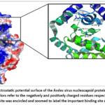 |
Figure 1: The electrostatic potential surface of the Andes virus nucleocapsid protein (NP) where the red and blue colors refer to the negatively and positively charged residues respectively. |
Molecular dynamics (MD) simulation study:
In this next level of the virtual screening, the best five docking hits were selected and subjected to the MD simulation study for 50 nanoseconds (ns) duration. Unlike the docking step, the MD study simulates the binding of a ligand to the target protein under physiological conditions by using dynamic forcefields. The MD simulation assesses the flexible motion and interactions of both drug and target protein by mathematically calculating forces like angle bending and bond stretching.27 To carry out the required simulation step, the program YASARA Dynamics version 20.12.24 was employed.28 For each of the selected top hits, the binding pose with the smallest energy was submitted to the MD simulation using options and parameters akin to what was employed in previously published virtual studies.29,30 Concisely, the current MD simulation methodology did encompass the addition of 0.9% of sodium chloride to the system. And to secure the neutralization of the complex, a surplus of either sodium or chloride ions were included. Also, the reductions of steepest descent and simulated annealing were applied to eliminate potential clashes inside simulation system. An optimization for the H-bonding was included to increase the stability of the solute, while a pKa anticipation was performed to adjust the protonation of all amino acids at pH limit of 7.4.31 Moreover, the simulation was performed with the help of the following forcefields: AMBER14 for solute, TIP3P for water, AM1BCC and GAFF2 for submitted ligands.32–34 The current MD study was performed under conditions similar to that of the biological environment with a temperature of 310 kelvin and a pressure of 1.0 atmosphere. The motion equations were employed in this simulation as 1.25 and 2.5 femtoseconds timesteps for bonded and non-bonded interactions respectively. After validation of the solute RMSD as a function of simulation duration, the 50 ns were regarded as an equilibration time and precluded from more analysis.35 By the end of the simulation duration, the results were analyzed by considering the ligand RMSD, Rg for the solute, kind of the per residue contact with the ligand, and the MMPBSA binding energy. It is worth mentioning that the following equation was employed by YASARA Dynamics to compute the MMPBSA binding energy:

Results
After finishing the virtual screening process, the hit drugs were arranged based on their docking energy calculated against hantavirus NP crystal. As can be seen in Table 1, only the best five hit drugs were presented with the least calculated energy of binding. This predicted docking energy in Table 1 seems to be ranging between -8.6 and -8.9 Kcal/ mol for these hits. According to Medscape database, the hit drugs in Table 1 appears to be prescription only medications with different clinical indications. In addition, the structural formula for these drugs reveals that the chemical nature of these hits is dissimilar.
Table 1: A tabular list of the molecular formula and medical indications for the best five hits generated by virtually screening FDA approved drugs against Andes virus nucleocapsid protein (NP). These listed hits were ranked based on their least docking energy.
|
No. |
Drug name | Chemical formula | Docking energy (Kcal/ mol) |
Indications |
|
1 |
Dihydroergotamine | C33H37N5O5 | -8.9 | Migraine headache |
| 2 | Conivaptan | C32H26N4O2 | -8.8 |
Hyponatremia |
|
3 |
Dutasteride | C27H30F6N2O2 | -8.8 | Benign prostate hyperplasia |
| 4 | Telmisartan | C33H30N4O2 | -8.8 |
Hypertension |
|
5 |
Lifitegrast | C29H24Cl2N2O7S | -8.6 |
Dry eye syndrome |
Later on, for each of these five hits, the docking complex was subjected to the MD simulation study for an interval of 50 ns. A concise summary for the results of this MD study is presented in Table 2 where the ligand movement RMSD, radius of gyration and binding energy were reported as an average parameter. It is evident from this table that the least ligand movement RMSD and MM-PBSA binding energy were computed to the drug Lifitegrast while the highest ones were reported to the hit Dutasteride. Generally, all the listed drugs in Table 2 were able to accomplish a ligand proximity to NP binding site that didn’t pass the level of 4 Å with the exception of Dutasteride. In the same direction, the most unfavorable MD binding energy was calculated for the drug Dutasteride with a value of -18.78 Kcal/ mol. Moreover, all the drugs presented in Table 2 are expected to form a stable complex with hantavirus NP crystal with an average radius of gyration (Rg) ranging between 18.12 and 18.33 Å. Of interest, the average Rg value was also calculated to the unbound (free) NP crystal and it was 18.50 Å which is slightly higher than that for the bound NP protein.
Table 2: Analysis of the molecular dynamics (MD) simulation outputs for the best hit drugs as a function of simulation time period.
|
No. |
Drug name | Average MD simulation outputs | ||
| Ligand movement RMSD (Å) | Radius of gyration Rg (Å) |
MM-PBSA binding energy (Kcal/ mol) |
||
|
1 |
Dihydroergotamine | 3.97 | 18.33 | -35.07 |
| 2 | Conivaptan | 3.86 | 18.28 |
-37.07 |
|
3 |
Dutasteride | 7.51 | 18.20 | -18.78 |
| 4 | Telmisartan | 3.51 | 18.12 |
-34.71 |
|
5 |
Lifitegrast | 3.03 | 18.20 |
-86.75 |
MD: Molecular dynamics; RMSD: Root mean square deviation; Å: Angstrom; Rg: Radius of gyration; MM-PBSA: Molecular mechanics Poisson Boltzmann Surface Area.
Furthermore, the inspection of the plot in Figure 2 provides a detailed anticipation for each drug movement RMSD away from target binding site throughout simulation. Based on this plot, it is easy to notice that the drug Dutasteride would deviate and move far from NP binding site as the simulation interval advances. And by the end of the simulation time interval, the Dutasteride would record a binding proximity of nearly 20 Å.
 |
Figure 2: A plot for each drug movement RMSD (Å) against the MD simulation time (ns). Click here to View Figure |
In addition, a thorough plot for the radius of gyration (Rg) parameter of each drug as a function of MD simulation time is given in Figure 3. For each drug, the Rg plot was compared to that of the free (unbound) NP target. Overall, as noted from Figure 3, the Rg plot for each of these five drugs was slightly lower than that for the unbound NP. But, the most remarkable difference in Rg plots was recognized between Telmisartan-NP complex and the free NP crystal as seen in Figure 3. Interestingly, the least Rg average value was also computed for the Telmisartan-NP complex as noted in MD summary results in Table 2.
 |
Figure 3: Detailed plots for the best screening drugs’ radius of gyration (Rg) versus simulation time. Click here to View Figure |
Then, a plot was generated for each drug to illustrate the interactions of the ligand with each residue in the NP binding site as seen in Figure 4. According to this figure, the interactions throughout simulation duration with each target residue are shown as red, green or blue colors for the hydrogen, hydrophobic and ionic interactions respectively. And when a residue is involved in more than one kind of interaction with the ligand, then a combination of these three colors can be employed as indicated in the Figure 4. Based on the per residue contacts analysis in this figure, it is evident that the drug Dutasteride will loss many of its contacts with target binding site as the simulation advances in duration. Moreover, both Telmisartan and Lifitegrast were able to maintain ionic and hydrogen bond interactions with NP binding site throughout 50 ns duration.
 |
Figure 4: A plot for the type of interaction between each residue in the nucleocapsid binding site and the drug molecule under investigation: (A) Dihydroergotamine, (B) Conivaptan, (C) Dutasteride (D) Telmisartan and (E) Lifitegrast. |
Lastly, a percentile presentation was created for the interaction of each drug with the Andes virus NP binding site residues as seen in Figure 5. In this figure, for each plot, the percentile and the type of contact are illustrated per each active site residue. As seen in this figure, the least contacts percentile was observed in plot (C) for the drug Dutasteride followed by Conivaptan in plot (B). Interestingly, both Telmisartan and Lifitegrast were able to participate in ionic and hydrogen bonds throughout most of the simulation time with Arg 367 and Arg 146 binding site residues respectively.
 |
Figure 5: A percentile plot for the per residue contact made with drugs under assessment: (A) Dihydroergotamine, (B) Conivaptan, (C) Dutasteride (D) Telmisartan and (E) Lifitegrast. |
Further, the per residue type of interaction was more assessed by examining the docking images for the Telmisartan and Lifitegrast poses with the least energy of binding to NP crystal. Based on the two-dimensional representation in Figure 6, both Telmisartan and Lifitegrast were engaged in a salt bridge interaction with Arg 367 and Arg 146 respectively in the binding site of NP crystal.
 |
Figure 6: A two-dimensional representation for the interaction between either (A) Telmisartan or (B) Lifitegrast with the Andes virus nucleocapsid protein binding site. Click here to View Figure |
Discussion
During the past years, the public interests in hantavirus disease was restricted and confined to particular endemic regions. However, the recent outbreak of Andes virus strain in the MV Hondius cruise ship has attracted global attention to this pathogen as a potential public health threat. This health challenge is emerging from the fact that no vaccine or therapy are clinically available to combat hantavirus. Besides, the Andes hantavirus reported onboard the cruise ship is the only strain with a human-to-human transmission risk. This hantavirus strain is clinically known to cause a pulmonary disease with a high fatality rate of 50%.36 Consequently, it is of interest to virtually explore and repurpose the approved drugs molecules as potential anti-hantavirus therapeutics. For this purpose, both docking tools and molecular dynamics (MD) simulation software were utilized in this study to screen the drugs library against Andes virus nucleocapsid protein (NP). The NP protein is a well-recognized molecular target in RNA viruses that plays a role in viral genomic encapsidation.13 As such, the aim of this computational study is to identify a small drug molecule that can interact with the RNA binding site of NP in a way that impedes the replication of viral particles.
At first, docking study indicated that the best five drugs with the least energy of binding are having dissimilar chemical nature and clinical indications. As provided in Table 1, these top drugs do have a docking energy of more than -8.5 Kcal/ mol when screened against Andes virus NP. Later on, these five hit drugs were subjected to 50 ns duration of MD simulation, the results of this simulation were reported as average values in Table 2. It is well-accepted that a low ligand movement RMSD gives an indication for a closer proximity of the drug to target binding site. In other words, a stronger interaction can be deduced when the ligand movement RMSD parameter is small.37 As such, all the drugs in Table 2 but Dutasteride were able to record average ligand movement RMSD of no more than 4.0 Å. These results for ligand proximity were further presented as a stepwise plot for the ligand movement RMSD as a function of MD simulation in Figure 2. Based on this plot, it is easy to notify that the drug Dutasteride would move away from the NP binding site as the simulation time progress. By the end of this simulation, Dutasteride would deviate to a distance of nearly 20 Å. This gradual deviation in Figure 2 would mean that the interaction between Dutasteride and NP crystal may be weaker than other four drugs. In the same trend, the most unfavorable MM-PBSA binding energy of -18.78 Kcal/ mol was computed for the drug Dutasteride as evident in Table 2. This high binding energy further emphasizes the possibility of weaker interaction between Dutasteride and Andes virus NP. In addition, the degree of NP compactness was reported as radius of gyration (Rg) for the five drugs in both Table 2 and Figure 3. It is accepted that a low Rg value would refer to a more compact and stable target protein.38 When considering data given in both Table 2 and Figure 3, it easy to conclude that all five drugs have Rg value that is slightly lower than the unbound or free NP crystal. As a result, it can be inferred that the binding of these top drugs can result in a stable ligand-protein complex.
Then, the kind of interaction between each drug molecule and NP binding site residues was studied as a function of simulation time in Figure 4. According to data reported in this figure, and in agreement with previous results, the least interaction was observed between Dutasteride and target binding site residues. As seen in Figure 4 (C), the drug Dutasteride would loss much of its contacts with NP binding site as the simulation advances in time. This may more affirm the possibility of weaker interaction between Dutasteride and NP binding site as compared to the other four drugs. On the contrast, the drugs Telmisartan and Lifitegrast were able to interact with NP binding site by ionic and hydrogen bonds throughout simulation time as seen in Figure 4 (D) and (E) respectively. Moreover, the per residue contacts analysis was visualized as a percentile plot for each drug as shown in Figure 5. Again, the least percentile interaction was noted for the drug Dutasteride as seen in Figure 5 (C). While in Figure 5 (D) and (E), the drugs Telmisartan and Lifitegrast were capable of being engaged in ionic and hydrogen bonds with with Arg 367 and Arg 146 residues respectively throughout most of MD simulation duration.
Finally, the examination of docking images in Figure 6 affirms the previous per residue contacts analysis. As evident in Figure 6, both Telmisartan and Lifitegrast were able to interact with Arg 367 and Arg 146 residues respectively by a salt bridge contact. It is known that the salt bridge contact is a combination of both ionic and hydrogen interactions between acidic and basic groups.39 In the case of Figure 6, the salt bridge interaction is formed between the positively charged (basic) Arginine residues and the negatively charged carboxyl groups found in Telmisartan and Lifitegrast.
All the above computational results must be further tested both in-vitro and in-vivo to validate its applicability. The data obtained by tools like docking and dynamics simulation does have some limitations. For example, the applied docking approach usually miss estimates the flexibility of target binding site.40 Additionally, the MD simulation software utilizes empirical forcefields to emulate the interaction between target and ligand. As such, the use of these theoretical forcefields may lead to possible flaws in calculating ligand binding affinity.41 Additionally, the researches related to pathogenic viruses like hantavirus require high biosafety levels which are available only in limited centers.
Conclusion
In this in silico research, both molecular docking and dynamics simulation were used to identify approved drugs that can target the RNA binding site of the Andes virus NP. Such NP inhibitor may have the capacity to limit viral replication and assembly. For this objective, the docking results have recognized five possible hits with energy of binding ranging between -8.6 and -8.9 Kcal/ mol and these hits are: Dihydroergotamine, Conivaptan, Dutasteride, Telmisartan, Lifitegrast. Of these five hits, the drug Lifitegrast was able to accomplish the closet proximity to target binding site and the best MM-PBSA binding energy. All the hits, except Dutasteride, were able to maintain ligand proximity to binding site that didn’t exceed 4.0 Å on average during simulation. Further, both docking and dynamics simulation results refer to the possibility that the drugs Telmisartan and Lifitegrast may have the ability to interact with Arg 367 and Arg 146 residues respectively in NP binding site by a salt bridge. Nonetheless, these initial computational results must be more assessed both in-vitro and in-vivo before antiviral activity can be inferred.
Acknowledgement
The author(s) would like to thanks the college of pharmacy, Al-Zahrawi University for their support of this work.
Funding Sources
The author(s) received no financial support for the research, authorship, and/or publication of this article.
Conflict of Interest
The author(s) do not have any conflict of interest.
Data Availability Statement
This statement does not apply to this article.
Ethics Statement
This research did not involve human participants, animal subjects, or any material that requires ethical approval.
Informed Consent Statement
This study did not involve human participants, and therefore, informed consent was not required.
Permission to reproduce material from other sources
This manuscript doesn’t contain any materials such as figures, tables, or text excerpts that have been previously published elsewhere.
Clinical Trial Registration
This research does not involve any clinical trials.
Authors’ Contribution
The sole author was responsible for the conceptualization, methodology, data collection, analysis, writing, and final approval of the manuscript.
References
- Muyangwa M, Martynova E V., Khaiboullina SF, Morzunov SP, Rizvanov AA. Hantaviral Proteins: Structure, Functions, and Role in Hantavirus Infection. Front Microbiol. 2015;6(NOV):1326.
CrossRef - Coelho R, Kehl S, Periolo N, et al. Virological characterization of a new isolated strain of Andes virus involved in the recent person-to-person transmission outbreak reported in Argentina. PLoS Negl Trop Dis. 2025;19(6):e0013205.
CrossRef - MacNeil A, Nichol ST, Spiropoulou CF. Hantavirus pulmonary syndrome. Virus Res. 2011;162(1-2):138-147.
CrossRef - Krautkrämer E, Zeier M, Plyusnin A. Hantavirus infection: An emerging infectious disease causing acute renal failure. Kidney Int. 2013;83(1):23-27.
CrossRef - Tortosa F, Carrasco G, Gallardo D, et al. [Prognostic factors for cardio-pulmonary syndrome and death by hantavirus Andes Sur: cohort study in San Carlos de Bariloche and health influence area]. Medicina (B Aires). 2022;82(3):351-360.
- Krüger DH, Schönrich G, Klempa B. Human pathogenic hantaviruses and prevention of infection. Hum Vaccin. 2011;7(6):685.
CrossRef - Martínez VP, Di Paola N, Alonso DO, et al. “Super-Spreaders” and Person-to-Person Transmission of Andes Virus in Argentina. N Engl J Med. 2020;383(23):2230-2241.
CrossRef - Afzal S, Ali L, Batool A, et al. Hantavirus: an overview and advancements in therapeutic approaches for infection. Front Microbiol. 2023;14:1233433.
CrossRef - Kruger DH, Figueiredo LTM, Song JW, Klempa B. Hantaviruses-Globally emerging pathogens. Journal of Clinical Virology. 2015;64:128-136.
CrossRef - Liu R, Ma H, Shu J, et al. Vaccines and Therapeutics Against Hantaviruses. Front Microbiol. 2020;10.
CrossRef - Watson DC, Sargianou M, Papa A, Chra P, Starakis I, Panos G. Epidemiology of Hantavirus infections in humans: a comprehensive, global overview. Crit Rev Microbiol. 2014;40(3):261-272.
CrossRef - Velavan TP, Schmidt-Chanasit J. When Rare Zoonoses Travel: Andes virus, Hantavirus Cardiopulmonary Syndrome, and the Preparedness Gap. International Journal of Infectious Diseases. Published online May 8, 2026:108778.
CrossRef - Guo Y, Wang W, Sun Y, et al. Crystal Structure of the Core Region of Hantavirus Nucleocapsid Protein Reveals the Mechanism for Ribonucleoprotein Complex Formation. J Virol. 2015;90(2):1048-1061.
CrossRef - Ganaie SS, Mir MA. The role of viral genomic RNA and nucleocapsid protein in the autophagic clearance of hantavirus glycoprotein Gn. Virus Res. 2014;187:72-76.
CrossRef - Cheng E, Wang Z, Mir MA. Interaction between hantavirus nucleocapsid protein (N) and RNA-dependent RNA polymerase (RdRp) mutants reveals the requirement of an N-RdRp interaction for viral RNA synthesis. J Virol. 2014;88(15):8706-8712.
CrossRef - Levine JR, Prescott J, Brown KS, Best SM, Ebihara H, Feldmann H. Antagonism of type I interferon responses by new world hantaviruses. J Virol. 2010;84(22):11790-11801.
CrossRef - Park SW, Han MG, Park C, Ju YR, Ahn BY, Ryou J. Hantaan virus nucleocapsid protein stimulates MDM2-dependent p53 degradation. J Gen Virol. 2013;94(Pt 11):2424-2428.
CrossRef - Mir MA, Panganiban AT. The Hantavirus Nucleocapsid Protein Recognizes Specific Features of the Viral RNA Panhandle and Is Altered in Conformation upon RNA Binding. J Virol. 2005;79(3):1824.
CrossRef - Reuter M, Krüger DH. The nucleocapsid protein of hantaviruses: much more than a genome-wrapping protein. Virus Genes. 2018;54(1):5-16.
CrossRef - Alshammari A. Identification of novel inhibitors against hantaviruses through 2D fingerprinting and molecular modeling approaches. Front Immunol. 2023;14:1113321.
CrossRef - Ahmed Z, Farzana B, Kazi M. In silico investigation of therapeutic activities of biochemical constituents found in nigella sativa against Sin nombre virus: A nucleocapsid inhibition approach. In Silico Research in Biomedicine. 2026;2:100301.
CrossRef - Meng XY, Zhang HX, Mezei M, Cui M. Molecular Docking: A powerful approach for structure-based drug discovery. Curr Comput Aided Drug Des. 2011;7(2):146.
CrossRef - Irwin JJ, Tang KG, Young J, et al. ZINC20-A Free Ultralarge-Scale Chemical Database for Ligand Discovery. J Chem Inf Model. 2020;60(12):6065-6073.
CrossRef - Morris GM, Huey R, Lindstrom W, et al. AutoDock4 and AutoDockTools4: Automated docking with selective receptor flexibility. J Comput Chem. 2009;30(16):2785-2791.
CrossRef - Eberhardt J, Santos-Martins D, Tillack AF, Forli S. AutoDock Vina 1.2.0: New Docking Methods, Expanded Force Field, and Python Bindings. J Chem Inf Model. 2021;61(8):3891-3898.
CrossRef - Gan J hong, Liu J xiang, Liu Y, et al. DrugRep: an automatic virtual screening server for drug repurposing. Acta Pharmacol Sin. 2023;44(4):888-896.
CrossRef - De Vivo M, Masetti M, Bottegoni G, Cavalli A. Role of Molecular Dynamics and Related Methods in Drug Discovery. J Med Chem. 2016;59(9):4035-4061.
CrossRef - Krieger E, Vriend G. YASARA View – molecular graphics for all devices – from smartphones to workstations. Bioinformatics. 2014;30(20):2981-2982.
CrossRef - Odhar HA. Screening of Traditional Chinese Medicine (TCM) Compounds for their Aromatase Inhibitory Potential by Applying Molecular Docking, Dynamics Simulation and Cytotoxicity Assay. Biomedical and Pharmacology Journal. 2025;18(3):2202-2213.
CrossRef - Odhar HA. Computer-based Screening of Compounds in the Traditional Chinese Medicine Database to Specify a Prospective Inhibitor of the New Delhi metallo-β-lactamase-1. Biomedical and Pharmacology Journal. 2026;19(1):468-478.
CrossRef - Krieger E, Dunbrack RL, Hooft RWW, Krieger B. Assignment of protonation states in proteins and ligands: combining pKa prediction with hydrogen bonding network optimization. Methods Mol Biol. 2012;819:405-421.
CrossRef - Maier JA, Martinez C, Kasavajhala K, Wickstrom L, Hauser KE, Simmerling C. ff14SB: Improving the Accuracy of Protein Side Chain and Backbone Parameters from ff99SB. J Chem Theory Comput. 2015;11(8):3696-3713.
CrossRef - Jakalian A, Jack DB, Bayly CI. Fast, efficient generation of high-quality atomic charges. AM1-BCC model: II. Parameterization and validation. J Comput Chem. 2002;23(16):1623-1641.
CrossRef - Wang J, Wolf RM, Caldwell JW, Kollman PA, Case DA. Development and testing of a general amber force field. J Comput Chem. 2004;25(9):1157-1174.
CrossRef - Krieger E, Vriend G. New ways to boost molecular dynamics simulations. J Comput Chem. 2015;36(13):996.
CrossRef - Kraft L, Latus J. Hantavirus Infections—Clinical Recognition and Diagnostic Challenges. The Lancet Regional Health – Europe. 2026;66.
CrossRef - Odhar HA, Odhar ZA, Muhi MR. Screening of Traditional Chinese Medicine Library Against Penicillin-binding Protein 2a for Methicillin-resistant Staphylococcus aureus by Molecular Docking, Dynamics Simulation and In vitro Antimicrobial Activity. Biomedical and Pharmacology Journal. 2025;18(1):823-834.
CrossRef - Bagewadi ZK, Yunus Khan TM, Gangadharappa B, Kamalapurkar A, Mohamed Shamsudeen S, Yaraguppi DA. Molecular dynamics and simulation analysis against superoxide dismutase (SOD) target of Micrococcus luteus with secondary metabolites from Bacillus licheniformis recognized by genome mining approach. Saudi J Biol Sci. 2023;30(9):103753.
CrossRef - Donald JE, Kulp DW, DeGrado WF. Salt Bridges: Geometrically Specific, Designable Interactions. Proteins. 2011;79(3):898.
CrossRef - Saikia S, Bordoloi M. Molecular Docking: Challenges, Advances and its Use in Drug Discovery Perspective. Curr Drug Targets. 2019;20(5):501-521.
CrossRef - Feig M, Nawrocki G, Yu I, Wang PH, Sugita Y. Challenges and opportunities in connecting simulations with experiments via molecular dynamics of cellular environments. J Phys Conf Ser. 2018;1036(1):012010.
CrossRef
- Abbreviations:
Å: Angstrom.
HPS: Hantavirus pulmonary syndrome
HFRS: Hemorrhagic fever renal syndrome
MD: Molecular dynamics
MMPBSA: Molecular mechanics Poisson Boltzmann Surface Area
ns: Nanosecond
NP: Nucleocapsid protein
Rg: Radius of gyration
RMSD: Root mean square deviation
 PlumX:
 Views: 299
  PDF Downloads:
3
PDF Downloads:
3