Antibacterial Activity of Calophyllum inophyllum L. Leaf Ethanolic Extract against Streptococcus mutans and Staphylococcus aureus and Its Acute Toxicity
 , I Nyoman Wirata2, Ni Komang Erny Astiti2, Ni Made Dwi Mahayati2and Ni Ketut Nuratni1
 1Department of Dental Health, Poltekkes Kemenkes Denpasar, Denpasar, Indonesia
2Department of Midwifery, Poltekkes Kemenkes Denpasar, Denpasar, Indonesia
Corresponding Author E-mail: niwayanarini@poltekkes-denpasar.ac.id
DOI : http://dx.doi.org/10.13005/bpj/3517
Download this article as:

The increasing prevalence of bacterial resistance to antibiotics has encouraged the exploration of natural products as potential sources of antibacterial agents. Calophyllum inophyllum L. has been traditionally used for various medicinal purposes, but evidence regarding the antibacterial activity and acute oral toxicity of its leaf extract remains limited. This study aimed to evaluate the antibacterial activity of ethanolic Calophyllum inophyllum L. leaf extract against Streptococcus mutans and Staphylococcus aureus and to assess its acute oral toxicity in mice. The leaf extract was prepared by maceration using ethanol. Antibacterial activity was evaluated using the disc diffusion method at extract concentrations of 10–50%, with 0.2% chlorhexidine and the extraction solvent serving as positive and negative controls, respectively. Each treatment was performed in quadruplicate. Acute oral toxicity was assessed in mice administered single oral doses of 250–2,000 mg/kg body weight and observed for 14 days. Data were expressed as mean ± standard deviation (SD), and inhibition zone diameters were analyzed using one-way analysis of variance (ANOVA), followed by Tukey’s honestly significant difference (HSD) post hoc test at a significance level of p < 0.05. The extraction yield was 11.248 ± 0.568%. Antibacterial activity against Streptococcus mutans was first observed at 20%, with the highest inhibition zone of 7.290 ± 0.796 mm at 30%. Against Staphylococcus aureus, antibacterial activity was detected from 10%, with the highest inhibition zone of 9.170 ± 0.893 mm. No mortality or apparent signs of acute toxicity were observed in mice at doses up to 2,000 mg/kg body weight during the 14-day observation period. These findings demonstrate antibacterial activity of the ethanolic Calophyllum inophyllum L. leaf extract against both bacterial species under the tested conditions, while no apparent acute toxic effects were observed at the tested doses.
KEYWORDS:Antibacterial Activity; Acute Toxicity; Ethanolic Leaf Extract; Calophyllum inophyllum L.; Streptococcus mutans; Staphylococcus aureus;
Introduction
Dental caries and skin infections remain major public health concerns worldwide and are predominantly associated with Gram-positive pathogenic bacteria, particularly Streptococcus mutans and Staphylococcus aureusI.1 Streptococcus mutans is the primary etiological agent of dental caries due to its ability to form dental biofilms and produce organic acids that demineralize tooth enamel. Meanwhile, Staphylococcus aureus is an opportunistic pathogen responsible for a wide range of infections, from superficial skin infections to severe systemic diseases. Although synthetic antibiotics are widely used to control these bacterial infections, their extensive and inappropriate use has contributed to the emergence of antimicrobial resistance, reduced therapeutic efficacy, and increased adverse effects.2
The increasing prevalence of antibiotic-resistant bacteria has intensified the search for natural antibacterial agents derived from medicinal plants. Indonesia possesses abundant biodiversity with numerous medicinal plants that have long been used in traditional medicine. One promising species is Calophyllum inophyllum L. (nyamplung), which has traditionally been utilized for wound healing, anti-inflammatory therapy, and the treatment of microbial infections.3 Phytochemical studies have demonstrated that its leaves contain a variety of bioactive secondary metabolites, including flavonoids, tannins, saponins, and phenolic compounds, all of which have been associated with antibacterial activity.4
Previous studies have shown that phenolic and flavonoid compounds exert antibacterial effects through multiple mechanisms. Phenolic compounds disrupt bacterial cell wall integrity by interacting with structural proteins and membrane components, resulting in increased membrane permeability and cell damage.5 Flavonoids inhibit essential bacterial enzymes, interfere with nucleic acid synthesis, and alter membrane permeability, ultimately suppressing bacterial growth and survival.6 These mechanisms suggest that Calophyllum inophyllum L. leaf extract may possess considerable antibacterial potential against clinically important Gram-positive pathogens.
Despite these promising findings, scientific evidence regarding the antibacterial activity of Calophyllum inophyllum L. leaf extract against both Streptococcus mutans and Staphylococcus aureus remains limited. Furthermore, most previous investigations have primarily focused on phytochemical characterization or antibacterial efficacy, whereas studies simultaneously evaluating antibacterial activity and safety through acute toxicity testing are still scarce.4 Safety evaluation is an essential prerequisite for the development of medicinal plant extracts as pharmaceutical or healthcare products because biological activity alone does not guarantee safe therapeutic use.
Acute toxicity assessment is commonly employed as an initial safety evaluation to determine the potential toxic effects of plant-derived extracts. Determination of the median lethal dose (LD₅₀) in experimental animals provides valuable information regarding the toxicological profile of a substance and supports the establishment of safe dosage ranges.7 In addition to mortality, acute toxicity studies also evaluate behavioral alterations, physiological responses, and clinical signs following administration, thereby providing comprehensive evidence of the extract’s safety.8
The novelty of the present study lies in the combined evaluation of the antibacterial activity and acute oral toxicity of Calophyllum inophyllum L. leaf ethanolic extract within the same experimental framework. Specifically, the study evaluates the antibacterial activity of the leaf extract against Streptococcus mutans and Staphylococcus aureus, two clinically relevant Gram-positive bacteria associated with dental caries and skin or opportunistic infections, respectively. In contrast to previous studies that investigated different bacterial targets and/or focused primarily on antibacterial activity, the present study additionally assesses the acute oral toxicity of the same leaf ethanolic extract in mice. This integrated approach provides complementary information on both antibacterial efficacy and preliminary safety, thereby contributing additional evidence for the potential application of Calophyllum inophyllum L. leaf extract as a natural antibacterial agent.
Therefore, this study aimed to evaluate the antibacterial activity of ethanolic leaf extract of Calophyllum inophyllum L. against Streptococcus mutans and Staphylococcus aureus using the disc diffusion method and to assess its acute oral toxicity in mice. By combining antibacterial and acute toxicity assessments, this study seeks to provide evidence of both the biological activity and preliminary safety of Calophyllum inophyllum L. leaf ethanolic extract. The findings are expected to support further investigation of this plant extract as a potential natural antibacterial agent for healthcare applications.
Materials and Methods
Study Design
This laboratory-based experimental study was conducted to evaluate the antibacterial activity of the ethanolic leaf extract of Calophyllum inophyllum L. against Streptococcus mutans and Staphylococcus aureus, as well as to assess its acute oral toxicity by determining the median lethal dose (LD₅₀) in mice. The study was carried out from March to August 2026 at the Agricultural Analysis Laboratory and the Agricultural Laboratory, Warmadewa University, Bali, Indonesia.
Plant Materials and Chemicals
Mature leaves of Calophyllum inophyllum L. were collected from Canggu Village, Badung Regency, Bali Province, Indonesia. The collected leaves were cleaned to remove adhering debris prior to processing.
The chemicals used in this study included technical-grade ethanol as the extraction solvent, ethyl acetate, n-hexane, distilled water, physiological saline (Oxoid, UK), Mueller–Hinton Agar (Oxoid, UK), Nutrient Broth, Nutrient Agar, and 0.2% chlorhexidine as the positive control. Cultures of Streptococcus mutans ATCC 25175 and Staphylococcus aureus ATCC 25923 were used as the test microorganisms.
Preparation of Plant Material
Fresh leaves were washed under running water, cut into small pieces, and dried in a hot-air oven at 50°C until a constant weight was achieved. The dried material was ground into powder using a laboratory blender and sieved through a 60-mesh sieve to obtain a homogeneous powdered simplicia. The dried material was kept in sealed, airtight containers at room temperature prior to extraction.
Extraction of Calophyllum inophyllum Leaves
The powdered leaves (480 g) were extracted by maceration using ethanol at room temperature for 24 h under light-protected conditions. After filtration, the residue was remacerated with fresh solvent until the filtrate became clear. All filtrates were pooled and concentrated under reduced pressure using a rotary evaporator at 45°C to obtain a viscous crude extract. The extraction yield was calculated as the percentage ratio of the dried extract weight to the initial dry plant material.
Antibacterial Assay
Antibacterial activity was evaluated using the disc diffusion method in accordance with the relevant Clinical and Laboratory Standards Institute (CLSI) guidelines. Fresh cultures of Streptococcus mutans and Staphylococcus aureus were propagated in Mueller–Hinton Broth (MHB) and incubated at 37 °C for 24 h under aerobic conditions. Following incubation, the bacterial suspensions were adjusted to a turbidity equivalent to a 0.5 McFarland standard, corresponding to approximately 1.5 × 10⁸ CFU/mL. Subsequently, 100 μL of each standardized bacterial suspension was uniformly spread over the surface of sterile Mueller–Hinton Agar (MHA) plates using sterile cotton swabs.
The ethanolic leaf extract of Calophyllum inophyllum L. was prepared at concentrations of 10%, 20%, 30%, 40%, and 50% (w/v), corresponding to 100, 200, 300, 400, and 500 mg/mL, respectively. Sterile paper discs (6 mm in diameter) were immersed in the respective extract solutions for 15 min to allow adequate impregnation. Following impregnation, the discs were removed aseptically using sterile forceps and allowed to dry at room temperature until the extraction solvent had completely evaporated. The dried extract-impregnated discs were subsequently placed onto the surface of the inoculated Mueller–Hinton Agar (MHA) plates.
Chlorhexidine (0.2%) was used as the positive control, while the extraction solvent was used as the negative control. The plates were incubated at 37 °C for 24 h under aerobic conditions. Antibacterial activity was evaluated by measuring the diameter of the inhibition zones surrounding the discs using a digital caliper. The inhibition zones were measured in millimeters, including the diameter of the paper disc. Each treatment was performed in quadruplicate.
Acute Oral Toxicity Test
Acute oral toxicity was conducted using a laboratory experimental design based on the general principles for acute toxicity assessment described in OECD Test Guideline 423. Healthy female mice (Mus musculus), aged 8–10 weeks and weighing 20–30 g, were acclimatized for seven days under standard laboratory conditions (22 ± 2°C, relative humidity 50–70%, and a 12 h light/12 h dark cycle) with free access to standard pellet diet and drinking water.
The animals were randomly assigned into five groups (n = 5 per group). The control group received the vehicle only, whereas the treatment groups received a single oral administration of the ethanolic leaf extract at doses of 250, 500, 1000, and 2000 mg/kg body weight using an oral gavage.
Animals were continuously observed during the first 4 h after administration, at 24 h, and subsequently once daily for 14 consecutive days. Clinical observations included mortality, locomotor activity, behavioral changes, physical appearance (fur, eyes, and posture), tremor, convulsion, salivation, diarrhea, respiratory abnormalities, lethargy, and other visible signs of toxicity. The acute toxicity profile was evaluated based on these clinical observations, and the LD₅₀ was interpreted according to the toxicity classification described in OECD Test Guideline 423.
Statistical Analysis
Data are presented as mean ± standard deviation (SD). The antibacterial inhibition zone diameters were analyzed using one-way analysis of variance (One-Way ANOVA), followed by Tukey’s honestly significant difference (HSD) post hoc test. Statistical significance was accepted at p < 0.05. Statistical analyses were performed using IBM SPSS Statistics version 26.
Results
Characteristics of Calophyllum inophyllum L. Leaf Extract
Extraction of Calophyllum inophyllum L. leaves by ethanol maceration produced a dark green, viscous extract with a characteristic odor. The extraction yield was 11.248 ± 0.568% (Table 1).
Table 1: Characteristics of the ethanolic leaf extract of Calophyllum inophyllum
|
Solvent |
Extraction yield (%) | Color | Odor |
Consistency |
|
Ethanol |
11.248 ± 0.568 | Dark green | Characteristic |
Viscous |
Antibacterial Activity against Streptococcus mutans
The ethanolic leaf extract exhibited antibacterial activity against Streptococcus mutans in a concentration-dependent manner only between 20% and 30% extract concentrations (Table 2). No inhibition zone was observed for the negative control or at the 10% extract concentration. Antibacterial activity was first detected at 20%, producing an inhibition zone of 6.250 ± 0.075 mm. The inhibition zone increased to 7.290 ± 0.796 mm at 30% extract concentration. However, no meaningful further increase was observed at 40% (7.227 ± 0.266 mm) and 50% (7.275 ± 0.345 mm), with the inhibition zones remaining relatively similar across these concentrations. Chlorhexidine (0.2%) exhibited the largest inhibition zone (13.332 ± 0.744 mm).
One-way ANOVA demonstrated significant differences among treatments (p < 0.05). Post hoc analysis revealed that extract concentrations of 20–50% produced significantly greater inhibition than the 10% extract and the negative control. Overall, these findings indicate that increasing the extract concentration from 20% to 30% enhanced the antibacterial activity against Streptococcus mutans, whereas further increases to 40–50% did not result in a corresponding increase in inhibition zone diameter.
Table 2: Inhibition zone diameters against Streptococcus mutans
|
Treatment |
Inhibition zone diameter (mm) |
|
Negative control |
0,000 ± 0,000 d |
| 0.2% Chlorhexidine |
13,332 ± 0,744 a |
|
Extract 10% |
0,000 ± 0,000 d |
| Extract 20% |
6.250 ± 0,075 c |
|
Extract 30% |
7.290 ± 0,796 bc |
| Extract 40% |
7.227 ± 0,266 b |
|
Extract 50% |
7,275 ± 0,345 b |
Antibacterial Activity against Staphylococcus aureus
The ethanolic leaf extract inhibited the growth of Staphylococcus aureus at all tested concentrations (Table 3). The inhibition zone increased from 6.160 ± 0.057 mm at the 10% concentration to 9.170 ± 0.893 mm at the 50% concentration. No inhibition was observed in the negative control, whereas chlorhexidine (0.2%) produced the largest inhibition zone (26.155 ± 0.211 mm). Statistical analysis showed significant differences among treatments (p < 0.05). Extract concentrations of 40% and 50% produced significantly larger inhibition zones than lower concentrations but remained significantly less effective than chlorhexidine.
Table 3: Inhibition zone diameters against Staphylococcus aureus
|
Treatment |
Inhibition zone diameter (mm) |
|
Negative control |
0,000 ± 0,000 e |
| 0.2% Chlorhexidine |
26.155 ± 0,211 a |
|
Extract 10% |
6.160 ± 0,057 d |
| Extract 20% |
6.385 ± 0,161 cd |
|
Extract 30% |
7.112 ± 0,069 c |
| Extract 40% |
8.582 ± 0,162 b |
|
Extract 50% |
9.170 ± 0,893 b |
Comparison of Antibacterial Activity
The ethanolic leaf extract inhibited both bacterial species. However, Staphylococcus aureus was more susceptible than Streptococcus mutans, as demonstrated by the larger inhibition zones obtained at comparable extract concentrations. Growth inhibition of Staphylococcus aureus was observed at all tested concentrations, whereas inhibition of Streptococcus mutans began only at the 20% concentration (Figure 1).
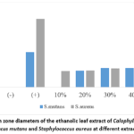 |
Figure 1: Inhibition zone diameters of the ethanolic leaf extract of Calophyllum inophyllum L. against Streptococcus mutans and Staphylococcus aureus at different extract concentrations. |
Acute Oral Toxicity
No mortality occurred in mice receiving single oral doses of 250, 500, 1000, or 2000 mg/kg body weight during the 14-day observation period. All animals remained active and maintained normal physical appearance throughout the experiment. Clinical observations revealed no signs of toxicity, including tremor, convulsions, excessive salivation, diarrhea, lethargy, respiratory abnormalities, abnormal posture, or changes in fur condition and eye appearance (Table 4).
Table 4: Acute oral toxicity observations following administration of the ethanolic leaf extract of Calophyllum inophyllum
|
Dose (mg/kg BW) |
n | Mortality | Activity | Physical condition | Clinical signs |
| Control | 5 | 0/5 | Normal | Normal |
None |
|
250 |
5 | 0/5 | Normal | Normal | None |
| 500 | 5 | 0/5 | Normal | Normal |
None |
|
1000 |
5 | 0/5 | Normal | Normal | None |
| 2000 | 5 | 0/5 | Normal | Normal |
None |
Discussion
The extraction yield of Calophyllum inophyllum L. leaf extract was 11.248 ± 0.568%, representing the proportion of crude extract recovered under the applied extraction conditions. Ethanol is widely recognized as an efficient extraction solvent because of its ability to dissolve a broad spectrum of polar and semi-polar phytochemicals, including phenolic compounds, flavonoids, tannins, coumarins, xanthones, triterpenoids, and certain alkaloids.9,10 The extraction yield may be influenced by several factors, including solvent polarity, solvent-to-material ratio, particle size, extraction duration, drying conditions, and the phytochemical composition of the plant material.11 The dark green color of the extract suggests the co-extraction of chlorophyll pigments together with secondary metabolites, whereas its viscous consistency indicates a relatively high concentration of dissolved plant constituents following solvent evaporation.12 These characteristics demonstrate that ethanol maceration successfully produced an extract suitable for subsequent biological evaluation.
The ethanolic leaf extract exhibited antibacterial activity against both Streptococcus mutans and Staphylococcus aureus. For Streptococcus mutans, the inhibition zone increased from 20% to 30% extract concentration but showed no meaningful further increase at 40% and 50%, indicating that the antibacterial response did not consistently increase across all tested concentrations. The increase observed between 20% and 30% may indicate that increasing the extract concentration provided a greater amount of bioactive compounds capable of diffusing through the agar medium and inhibiting bacterial growth.13 At concentrations above 30%, the inhibition zone against S. mutans showed no meaningful further increase, suggesting that the diffusion of active compounds within the agar medium may have approached saturation under the experimental conditions.14
The antibacterial activity observed in this study is likely attributable to the synergistic effects of several phytochemicals previously reported in Calophyllum inophyllum L, including flavonoids, phenolic compounds, tannins, xanthones, coumarins, and triterpenoids.12 Flavonoids have been reported to disrupt bacterial cell membranes, inhibit nucleic acid synthesis, and interfere with biofilm formation.15,16 Phenolic compounds increase membrane permeability and induce protein denaturation, whereas tannins inhibit bacterial enzymes and precipitate cell wall proteins.17,18 In addition, xanthones and coumarins, which are characteristic constituents of the genus Calophyllum, have demonstrated antibacterial activity against Gram-positive bacteria through disruption of bacterial cell structure and inhibition of microbial growth. The combined action of these metabolites most likely contributed to the antibacterial activity observed against both bacterial species.4
Although the extract inhibited both test organisms, Staphylococcus aureus exhibited greater susceptibility than Streptococcus mutans, as indicated by the larger inhibition zones observed at comparable extract concentrations. This difference may be associated with differences in bacterial physiology rather than differences in Gram staining, since both organisms are Gram-positive. Streptococcus mutans possesses a remarkable ability to synthesize extracellular glucans through glucosyltransferase activity, resulting in a dense biofilm matrix that limits the penetration of antimicrobial compounds.19,20 In contrast, under disc diffusion conditions, Staphylococcus aureus is predominantly present as planktonic cells, allowing antibacterial compounds to interact more readily with the bacterial cell wall and membrane.21 Consequently, S. aureus appears to be more susceptible to the antibacterial constituents of the extract.
Despite the observed antibacterial activity, the inhibition zones produced by the extract remained considerably smaller than those produced by 0.2% chlorhexidine, indicating that the crude extract was less potent than the standard antiseptic. Nevertheless, these findings demonstrate that Calophyllum inophyllum L. leaves possess promising antibacterial properties and may serve as a potential source of natural antibacterial agents. Further improvement in antibacterial efficacy may be achieved through optimization of extraction procedures, fractionation of active constituents, isolation of bioactive compounds, and formulation development to enhance stability and bioavailability.
Besides antibacterial efficacy, the safety profile of medicinal plant extracts is an essential consideration for their pharmaceutical development. In the present study, no mortality or observable clinical signs of toxicity were detected following single oral administration of the extract at doses up to 2000 mg/kg body weight, indicating a favorable acute oral safety profile. According to the toxicity classification described in OECD Test Guideline 423, the absence of mortality at the highest tested dose indicates that the median lethal dose (LD₅₀) is estimated to be greater than 2000 mg/kg body weight, suggesting relatively low acute toxicity.
The low acute toxicity observed in this study may be associated with the presence of secondary metabolites previously reported in Calophyllum inophyllum L. leaves, including flavonoids, phenolic compounds, tannins, xanthones, and coumarins, which have generally demonstrated favorable safety profiles within appropriate dosage ranges.22 However, acute toxicity assessment alone is insufficient to establish the overall safety of herbal extracts. Comprehensive toxicological evaluation should include subacute, subchronic, and chronic toxicity studies, as well as biochemical and histopathological examinations of major organs such as the liver and kidneys, to determine potential adverse effects associated with repeated or long-term administration.23
Taken together, the present findings demonstrate that the ethanolic leaf extract of Calophyllum inophyllum L. possesses moderate antibacterial activity against two clinically important Gram-positive pathogens while exhibiting low acute oral toxicity. These combined biological properties support its potential as a natural antibacterial candidate for further pharmaceutical development. Nevertheless, several limitations should be acknowledged. The antibacterial activity was evaluated using the disc diffusion method without determination of the minimum inhibitory concentration (MIC) and minimum bactericidal concentration (MBC). In addition, comprehensive phytochemical characterization was not performed to identify or quantify the specific compounds potentially responsible for the observed antibacterial activity. Further analyses using total phenolic and flavonoid assays, chromatographic fingerprinting, and advanced analytical techniques such as HPLC-DAD, LC-MS, or LC-MS/MS are therefore needed to characterize the chemical composition of the extract. The safety evaluation was also limited to acute oral toxicity in mice, without in-vitro cytotoxicity, biochemical, or histopathological assessments. Future studies should therefore focus on comprehensive phytochemical characterization, determination of MIC and MBC values, in-vitro cytotoxicity assessment, elucidation of the underlying antibacterial mechanisms, and comprehensive toxicity evaluation to further establish the safety and antibacterial potential of Calophyllum inophyllum L. leaf extract.
Conclusion
Extraction of Calophyllum inophyllum L. leaves using ethanol produced a crude extract with a yield of 11.248 ± 0.568%. The ethanolic leaf extract exhibited antibacterial activity against both Streptococcus mutans and Staphylococcus aureus in a concentration-dependent manner. Staphylococcus aureus showed greater susceptibility than Streptococcus mutans, as indicated by antibacterial activity observed at lower extract concentrations and larger inhibition zones. Acute oral administration of the extract at doses up to 2000 mg/kg body weight produced no mortality or observable clinical signs of toxicity in mice during the 14-day observation period, indicating that the estimated LD₅₀ exceeded 2000 mg/kg body weight. These findings suggest that the ethanolic leaf extract of Calophyllum inophyllum L. possesses promising antibacterial activity against clinically relevant Gram-positive bacteria while demonstrating a favorable preliminary safety profile. Further studies are required to identify the active antibacterial constituents, determine the minimum inhibitory and bactericidal concentrations, elucidate the mechanisms of action, and evaluate long-term toxicity before pharmaceutical or oral healthcare applications can be considered.
Acknowledgement
The authors would like to express their sincere gratitude to the Director of Poltekkes Kemenkes Denpasar and to all individuals who contributed to and supported the successful completion of this research.
Funding Source
This research was funded by Poltekkes Kemenkes Denpasar (Grant No. BJ.01.03/F.XXIV. 24/0807.19/2026)
Conflict of Interest
The author(s) do not have any conflict of interest.
Data Availability Statement
This statement does not apply to this article.
Ethics statement
This study had been approved by The Ethical Committee for Research from Wangaya Hospital No. 000.9.2/980/RSUDW.
Informed Consent Statement
This study did not involve human participants, and therefore, informed consent was not required.
Clinical Trial Registration
This research does not involve any clinical trials.
Permission to reproduce material from other sources
Not Applicable
Author contributions
- Ni Wayan Arini: Writing, Data collection, Funding Acquisition, Supervision.
- I Nyoman Wirata: Data Collection, Analysis, Writing, Review & Editing.
- Ni Komang Erny Astiti: Data Collection, Analysis, Writing, Review & Editing.
- Ni Made Dwi Mahayati: Data Collection, Analysis, Writing, Review & Editing.
- Ni Ketut Nuratni: Data Collection, Analysis, Writing, Review & Editing.
References
- Rajasekaran JJ, Krishnamurthy HK, Bosco J, et al. Oral Microbiome: A Review of Its Impact on Oral and Systemic Health. Microorganisms. Multidisciplinary Digital Publishing Institute (MDPI). 2024;12(9). doi:10.3390/microorganisms12091797
CrossRef - Spatafora G, Li Y, He X, Cowan A, Tanner ACR. The Evolving Microbiome of Dental Caries. Microorganisms. Multidisciplinary Digital Publishing Institute (MDPI). 2024;12(1). doi:10.3390/microorganisms12010121
CrossRef - Fathani IJ, Miladiyah I, Kedokteran J, Indonesia K. Cytotoxicity of ethanolic extract of fruit shells and seeds of Nyamplung (Calophyllum inophyllum L.) on WiDr colorectal cancer cells 166. Indonesian Journal of Medicine and Health. 2021;12(2):166-174. doi:10.20885/JKKI.Vol12.Iss2art10
CrossRef - Vittaya L, Chalad C, Janyong S, Leesakul N. Bioactive Potential of Calophyllum inophyllum: Phytochemical Profiles, Biological Activities, and In Silico Pharmacokinetic Predictions. Phyton-International Journal of Experimental Botany. 2025;94(12):4091-4115. doi:10.32604/phyton.2025.074891
CrossRef - De Rossi L, Rocchetti G, Lucini L, Rebecchi A. Antimicrobial Potential of Polyphenols: Mechanisms of Action and Microbial Responses—A Narrative Review. Antioxidants. Multidisciplinary Digital Publishing Institute (MDPI). 2025;14(2). doi:10.3390/antiox14020200
CrossRef - Rodríguez B, Pacheco L, Bernal I, Piña M. Mechanisms of Action of Flavonoids: Antioxidant, Antibacterial and Antifungal Properties. Ciencia, Ambiente y Clima. 2023;6(2):33-66. doi:10.22206/cac.2023.v6i2.3021
CrossRef - Erhirhie EO, Ihekwereme CP, Ilodigwe EE. Advances in acute toxicity testing: strengths, weaknesses and regulatory acceptance. Interdiscip Toxicol. 2018;11(1):5-12. doi:10.2478/intox-2018-0001
CrossRef - Falya Y, Sumiwi A, Levita J. Mini Review:Toxicity Study Of Plant Extracts. IOSR Journal Of Pharmacy And Biological Sciences (IOSR-JPBS). 2020;15(2):25-32. doi:10.9790/3008-1502012532
- Ferdosh S. The Extraction of Bioactive Agents from Calophyllum inophyllum L., and Their Pharmacological Properties. Sci Pharm. Multidisciplinary Digital Publishing Institute (MDPI). 2024;92(1). doi:10.3390/scipharm92010006
CrossRef - Alara OR, Abdurahman NH, Ukaegbu CI. Extraction of phenolic compounds: A review. Curr Res Food Sci. 2021;4:200-214. doi:https://doi.org/10.1016/j.crfs.2021.03.011
CrossRef - Bitwell C, Indra S Sen, Luke C, Kakoma MK. A review of modern and conventional extraction techniques and their applications for extracting phytochemicals from plants. Sci Afr. 2023;19:e01585. doi:https://doi.org/10.1016/j.sciaf.2023.e01585
CrossRef - Ardhyni DH, Aparamarta HW, Widjaja A, Ibrahim R, Gunawan S. The extraction and purification of squalene from Nyamplung (Calophyllum Inophyllum L) leaves. In: IOP Conference Series: Earth and Environmental Science. Vol 963. IOP Publishing Ltd; 2022. doi:10.1088/1755-1315/963/1/012042
CrossRef - Ribeiro J, Silva V, Aires A, et al. Platanus hybrida’s Phenolic Profile, Antioxidant Power, and Antibacterial Activity against Methicillin-Resistant Staphylococcus aureus (MRSA). Horticulturae. 2022;8(3). doi:10.3390/horticulturae8030243
CrossRef - Shen W, Xue R, Liu Y, et al. Principle Investigation and Method Standardization of Inhibition Zone Assay Based on Antimicrobial Peptides Extracted from Black Soldier Fly Larvae. BioTech. 2024;13(3). doi:10.3390/biotech13030031
CrossRef - Shamsudin NF, Ahmed QU, Mahmood S, et al. Antibacterial Effects of Flavonoids and Their Structure-Activity Relationship Study: A Comparative Interpretation. Molecules. MDPI. 2022;27(4). doi:10.3390/molecules27041149
CrossRef - Zhang Z, Cao M, Shang Z, et al. Research Progress on the Antibacterial Activity of Natural Flavonoids. Antibiotics. Multidisciplinary Digital Publishing Institute (MDPI). 2025;14(4). doi:10.3390/antibiotics14040334
CrossRef - Czerkas K, Olchowik-Grabarek E, Łomanowska M, Abdulladjanova N, Sękowski S. Antibacterial Activity of Plant Polyphenols Belonging to the Tannins against Streptococcus mutans—Potential against Dental Caries. Molecules. 2024;29(4). doi:10.3390/molecules29 040879
CrossRef - Huang J, Zaynab M, Sharif Y, et al. Tannins as antimicrobial agents: Understanding toxic effects on pathogens. Toxicon. 2024;247:107812. doi:https://doi.org/10.1016/j.toxicon.2024.107812
CrossRef - Gao Z, Chen X, Wang C, et al. New strategies and mechanisms for targeting Streptococcus mutans biofilm formation to prevent dental caries: A review. Microbiol Res. 2024;278:127526. doi:https://doi.org/10.1016/j.micres.2023.127526
CrossRef - Fitri DK, Tuygunov N, Wan Harun WHA, Purwasena IA, Cahyanto A, Zakaria MN. Key virulence genes associated with Streptococcus mutans biofilm formation: a systematic review. Frontiers in Oral Health. Frontiers Media SA. 2025;6. doi:10.3389/froh.2025.1654428
CrossRef - Tuon FF, Suss PH, Telles JP, Dantas LR, Borges NH, Ribeiro VST. Antimicrobial Treatment of Staphylococcus aureus Biofilms. Antibiotics. MDPI. 2023;12(1). doi:10.3390/antibiotics12010087
CrossRef - Farida S, Jenie RI, Fakhrudin N. Calophyllum inophyllum: A Comprehensive Analysis of its Ethnobotanical, Phytochemical, and Pharmacological Properties. Majalah Obat Tradisional. 2024;29(2):121-142. doi:10.22146/mot.87488
CrossRef - Organisation for Economic Co-operation and Development. Test No. 407: Repeated Dose 28-Day Oral Toxicity Study in Rodents. 2025. http://www.oecd.org/termsandconditions/
 PlumX:
 Views: 366
  PDF Downloads:
8
PDF Downloads:
8 
Accepted on: 21-09-2026
Second Review by: Dr. Faheema Jabbar Aboalhor and Dr. Teshome Geremew
Final Approval by: Dr. Mariia Shanaida