
Manuscript accepted on :10-04-2026
Published online on: 19-05-2026
Plagiarism Check: Yes
Reviewed by: Dr. Ramya Sri and Dr. Ban Ali Ahmed
Second Review by: Dr. Koteswara Rao Inabathina
Final Approval by: Dr. Mariia Shanaida
Mazlin Mohideen1* , Dhia Ali Fudail Che Rosli1, Wan Ahmad Irfan Hakimi Wan Abd Malik1 and Nur Azzalia Kamaruzan2
1Faculty of Pharmacy and Health Sciences, Universiti Kuala Lumpur Royal College of Medicine Perak, Ipoh Perak, Malaysia.
2National Poison Centre, Universiti Sains Malaysia, Minden, Pulau Pinang, Malaysia.
Corresponding Author E-mail:mazlin.mohideen@unikl.edu.my
DOI : https://dx.doi.org/10.13005/bpj/3473
Abstract
Oral infections remain a significant public health concern, and prolonged use of chlorhexidine mouthwashes may cause adverse effects such as tooth staining and mucosal irritation. Consequently, increasing attention has been directed toward the development of plant-based oral healthcare products with antimicrobial properties and improved safety profiles. This study evaluated the antimicrobial activity of an ethanolic extract of Piper betle L. leaves and its application in a herbal mouthwash formulation. The extract was prepared by maceration and subjected to qualitative phytochemical screening. Antimicrobial activity was assessed using the disc diffusion method against Staphylococcus aureus, Pseudomonas aeruginosa, and Candida albicans. The extract exhibited measurable antimicrobial activity, with inhibition zones ranging from 24.00 ± 2.00 mm to 26.00 ± 1.00 mm against S. aureus, 16.33 ± 1.53 mm to 18.00 ± 1.00 mm against P. aeruginosa, and 26.00 ± 1.73 mm to 27.33 ± 1.53 mm against C. albicans. Two mouthwash formulations containing 150 mg and 250 mg extract per 100 mL were prepared and evaluated for physicochemical characteristics and short-term stability. Both formulations demonstrated acceptable characteristics, with pH values ranging from 5.90 to 6.58, good clarity, moderate foaming capacity, and stability over a seven-day storage period. These findings suggest that Piper betle extract has potential as a natural antimicrobial ingredient for oral healthcare formulations, although further studies including quantitative antimicrobial analysis, long-term stability testing, and clinical evaluation are required.
Keywords
Antifungal Activity; Antimicrobial Activity; Herbal Mouthwash; Oral Pathogens; Piper betle; Phytochemical Screening
Download this article as:| Copy the following to cite this article: Mohideen M, Rosli D. A. F. C, Malik W. A. I. H. W. A, Kamaruzan N. A. Formulation and Preliminary Antimicrobial Evaluation of an Ethanolic Piper betle L. Leaf Extract Herbal Mouthwash. Biomed Pharmacol J 2026;19(2). |
| Copy the following to cite this URL: Mohideen M, Rosli D. A. F. C, Malik W. A. I. H. W. A, Kamaruzan N. A. Formulation and Preliminary Antimicrobial Evaluation of an Ethanolic Piper betle L. Leaf Extract Herbal Mouthwash. Biomed Pharmacol J 2026;19(2). Available from: https://bit.ly/42JRjzF |
Introduction
Oral health is an essential component of overall well-being, influencing important functions such as mastication, speech, and facial appearance. Despite improvements in preventive dental care and public health awareness in recent decades, oral diseases remain among the most prevalent health problems worldwide, affecting billions of individuals and placing a substantial burden on healthcare systems.1,2Many oral conditions, including dental caries, periodontal diseases, and oral fungal infections, are closely associated with the accumulation and persistence of pathogenic microorganisms within dental biofilms.1-3 Therefore, effective control of microbial populations within the oral cavity is essential for maintaining oral health and preventing the progression of oral diseases.1-3
Chlorhexidine-containing mouthwashes are widely used in clinical dentistry due to their broad antimicrobial spectrum and prolonged substantivity on oral tissues.4-6 However, long-term use of chlorhexidine has been associated with several undesirable effects, including tooth discoloration, taste disturbances, mucosal irritation, and alterations in the normal oral microbiota.4-6 These limitations have encouraged increasing interest in the exploration of alternative oral care agents derived from natural products that may provide comparable antimicrobial activity with improved safety and patient acceptability.6-7
Medicinal plants represent an important source of biologically active secondary metabolites, including flavonoids, tannins, phenolic compounds, and saponins. These phytochemicals have been widely reported to possess antimicrobial, anti-inflammatory, and antioxidant properties that are beneficial for oral health applications.8Consequently, plant extracts and essential oils have attracted increasing attention as potential ingredients in herbal oral care products. Previous studies have shown that plant-derived compounds may inhibit microbial growth through several mechanisms, including disruption of cell membrane integrity, interference with essential metabolic pathways, and inhibition of microbial biofilm formation.9-10
Among medicinal plants traditionally used in oral hygiene practices, Piper betle L. (Piperaceae) has received considerable attention due to its reported antimicrobial properties. According to the Plants of the World Online (POWO), Royal Botanic Gardens, Kew, Piper betle L. is a well-documented species widely distributed in tropical regions of Asia and recognized for its ethnomedicinal importance.11 The leaves of P. betle contain various bioactive compounds, particularly phenolic constituents and flavonoids, which contribute to antibacterial and antifungal activity.12-15
Recent studies (2024–2025) have further confirmed that P. betle exhibits broad-spectrum antimicrobial and antibiofilm activity against oral pathogens, including its ability to disrupt microbial adhesion and quorum-sensing mechanisms.16-19 These findings highlight its increasing relevance as a natural therapeutic agent in oral healthcare applications. Several studies have demonstrated the effectiveness of P. betle extracts against a range of microbial pathogens associated with oral infections.12-15,20-23However, many of these investigations have primarily focused on in vitro antimicrobial screening of crude extracts, with limited emphasis on the development and physicochemical evaluation of practical oral healthcare formulations.
Despite the reported antimicrobial properties of P. betle extracts, limited studies have investigated their incorporation into formulated oral healthcare products together with the evaluation of physicochemical characteristics and formulation stability.Therefore, the present study aimed to evaluate the antimicrobial activity of an ethanolic extract of P. betle leaves against selected oral pathogens and to develop a herbal mouthwash formulation incorporating the extract. In addition to antimicrobial screening, the study also assessed key physicochemical properties of the formulated mouthwash, including pH, foaming characteristics, and short-term stability. By combining antimicrobial evaluation with formulation development and physicochemical characterization, this study provides a preliminary translational framework for incorporating Piper betle extract into herbal oral healthcare formulations.
Materials and Methods
Plant Material Preparation
Fresh leaves of Piper betle were obtained from a local supplier in Ipoh, Perak, Malaysia. A qualified taxonomist at the Faculty of Pharmacy and Health Sciences, Universiti Kuala Lumpur Royal College of Medicine Perak (UniKL RCMP), identified the plant based on morphological examination and established taxonomic references. The collected leaves were washed with running water to remove surface contaminants and then air-dried under laboratory conditions. They were then dried in a hot-air oven at 40-50 °C for 24 hours to ensure adequate moisture reduction. The dried leaves were milled into a fine powder using a laboratory grinder and stored in airtight containers at room temperature until further extraction procedures were performed.24
Extraction by Maceration
A total of 50 g of powdered P. betle leaves was soaked in 500 mL of 95% ethanol using a 1:10 (w/v) plant material to solvent ratio. The mixture was agitated continuously at 200 rpm for 24 hours under ambient laboratory conditions to extract bioactive constituents. After extraction, the solution was filtered through Whatman No. 1 filter paper to remove plant residues, and the filtrate was concentrated under reduced pressure using a rotary evaporator at 50 °C. The resulting semi-solid extract was dried to constant weight before determining the percentage yield.25 The dried crude extract was stored at 4 °C until further experimental use. To ensure reproducibility, the entire extraction process was performed in duplicate under identical conditions. Qualitative phytochemical screening was conducted in triplicate using established procedures to identify tannins, flavonoids, saponins, and steroids.24
Tannins
A 0.5 mL aliquot of the extract was treated with 10% alcoholic ferric chloride solution. The development of a blue or greenish-black coloration indicated the presence of tannin compounds.26
Flavonoids
A few drops of concentrated sulfuric acid were carefully added to the extract solution. The transient appearance of a yellow coloration indicated the presence of flavonoid compounds.26
Saponins
The extract was diluted with distilled water and vigorously shaken. The formation of stable and persistent froth indicated the presence of saponin compounds.26
Steroids
The extract was first dissolved in chloroform, followed by the sequential addition of acetic acid and concentrated sulfuric acid. The development of a blue-green coloration indicated the presence of steroidal compounds.26
Antimicrobial Activity
Microorganisms
Standard reference microorganisms were used in this study: Staphylococcus aureus (ATCC 25923), Pseudomonas aeruginosa (ATCC 27853), and Candida albicans (ATCC 10231). These strains were obtained from the culture repository of the Microbiology Laboratory at Universiti Kuala Lumpur Royal College of Medicine Perak (UniKL RCMP). These microorganisms were selected because they are commonly associated with oral infections and are frequently used as representative bacterial and fungal pathogens in antimicrobial studies related to oral healthcare. All assays were performed in triplicate and repeated in three independent experimental runs to ensure reproducibility of the results.
Disc Diffusion Assay
The antimicrobial activity of the extract was evaluated using the agar disc diffusion technique on Mueller-Hinton agar plates according to standard microbiological procedures.25-28Agar plates were prepared to a uniform depth of 4 mm. Microbial suspensions were adjusted to a 0.5 McFarland standard (OD 0.08-0.12 at 625 nm). The standardized inoculum was evenly spread over the agar surface using a sterile cotton swab to obtain uniform lawn growth.25-28Sterile 10 mm filter paper discs were impregnated with 20 µL of each extract solution. The selected extract concentrations (150-250 mg/mL) were chosen based on preliminary screening experiments and previously reported concentration ranges commonly used in antimicrobial evaluation of plant extracts. The discs were dried under aseptic conditions for 15 minutes before being placed on the agar surface to ensure solvent evaporation and minimize interference. Residual ethanol was considered negligible and unlikely to contribute to antimicrobial inhibition, as confirmed by the absence of inhibition zones in the negative control. Tetracycline and fluconazole discs served as positive controls for bacterial and fungal strains, respectively, while discs impregnated with 95% ethanol were used as negative controls. Plates were incubated at 37 °C for 24 hours, after which the diameters of inhibition zones were measured in millimeters. All assays were performed in triplicate.
Statistical Analysis
Results are expressed as mean ± standard deviation (SD) from three independent experiments (n = 3). Statistical analysis was performed using one-way analysis of variance (ANOVA) followed by Tukey’s post hoc test using IBM SPSS Statistics version 26.0. A p-value < 0.05 was considered statistically significant.
Development of Ethanolic Piper betle-Based Mouthwash Formulations
Two mouthwash formulations were developed, containing 250 mg (F1) and 150 mg (F2) of P. betle ethanolic extract per 100 mL of final preparation (Table 1). The extract was first dispersed in purified water, followed by the sequential addition of PEG-40 (4 g), glycerol (8.5 mL), saccharin (50 mg), peppermint oil (3 drops), sodium chloride (500 mg), and lemon yellow colorant (1-2 drops) with continuous stirring. The final volume was adjusted to 100 mL with purified water, and the mixture was maintained at 40 °C with agitation until a transparent and homogeneous solution was obtained.29-30 A commercially available mouthwash (Listerine®) was included as a reference formulation for comparison.
Table 1: Composition of Herbal Mouthwash Formulations
| Ingredient | Function | F1 (250 mg) | F2 (150 mg) |
| Piper betle extract | Active ingredient | 250 mg | 150 mg |
| PEG-40 | Surfactant | 4 g | 4 g |
| Glycerol | Co-surfactant | 8.5 mL | 8.5 mL |
| Saccharin | Sweetener | 50 mg | 50 mg |
| Peppermint oil | Flavoring agent | 3 drops | 3 drops |
| Sodium chloride | Tonicity agent | 500 mg | 500 mg |
| Lemon yellow | Colouring agent | 1–2 drops | 1–2 drops |
| Purified water | Vehicle | up to 100 mL | up to 100 mL |
Physicochemical Characterization and Stability Assessment of Mouthwash Formulations
The prepared formulations were evaluated for organoleptic characteristics, including appearance, color, and odor, as well as pH, foaming capacity, and short-term stability.31 pH was measured using a digital pH meter calibrated in advance with standard buffer solutions at pH 4.0 and 7.0. Foam height was determined after 10 vigorous manual shakes in a graduated cylinder, with the foam height recorded in centimeters after 1 minute. Stability studies were conducted under controlled ambient conditions (25 ± 2°C) for seven days according to standard preliminary stability assessment protocols.29,32
Results
Extraction Yield
Ethanolic extraction of Piper betle leaves produced crude extract yields of 7.60% and 8.24% for batches 1 and 2, respectively (Table 2).
Table 2: Extraction Yield of Piper betle L. Leaves
| Batch | Extract Weight (g) | Percentage Yield (%) |
| 1 | 18.72 | 7.60 |
| 2 | 9.57 | 8.24 |
Phytochemical Screening
Preliminary qualitative phytochemical screening of the ethanolic P. betle extract revealed the presence of several secondary metabolites, including saponins, flavonoids, tannins, and steroidal compounds (Table 3, Figure 1). The characteristic color changes and persistent froth observed during the tests confirmed the presence of these phytochemical constituents.
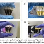 |
Figure 1: Representative qualitative phytochemical reactions of Piper betle L. ethanolic extract showing (a) saponins, (b) flavonoids, (c) steroids, and (d) tannins. |
Table 3: Qualitative Phytochemical Screening of Piper betle L. Extract
| Phytochemical | Test Method | Observation | Result |
| Tannins | Ferric chloride test | Blue/greenish-black | + |
| Flavonoids | Ammonia test | Yellow coloration | + |
| Saponins | Froth test | Persistent foam | + |
| Steroids | Liebermann–Burchard test | Blue–green color | + |
Qualitative phytochemical screening results are expressed as presence (+) of constituents based on standard test observations.
Antimicrobial Activity
The antimicrobial activity of the ethanolic P. betle leaf extract against Staphylococcus aureus, Pseudomonas aeruginosa, and Candida albicans is presented in Table 4 and Figures 2-4. The results are expressed as mean inhibition zone diameter ± standard deviation (SD) obtained from three independent experiments (n = 3). One-way analysis of variance (ANOVA) indicated that differences among the tested concentrations were not statistically significant for S. aureus and P. aeruginosa (p > 0.05), whereas a statistically significant increase in inhibition zone diameter was observed for C. albicans at the highest concentration (p < 0.05). Inhibitory activity was observed for all tested microorganisms at extract concentrations of 150, 200, and 250 mg/mL. The negative control (95% ethanol) produced no detectable inhibition zone, confirming that the antimicrobial activity observed was attributable to the extract rather than the residual solvent.
For S. aureus, inhibition zones ranged from 24.00 ± 2.00 mm to 26.00 ± 1.00 mm, showing a modest concentration-related increase; however, statistical analysis revealed no significant differences among the tested concentrations (p > 0.05). The positive control, tetracycline, produced a substantially larger inhibition zone (35.33 ± 1.53 mm). For P. aeruginosa, the extract showed comparatively lower antibacterial activity, with inhibition zone diameters ranging from 16.33 ± 1.53 mm to 18.00 ± 1.00 mm. Similar to S. aureus, the differences among concentrations were not statistically significant (p > 0.05), while tetracycline produced markedly greater inhibition (27.67 ± 1.15 mm). Notably, strong antifungal activity was observed against C. albicans, with inhibition zones ranging from 26.00 ± 1.73 mm to 27.33 ± 1.53 mm. The highest tested concentration (250 mg/mL) produced a significantly larger inhibition zone compared with the reference antifungal agent fluconazole (23.67 ± 2.31 mm) (p < 0.05).
Overall, the ethanolic P. betle leaf extract showed broad-spectrum antimicrobial activity, with stronger effects against S. aureus and C. albicans than against P. aeruginosa under the present experimental conditions.One-way analysis of variance (ANOVA) showed that differences among concentrations were not statistically significant for S. aureus and P. aeruginosa (p > 0.05), while a statistically significant increase was observed for C. albicans at the highest concentration (p < 0.05).
Table 4: Antimicrobial activity of ethanolic Piper betle L. leaf extract against selected oral pathogens determined by the disc diffusion assay.
| Sample | Concentration | Staphylococcus aureus (mm) | Pseudomonas aeruginosa (mm) | Candida albicans (mm) |
| Extract | 150 mg/mL | 24.00 ± 2.00 | 16.33 ± 1.53 | 26.00 ± 1.73 |
| Extract | 200 mg/mL | 24.33 ± 1.53 | 17.67 ± 0.58 | 26.00 ± 1.00 |
| Extract | 250 mg/mL | 26.00 ± 1.00 | 18.00 ± 1.00 | 27.33 ± 1.53 |
| Positive control | — | 35.33 ± 1.53 | 27.67 ± 1.15 | 23.67 ± 2.31 |
Inhibition zones are expressed as mean diameter (mm) ± standard deviation (SD) from three independent experiments (n = 3). Statistical analysis was performed using one-way ANOVA, with p < 0.05 considered statistically significant.
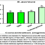 |
Figure 2: Mean inhibition zone diameters for Staphylococcus aureus after exposure to various concentrations (150-250 mg/mL) of Piper betle L. ethanolic extract and tetracycline as the reference control. |
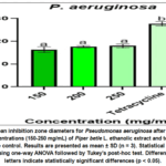 |
Figure 3: Mean inhibition zone diameters for Pseudomonas aeruginosa after exposure to varying concentrations (150-250 mg/mL) of Piper betle L. ethanolic extract and tetracycline as the reference control. |
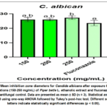 |
Figure 4: Mean inhibition zone diameters for Candida albicans after exposure to various concentrations (150-250 mg/mL) of Piper betle L. ethanolic extract and fluconazole as the reference antifungal control. |
Evaluation of Herbal Mouthwash
Both formulations (F1 and F2) appeared transparent and homogeneous, with a characteristic peppermint odor (Table 5). The pH values ranged from 5.90 to 6.58. Foam height measurements indicated moderate foaming capacity. No phase separation or visible microbial growth was observed after seven days of storage at room temperature, indicating acceptable short-term stability of the formulations.
Table 5: Physicochemical Evaluation of Herbal Mouthwash
| Parameter | F1 (250 mg) | F2 (150 mg) | Listerine® |
| Appearance | Clear, golden yellow | Clear, light yellow | Clear, pastel yellow |
| Odor | Peppermint | Peppermint | Minty |
| pH | 5.90 | 6.58 | 5.66 |
| Foam Height (cm) | 2.9 | 2.3 | 0.8 |
| Stability (7 days) | Stable | Stable | Stable |
Stable = No phase separation or microbial growth observed.
Discussion
The present study demonstrated that the ethanolic extract of Piper betle exhibited measurable antibacterial activity against Staphylococcus aureus and Pseudomonas aeruginosa, as well as notable antifungal activity against Candida albicans, and could be successfully incorporated into a stable herbal mouthwash formulation.
Oral infections remain a major global health concern and are closely associated with the persistence of pathogenic microorganisms within dental biofilms. Although chlorhexidine is widely recognized as a gold-standard antimicrobial mouthwash, prolonged use has been associated with several undesirable effects, including tooth staining, dysgeusia, mucosal irritation, and disruption of the natural oral microbiota.33-34 Recent clinical evaluations have further highlighted concerns regarding microbiome imbalance and reduced patient compliance following long-term chlorhexidine exposure.35-36 These limitations have stimulated growing interest in the development of plant-based oral formulations that can provide antimicrobial activity with improved safety and tolerability.
In the present study, P. betle was selected due to its well-documented traditional use and reported antimicrobial potential. This is further supported by its recognition in global botanical databases such as POWO, which confirms its taxonomic identity and ethnomedicinal relevance.11 Previous investigations have confirmed the antibacterial and antifungal properties of P. betle leaf extracts; however, many studies have primarily focused on in vitro antimicrobial screening without progressing toward formulation development.20-23
The antimicrobial activity observed in the present study is consistent with earlier reports demonstrating the inhibitory effects of P. betle extracts against various microbial pathogens associated with oral infections.12-15,20-23Recent studies (2024–2025) have further strengthened this evidence by demonstrating that P. betle exhibits significant antibiofilm activity and the ability to disrupt microbial adhesion and quorum-sensing pathways, which are critical in oral biofilm-associated infections.18-19 By integrating antimicrobial screening with formulation design and physicochemical characterization, the present work adopts a more translational approach aimed at bridging laboratory findings with potential product development. This approach provides additional insight into the potential application of P. betle in herbal oral healthcare products.
The extraction yield obtained using 95% ethanol ranged from 7.60% to 8.24%, which is consistent with previously reported yields for ethanol-based extraction of medicinal plants.37-38 Ethanol was selected as the extraction solvent due to its ability to efficiently dissolve phenolic compounds while remaining acceptable for pharmaceutical and oral formulations.30 Compared with methanol, ethanol is considered safer for human use and is commonly employed in herbal product preparation. Previous studies have also demonstrated that ethanolic extraction of P. betle effectively concentrates phenolic compounds, flavonoids, and tannins that contribute to antimicrobial activity.23
Phytochemical screening confirmed the presence of saponins, flavonoids, tannins, and steroids in the extract, which is consistent with earlier phytochemical analyses of P. betle.39-42 These compounds are known to exert antimicrobial effects through multiple mechanisms. Flavonoids and phenolic compounds may suppress microbial growth through several mechanisms, including damage to cell membrane integrity, increased membrane permeability, and disruption of essential intracellular processes such as nucleic acid synthesis.39-42 Tannins can precipitate proteins and interfere with microbial cell wall integrity, while saponins interact with membrane sterols and increase membrane permeability.41-42
Recent investigations (2024–2025) have further identified key phenolic constituents of P. betle, such as hydroxychavicol, eugenol, and chavibetol, as major contributors to its antimicrobial and antibiofilm activities.18-19 In addition, recent studies have shown that plant-derived phenolics may interfere with quorum-sensing pathways and inhibit microbial biofilm formation, which is particularly relevant for oral pathogen control.43These mechanisms collectively support the observed antimicrobial activity and suggest a synergistic interaction among multiple phytochemical constituents present in the extract.19
To further elucidate the chemical basis of the observed antimicrobial activity, future studies should incorporate quantitative phytochemical analyses, including determination of total phenolic content (TPC) and total flavonoid content (TFC). In addition, advanced analytical techniques such as high-performance liquid chromatography (HPLC) or gas chromatography-mass spectrometry (GC-MS) are recommended to identify and characterize specific bioactive compounds (e.g., eugenol, chavibetol) responsible for the antimicrobial effects. Recent studies (2024–2025) have emphasized the importance of correlating specific phenolic constituents with antimicrobial and antibiofilm activities to better understand structure-activity relationships in plant-derived therapeutics.18-19 Such approaches would provide a more comprehensive understanding of the extract composition and strengthen the correlation between phytochemical constituents and biological activity.
The ethanolic extract demonstrated antibacterial activity against Staphylococcus aureus, although the inhibition zones did not significantly increase with increasing extract concentration. This plateau effect may be attributed to diffusion limitations associated with the disc diffusion method, particularly when evaluating crude plant extracts containing complex phytochemical mixtures.44 In contrast, the comparatively lower susceptibility of Pseudomonas aeruginosa may be explained by its intrinsic resistance mechanisms. This Gram-negative bacterium possesses a restrictive outer membrane and active efflux pump systems that reduce antimicrobial penetration, making it less susceptible to many plant-derived antimicrobial agents.44-46
Notably, strong antifungal activity was observed against Candida albicans, with the highest extract concentration producing inhibition zones larger than those generated by the reference antifungal agent under the present experimental conditions. However, such comparisons should be interpreted cautiously because diffusion-based assays may be influenced by compound diffusion rates and concentration gradients within the agar medium. The increasing incidence of oral candidiasis and the emergence of antifungal resistance among Candida species highlight the importance of identifying alternative antifungal agents from natural sources.47-50Recent findings have further demonstrated that phenolic compounds from P. betle can disrupt fungal membrane integrity through interaction with ergosterol and inhibit hyphal development and biofilm formation, thereby enhancing antifungal efficacy.18
Despite the promising antimicrobial findings, the disc diffusion method used in this study provides only preliminary screening data and does not directly reflect intrinsic antimicrobial potency. Recent literature (2024–2025) strongly recommends the inclusion of quantitative antimicrobial assays, such as minimum inhibitory concentration (MIC), minimum bactericidal concentration (MBC), and minimum fungicidal concentration (MFC), as well as antibiofilm assays, to improve the clinical relevance of plant-based antimicrobial studies.18-19Additionally, evaluation of antibiofilm activity would provide further insight into the potential effectiveness of the extract in oral healthcare applications. In addition, inclusion of chlorhexidine as a positive control in future studies would enable direct clinical comparison with standard oral antimicrobial formulations and strengthen the translational relevance of the findings.
The stability assessment of the formulated mouthwash was limited to a seven-day observation period under ambient conditions. More comprehensive stability evaluations, including accelerated and long-term stability studies, are necessary to ensure formulation stability, shelf-life determination, and product quality. Further investigations involving cytotoxicity testing and in vivo validation are also required to confirm safety and therapeutic applicability. Overall, these findings indicate that P. betle extract has promising potential as a natural antimicrobial ingredient in herbal mouthwash formulations and may contribute to the development of plant-based oral healthcare products.
Conclusion
This study demonstrated that the ethanolic extract of Piper betle L. leaves contains bioactive phytochemicals, including flavonoids, tannins, saponins, and steroidal compounds, which contribute to its antimicrobial activity against selected oral pathogens. The extract exhibited measurable antibacterial activity against Staphylococcus aureus and Pseudomonas aeruginosa and notable antifungal activity against Candida albicans. Furthermore, the extract was successfully incorporated into herbal mouthwash formulations that showed acceptable physicochemical properties, including appropriate pH, clarity, foaming capacity, and short-term stability. These findings suggest that P. betle extract may have potential as a natural antimicrobial ingredient for oral healthcare formulations; however, further studies involving quantitative antimicrobial assays (MIC/MBC/MFC), toxicity evaluation, long-term stability testing, and clinical validation are required before practical therapeutic application can be confirmed.
Acknowledgement
The authors express their appreciation to Universiti Kuala Lumpur Royal College of Medicine Perak (UniKL RCMP) for providing the necessary research facilities and institutional support.
Funding Sources
The author(s) received no financial support for the research, authorship, and/or publication of this article.
Conflict of Interest
The author(s) do not have any conflict of interest.
Data Availability Statement
This statement does not apply to this article.
Ethics Statement
This research did not involve human participants, animal subjects, or any material that requires ethical approval.
Informed Consent Statement
This study did not involve human participants, and therefore, informed consent was not required.
Clinical Trial Registration
This research does not involve any clinical trials.
Permission to reproduce material from other sources
Not Applicable
Author Contributions
- Mazlin Mohideen: Conceptualization, Project supervision, Methodology design, Writing – review and editing, Final approval of the manuscript.
- Dhia Ali Fudail Che Rosl): Investigation, Data collection, Laboratory experiments, Writing – original draft preparation.
- Wan Ahmad Irfan Hakimi Wan Abd Malik: Methodology, Laboratory analysis, Data validation.
- Nur Azzalia Kamaruzaman: Supervision, Scientific review, Writing – review and editing.
References
- Shekar BC, Nagarajappa R, Suma S, Thakur R. Herbal extracts in oral health care. Pharmacogn Rev. 2015;9(18):87-92.https://doi.org/10.4103/0973-7847.162101
CrossRef - World Health Organization. Global Oral Health Status Report: Towards Universal Health Coverage for Oral Health by 2030. WHO, 2022.
- World Health Organization. Oral health. WHO, 2024.
- Manipal S. The mouthwash war – chlorhexidine vs herbal mouth rinses: a meta-analysis. J Clin Diagn Res. 2016;10(5):ZE01-ZE05.https://doi.org/10.7860/JCDR/2016/17529.7764
CrossRef - Sheik R, Lakshmi T. Awareness of herbal mouthwash among schoolchildren. J Adv Pharm Educ Res. 2017;7(3):282-285.
- Tidke S, Chhabra GK, Madhu PP, et al. Herbal versus non-herbal mouthwash effectiveness. Cureus. 2022;14(8):e28409.https://doi.org/10.7759/cureus.28409
CrossRef - James P, Worthington HV, Parnell C, et al. Chlorhexidine mouthrinse as an adjunctive treatment for gingival health. Cochrane Database Syst Rev. 2022.https://doi.org/10.1002/14651858.CD008676.pub3
- Agbor MA, Naidoo S. Ethnomedicinal plants used by traditional healers to treat oral health problems. Evid Based Complement Alternat Med. 2015;2015:1-10.https://doi.org/10.1155/2015/649832
CrossRef - Mohideen M, Mahadi NNSJ, Suhaimi NAN, Kamaruzaman NA. Antibacterial properties of essential oil extracted from kaffir lime (Citrus hystrix) peel. Orient J Chem. 2022.
CrossRef - Mohideen M, Mahadi NNSJ, Suhaimi NAN, Kamaruzaman NA. Antioxidant and antibacterial activities of kaffir lime essential oil. Mater Innov Sci Technol. 2023:143-152.
CrossRef - Plants of the World Online (POWO). Piper betle L. Royal Botanic Gardens, Kew; 2024 [cited 2026 Apr 4]. Available from: https://powo.science.kew.org/taxon/urn:lsid:ipni.org:names:680605-1
- Valle DL, Cabrera EC, Puzon JJM, Rivera WL. Antimicrobial activities of Piper betle extracts. PLoS One. 2016;11(1):e0146349.https://doi.org/10.1371/journal.pone.0146349
CrossRef - Baviskar HP, Dhake GT, Kasai MA, et al. Review of Piper betle. Res J Pharmacogn Phytochem. 2017;9(2):128-134.
CrossRef - Andrianto D, Husnawati N, Hermita S, et al. Phytochemical classification of Piper betle. Biodiversitas. 2019;21(1):102-111.https://doi.org/10.13057/biodiv/d210115
CrossRef - Nayaka NMDMW, Sasadara MMV, Sanjaya DA, et al. Piper betle L.: recent review of antibacterial and antifungal properties. Molecules. 2021;26(8):2321.https://doi.org/10.3390/molecules26082321
CrossRef - Sari RK, Putri DA, Nugroho AE, Pratiwi SU. Antimicrobial and antibiofilm activity of Piper betle leaf extract. J Herbal Med. 2024;45:100789. https://doi.org/10.1016/j.hermed.2024.100789
- Khan M, Ahmad A, Rahman S, Ullah R. Advances in antimicrobial applications of Piper betle. Front Pharmacol. 2024;15:1289456. https://doi.org/10.3389/fphar.2024.1289456
- Rahman AFM, Karim MM, Hossain MS, Islam MT. Plant phenolics as antibiofilm agents in oral infections. Phytomedicine. 2024;120:155045. https://doi.org/10.1016/j.phymed.2024.155045
- Lim XY, Tan WS, Lee SY, Wong KY. Bioactive compounds and oral health applications of Piper betle. Molecules. 2025;30(2):455. https://doi.org/10.3390/molecules30020455
- Prabuseenivasan S, Jayakumar M, Ignacimuthu S. Antibacterial activity of plant essential oils. BMC Complement Altern Med. 2006;6:39.https://doi.org/10.1186/1472-6882-6-39
CrossRef - Nalina T, Rahim ZHA. Antibacterial effect of Piper betle against Streptococcus mutans. Am J Biochem Biotechnol. 2007;3(1):10-15.
CrossRef - Arambewela LSR, Arawwawala LDAM. Piper betle: a review of its ethnopharmacology, phytochemistry, and pharmacology. Int J Pharm Sci Res. 2019;10(4):1596-1603.https://doi.org/10.13040/IJPSR.0975-8232.10(4).1596-03
- Patil RS, Sapkale GN, Surwase SN, Bhangale JO. Phytochemical and antimicrobial evaluation of Piper betle leaf extracts. Int J Pharm Sci Res. 2020;11(5):2150-2156.https://doi.org/10.13040/IJPSR.0975-8232.11(5).2150-56
- Chaudhary P, Sharma R, Rupanar S, et al. Preparation and evaluation of herbal mouthwash formulations. Asian J Biol Life Sci. 2023;12(1):172-178.
CrossRef - Ngamsurach P, Praipipat P. Antibacterial activities of extracted Piper betle leaf materials. RSC Adv. 2022;12(40):26435-26454.https://doi.org/10.1039/D2RA04716A
CrossRef - Britto M, Thenmozhi S, Malindevi M, Vijayasimha M, Jayarajan D. Assessment of phytoconstituents and in-vitro antimicrobial screening of Piper betle extracts. J Popul Ther Clin Pharmacol. 2023;30(18):248-262.
- Clinical and Laboratory Standards Institute. Performance Standards for Antimicrobial Susceptibility Testing. 33rd ed. CLSI supplement M100. CLSI; 2023.
- Adrian. Mueller-Hinton agar and Mueller-Hinton broth: composition and preparation. LabMal. 2019.
- Tadros TF. Emulsion Formation, Stability, and Rheology. Wiley-VCH; 2013.
CrossRef - Aulton ME, Taylor K. Aulton’s Pharmaceutics: The Design and Manufacture of Medicines. 5th ed. Elsevier; 2018.
- Routray W, Orsat V. Solubility and stability of plant bioactive compounds. Food Bioprocess Technol. 2012;5(2):409-424.https://doi.org/10.1007/s11947-010-0360-4
CrossRef - McClements DJ. Emulsion design for delivery of bioactive components. Food Hydrocoll. 2010;24(2-3):209-221.https://doi.org/10.1016/j.foodhyd.2009.09.010
CrossRef - Brookes ZLS, Bescos R, Belfield LA, Ali K, Roberts A. Current uses of chlorhexidine for management of oral disease: a narrative review. J Dent. 2020;103:103497.https://doi.org/10.1016/j.jdent.2020.103497
CrossRef - Van Strydonck DA, Slot DE, Van der Velden U, Van der Weijden F. Effect of chlorhexidine mouthrinse on plaque, gingival inflammation and staining in gingivitis patients: a systematic review. J Clin Periodontol. 2012;39(11):1042-1055.https://doi.org/10.1111/j.1600-051X.2012.01883.x
CrossRef - Bescos R, Ashworth A, Cutler C, et al. Effects of chlorhexidine mouthwash on the oral microbiome. Sci Rep. 2020;10:5254.https://doi.org/10.1038/s41598-020-61912-4
CrossRef - Maltz M, Jardim JJ, Alves LS. Health outcomes associated with long-term antimicrobial mouthrinse use. J Dent Res Clin Transl Res. 2019;4(3):203-210.https://doi.org/10.1177/2380084419849101
- Do QD, Angkawijaya AE, Tran-Nguyen PL, et al. Effect of extraction solvent on phenolic content and antioxidant activity. J Food Drug Anal. 2014;22(3):296-302.https://doi.org/10.1016/j.jfda.2013.11.001
CrossRef - Azwanida NN. A review on extraction methods used in medicinal plants. Med Aromat Plants. 2015;4(3):196.https://doi.org/10.4172/2167-0412.1000196
CrossRef - Daglia M. Polyphenols as antimicrobial agents. Curr Opin Biotechnol. 2012;23(2):174-181.https://doi.org/10.1016/j.copbio.2011.08.007
CrossRef - Cushnie TPT, Lamb AJ. Antimicrobial activity of flavonoids. Int J Antimicrob Agents. 2005;26(5):343-356.https://doi.org/10.1016/j.ijantimicag.2005.09.002
CrossRef - Scalbert A. Antimicrobial properties of tannins. Phytochemistry. 1991;30(12):3875-3883.https://doi.org/10.1016/0031-9422(91)83426-L
CrossRef - Sparg SG, Light ME, Van Staden J. Biological activities of plant saponins. J Ethnopharmacol. 2004;94(2-3):219-243.https://doi.org/10.1016/j.jep.2004.05.016
CrossRef - Ribeiro I, Rocha J, Sepodes B, Mota-Filipe H. Plant phenolics as quorum-sensing inhibitors. Front Microbiol. 2020;11:2109.https://doi.org/10.3389/fmicb.2020.02109
CrossRef - Balouiri M, Sadiki M, Ibnsouda SK. Methods for in vitro evaluating antimicrobial activity. J Pharm Anal. 2016;6(2):71-79.https://doi.org/10.1016/j.jpha.2015.11.005
CrossRef - Nikaido H. Multidrug resistance in bacteria. Annu Rev Biochem. 2009;78:119-146.https://doi.org/10.1146/annurev.biochem.78.082907.145923.
CrossRef - Breidenstein EBM, de la Fuente-Núñez C, Hancock REW. Pseudomonas aeruginosa: all roads lead to resistance. Trends Microbiol. 2011;19(8):419-426.https://doi.org/10.1016/j.tim.2011.05.004
CrossRef - Raut JS, Karuppayil SM. Antifungal resistance in Candida species. J Mycol. 2014;2014:164967.https://doi.org/10.1155/2014/164967
- Rodrigues CF, Silva S, Henriques M. Candida glabrata: features and resistance. Eur J Clin Microbiol Infect Dis. 2014;33(5):673-688.https://doi.org/10.1007/s10096-013-2009-5
CrossRef - Williams DW, Lewis MAO. Pathogenesis and treatment of oral candidosis. J Oral Microbiol. 2011;3:5771.https://doi.org/10.3402/jom.v3i0.5771
CrossRef - Pappas PG, Kauffman CA, Andes DR, et al. Clinical practice guideline for management of candidiasis. Clin Infect Dis. 2016;62(4):e1-e50.https://doi.org/10.1093/cid/civ933
CrossRef
Abbreviations
P. betle–Piper betle;
ATCC – American Type Culture Collection;
CLSI – Clinical and Laboratory Standards Institute;
PEG – Polyethylene Glycol;
MIC – Minimum Inhibitory Concentration;
MBC – Minimum Bactericidal Concentration;
MFC – Minimum Fungicidal Concentration;
SD – Standard Deviation.

