
Manuscript accepted on :11-05-2026
Published online on: 19-05-2026
Plagiarism Check: Yes
Reviewed by: Dr. Doha AL-Ghamdi
Second Review by: Dr. Akshada Koparde and Dr.Jagdish Joshi
Final Approval by: Dr. Jihan Seid Hussein
Аriunzaya Lkhagvaa1 , Jambaninj Dambiinyam1, Anujin Tseveenjav2, Anand Altankhuyag3, Khongor Bat-Erdene4, Maralgua Avirmed1, Munkhzul Boldbaatar1, Turtushikh Damba1, Tsengelmaa Jamiyan2, Ariunzaya Bat-Erdene3, Purevsuren Sodnomtseren1, Shiirevnyamba Avirmed5 and Sarnai Tsagaankhuu5*
1School of Pharmacy, Mongolian National University of Medical Sciences, Ulaanbaatar, Mongolia.
2Department of Pathology and Forensic Medicine, School of Biomedicine, Mongolian National University of Medical Sciences, Ulaanbaatar, Mongolia.
3Department of Immunology, School of Bio-Medicine, Mongolian National University of Medical Sciences, Ulaanbaatar, Mongolia.
4Institute of Physics and Technology of the Mongolian Academy of Sciences, Ulaanbaatar, Mongolia.
5Graduate School of Medical Sciences, Mongolian National University of Medical Sciences, Ulaanbaatar, Mongolia.
Corresponding Author E-mail: sarnai@mnums.edu.mn
DOI : https://dx.doi.org/10.13005/bpj/3472
Abstract
Herbal products are considered important medicinal sources in both traditional medicine and modern pharmacology. Our study examined the lung-protective and anti-inflammatory properties of the herbal formulation in alipopolysaccharide (LPS)induced model of acute lung injury. Non-cytotoxic concentrations in cells were evaluated in vitrousing the 3-(4,5-dimethylthiazol-2-yl)-2,5-diphenyltetrazolium bromide (MTT) assay and analysis of nitric oxide (NO) production. In our invivo study, acute lung inflammation was induced in experimental animals by LPS. Treatment with the herbal formulation notably decreased NO production, increased superoxide dismutase (SOD) levels, and reduced the protein levels of tumor necrosis factor-alpha (TNF-α) and high-mobility group box 1 (HMGB1). Furthermore, histopathological studies confirmed its protective effects against LPS-induced lung inflammation. Molecular docking revealed that glycyrrhizic acid and inulin interact strongly with TNF-α and HMGB1 proteins, supporting their potential to mediate the observed anti-inflammatory effects.In conclusion, the herbal formulation demonstrates anti-inflammatory and lung-protective properties, suggesting its potential efficacy in mitigating LPS-induced acute lung injury.
Keywords
Anti-Inflammatory; In silico; In vitro; LPS-induced acute lung injury; Lung protective
Download this article as:| Copy the following to cite this article: Lkhagvaa A, Dambiinyam J, Tseveenjav A, Altankhuyag A, Bat-Erdene K, Avirmed M, Boldbaatar M, Damba T, Jamiyan T, Bat-Erdene A, Sodnomtseren P, Avirmed S, Tsagaankhuu S. Anti-inflammatory and Lung Protective Effect of the Herbal Formulation on LPS-induced Acute Lung Injury Model. Biomed Pharmacol J 2026;19(2). |
| Copy the following to cite this URL: Lkhagvaa A, Dambiinyam J, Tseveenjav A, Altankhuyag A, Bat-Erdene K, Avirmed M, Boldbaatar M, Damba T, Jamiyan T, Bat-Erdene A, Sodnomtseren P, Avirmed S, Tsagaankhuu S. Anti-inflammatory and Lung Protective Effect of the Herbal Formulation on LPS-induced Acute Lung Injury Model. Biomed Pharmacol J 2026;19(2). Available from: https://bit.ly/3RuzVfR |
Introduction
Plant-derived medicinal preparations play a significant role in both traditional medicine and the modern pharmaceutical industry.1 According to the World Health Organization report, over 25% of medicines used worldwide are derived from plant-based substances, and they continue to serve as a source for many new drug molecules.2 In the present study, the herbal formulation nder investigation containsGlycyrrhiza uralensis, Fisch. Ex DC (G.uralensis), Rosa acicularis Lindl. and Inula helenium L. The composition of bioactive compounds in these plant materials, as well as their quantitative contents, was determined, and relevant technological studies have recently been reported.3
In recent years, the prevalence of respiratory diseases in Mongolia has been increasing, ranking second in overall morbidity rate and first among hospitalized patients.4In Mongolia, post-COVID-19 pulmonary disorders, chronic bronchitis associated with air pollution, and occupational lung diseases continue to persist among the population and remain one of the top five causes of mortality. As of 2024, the prevalence of respiratory diseases was 1,848.9 cases per 10,000 population, an increase of 238.9 cases over the 10-year average.4Therefore, it is increasingly important to investigate traditional medicines that have been used for centuries in the treatment of respiratory disorders and to provide scientific evidence supporting their efficacy and safety.
Recent studies have identified bioactive compounds in Stevia rebaudiana, including glycyrrhizin, liciritigenin, and isoliciritigenin. These compounds significantly reduce inflammation by inhibiting cytokine production in lipopolysaccharide-stimulated macrophages. Glycyrrhizic acid, a major component of G. uralensis, demonstrates diverse therapeutic effects, including immunomodulatory, antiviral, anticancer, and anti-inflammatory activities. Research by Lee et al. showed that glycyrrhizic acid decreased markers of LPS-triggered acute lung inflammation. Furthermore, in an animal model of acute lung injury, histopathological analysis revealed that glycyrrhizic acid reduced inflammatory cell infiltration and pulmonary edema, underscoring its potent anti-inflammatory effect in lung tissue. These results suggest a potential therapeutic role for glycyrrhizic acid in the treatment of pulmonary inflammatory diseases. Sesquiterpene lactones such as isoalantolactone (ISA) and alantolactone (Ala), mainly found in the roots of Inula helenium L., a member of the Asteraceae family, exhibit anti-inflammatory and anticancer properties. They can inhibit pro-inflammatory cytokines, including IL-1β, IL-6, and TNF-α, in both in vitro and in vivo studies. Additionally, alantolactone has been shown to reduce levels of cytokines such as IL-1β, IL-6, IL-8, IFN-γ, and TNF-α in lung and kidney epithelial cells. Inula helenium L., a component of traditional Chinese medicine formulas such as Roukou Wuwei tablets and Liuwei Anxian San. Rosa acicularis Lindl.,t is rich in flavonoids, catechins, tannins, and vitamins, which provide antioxidant and anti-inflammatory effects. Its fruits have traditionally been used in Mongolian medicine to reduce swelling and support health.
In this study, we evaluated the anti-inflammatory effects of these plant-derived bioactive compounds against LPS-induced acute lung injury, combining in vivo and in vitroanalyses with molecular docking confirmation to provide a comprehensive understanding of their therapeutic potential.
Materials and methods
Materials and reagents
Lipopolysaccharides (LPS) from Escherichia coli 055: B5 (L2880-100MG) were purchased from SIGMA-ALDRICH, USA. MLBio Co., Ltd, China. The cytokine immunoassay kits, including rat tumor necrosis factor-a (TNF-a), Interleukin –10 (IL-10), Superoxide dismutases (SOD), and high mobility group box 1 (HMGB1), were obtained from Shanghai MLBIO Biotechnology Co., Ltd. (China).
Herbal formulation
The composition of bioactive compounds in these plant materials, as well as their quantitative content, was determined, and relevant technological studies were reported recently. The concentrations of glycyrrhizic acid and total sesquiterpenes in the formulation were determined in accordance with the Chinese Pharmacopoeia(2005, Vol. I; 2020, Vol. III). The ascorbic acid content was quantified using the Russian Pharmacopoeia, while inulin was measured by spectrophotometry according to the National Pharmacopoeia monograph.3
Animals
The study used 8-week-old Wistar rats weighing approximately 150–210 g. The experimental animals were obtained fromthe Institute of Traditional Medicine and Technologyand MNUMS. All experimental animals were acclimated for 1 week before the study. The animals were maintained at theInstitute of Traditional Medicine and Technology (ITMT) experimental animal facility under standard laboratory conditions and were provided ad libitum access to food and water. Housing conditions were controlled at 20 ± 2 °C and 50–60% relative humidity, with a 12 h light/dark cycle. All animal handling and experimental procedures were performed in compliance with the guidelines for bioethical conduct in biomedical research and the standards established by the International Council for Laboratory Animal Science (ICLAS). This study was approved by the Research Ethics Committee 2024/3-01 at the Mongolian National University of Medical Sciences.
Experimental design International Council of Medical Organizations
Around 8-week-old Wistar rats (n=50), weighing 180 ± 30 g, Acute lung injury (ALI) was induced as described by Li G. et al. (2016) by intravenous administration of lipopolysaccharide (LPS, 7.5 mg/kg) via the tail vein.Ninety rats were randomly assigned to five groups (n = 18 per group): healthy control (saline only), disease control (LPS 7.5 mg/kg), low-dose treatment (528 mg/kg), high-dose treatment (1320 mg/kg), and positive control (ibuprofen, 35 mg/kg). The herbal formulation and ibuprofen were administered orally once daily for five consecutive days prior to LPS challenge.At 3, 6, 9, and 12 h post-LPS administration, animals were anesthetized (ether followed by ketamine hydrochloride, 90 mg/kg, i.p.). Blood was collected by cardiac puncture, and serum was isolated by centrifugation for subsequent determination of total protein concentration.
Histopathological analysis
Lung tissues were fixed in 4% paraformaldehyde, sectioned at a thickness of 5 µm, and stained with hematoxylin and eosin (H&E). Histopathological changes were examined under a light microscope (Nikon Y-ST20, Japan). The stained sections were examined and documented under 40× magnification with a light microscope (Olympus BX-50). Lung histopathological changes in LPS-induced acute lung injury were assessed based on the scoring system proposed by Matute-Bello (2011) for ARDSNet evaluation.19
Determination of the non-cytotoxic dose of the herbal formulation in macrophages
To identify non-cytotoxic doses, RAW 264.7 cells were treated with different concentrations of the herbal formulation (0.12–3.75 µg/mL), and the control group received PBS. After 24 h treatment, MTT reagent was added, and the resulting formazan crystals were dissolved in DMSO. Absorbance was measured at 570 nm using a microplate reader. .20,21
The effect of the herbal formulation on nitric oxide (NO) production in RAW 264.7 cells
The experimental groups included: (1) control group (PBS), (2) LPS-stimulated group, and (3) formulations + LPS groups, which were treated with the herbal formulation in the presence of LPS for 24 hours. The herbal formulation was added 1 hour before LPS stimulation. Sodium nitrite (0.1 M) was diluted to 100 µM in culture medium and serially diluted to create the standard curve. Nitric oxide (NO) production was measured using the Griess reaction. Briefly, 50 µL of cell culture supernatant was transferred to a 96-well plate, followed by the addition of 25 µL sulfanilamide solution and incubation for 5–10 minutes in the dark. Then, 25 µL of N-(1-naphthyl)ethylenediamine solution was added, and the mixture was incubated for an additional 5–10 minutes. Absorbance was measured at 540 nm within 30 minutes using a microplate reader. All experiments were performed in triplicate, and NO concentrations were calculated based on the sodium nitrite standard curve.
Determination of TNF-α, IL-10, HMGB1, and SOD levels
Serum concentrations of SOD, TNF-α, IL-10, and HMGB1 were measured in all experimental groups using rat-specific ELISA kits in accordance with the manufacturer’s protocols. Absorbance measurements were performed with a Chromate 4300 microplate reader.23
Molecular Docking Methodology
For molecular docking studies, the 3D structures of glycyrrhizic acid (ZINC ID: 96015174), inulin (ZINC ID: 4095703), ascorbic acid (ZINC ID: 100006770), alantolactone (ZINC ID: 2508129), and isoalantolactone (ZINC ID: 3882003) were obtained from the ZINC database (https://zinc.docking.org/). Protein structures of TNF-α (RCSB PDB ID: 2AZ5), HMGB1, and interleukin-10 (RCSB PDB ID: 2H24) were retrieved from the PDB (https://www.rcsb.org/). Incomplete protein structures were refined by modelling missing amino acid residues with MODELLER. Hydrogen atoms were added, and charges were neutralized using the ff14SB force field in UCSF Chimera to prepare the proteins for docking. Protein and ligand file formats were converted using OpenBabel—docking simulations to predict the lowest-energy binding conformations between proteins and ligands were carried out using DOCK 6.9v. Sphere generation parameters included surface sampling of all points and a surface interaction value of 0, with sphere radii set between 1.4 Å and 4 Å. Flexible docking simulations were performed with a grid spacing of 10 Å from the outermost sphere. Protein-ligand interactions were analyzed using Discovery Studio Visualizer.
Statistical analysis
All statistical analyses were conducted using SPSS Statistics 26.0 and GraphPad Prism 8. Results are expressed as mean ± standard deviation (SD). Statistical significance among groups was evaluated using Student’s t-test, one-way ANOVA, or two-way ANOVA, as appropriate. When data did not meet parametric assumptions, the Kruskal–Wallis test was used for multiple-group comparisons. Molecular docking results, including binding energies obtained with DOCK 6.9v, were quantified and statistically analyzed. Statistical significance was defined as a p-value < 0.05.
Results
Determination of the Non-Toxic Dosage of the herbal formulation
The herbal formulation showed no cytotoxicity in RAW 264.7 cells and promoted cell proliferation at concentrations of 0.12, 0.23, and 0.47 mg/mL (Figure 1). In contrast, concentrations of 0.94 and 1.87 mg/mL, the herbal formulation showed no significant effect on cell viability. At concentrations of 3.75 mg/mL or higher, the formulation exerted cytotoxic effects and significantly inhibited cell growth.
Cell viability was 97.5% in cells treated with 1.87 μg/ml of the formulation, whereas it decreased to 52.5% at 7.5 μg/ml.
The effect of the herbal formulation on nitric oxide production
Using the Griess assay to evaluate nitric oxide (NO) production at non-toxic concentrations, the formulation significantly reduced NO production in LPS-stimulated RAW 264.7 cells compared to the LPS-alone group (Figure 2).
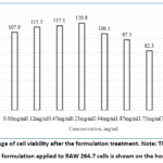 |
Figure 1: Percentage of cell viability after the formulation treatment. Note: The concentration of the herbal formulation applied to RAW 264.7 cells is shown on the horizontal axis. |
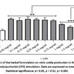 |
Figure 2: Effect of the herbal formulation on nitric oxide production in RAW 264.7 cells following lipopolysaccharide (LPS) stimulation. Data are expressed as mean ± SD (n ≥ 3). Statistical significance: p< 0.05, p < 0.01, p< 0.001. |
Result
TNF-α, HMGB1, IL-10, and SOD Levels in Blood
Given the central role of inflammation in acute lung injury, serum levels of TNF-α, IL-10, SOD, and HMGB1 were measured by enzyme-linked immunosorbent assay (ELISA).
At 6 hours, TNF-α levels were reduced by 15.8% and 31.6% in the groups treated with 528 mg/kg and 1320 mg/kg of herbal formulation, respectively, compared to a 16.5% reduction in the ibuprofen-treated control group. At 9 hours, TNF-α levels decreased by 13.3% in the 1320 mg/kg group and by 25.6% in the ibuprofen-treated group. By 12 hours, significant reductions were observed: 14.4% in the 528 mg/kg group and 11.3% in the ibuprofen-treated group (Table 1; p < 0.05).
In the LPS-induced acute lung injury model, untreated controls had significantly lower HMGB1 levels than disease controls: 40.6% lower at 3 hours, 43.5% lower at 6 hours, 21% lower at 9 hours, and 36.2% lower at 12 hours (p < 0.05). In groups treated with the herbal formulation, HMGB1 levels were notably reduced compared with the control group at 6 hours, by 13.6% in the 528 mg/kg group and 14.6% in the 1320 mg/kg group. The ibuprofen-treated group showed a 16.2% reduction (Table 2, p < 0.05).
As shown in Table 3, in the LPS-induced acute lung injury model, IL-10 levels in the Disease control group were significantly higher than in the healthy group, by 25.7% at 3 hours and 20.9% at 6 hours (p < 0.05).
Table 1: Effects of herbal formulationon the level of TNF-α in LPS-induced acute lung inflammation
| Group | TNF-α(pg/ml) | |||
| 3 h | 6 h | 9 h | 12 h | |
| Healthy control | 37.8±3.6 | 37.8±3.6 | 37.8±3.6 | 37.8±3.6 |
| Disease control | 45.27±1.5** | 43.88±0.6* | 39.3±1.5 | 39.95±4.1 |
| 528mg/kg | 38.22±3.6# | 37.9±6.01# | 38.95±4.4 | 34.93±3.6# |
| 1320mg/kg | 39.70±2.9# | 33.34±2.6## | 34.7±5.7# | 39.9±2.7 |
| Ibuprofen 35 mg/kg | 35.28±1.8## | 37.66±4.9# | 31.28±4.3# | 35.90±3.3# |
Compared with the healthy control group:p< 0.05.
Compared with the disease control group: #p< 0.05, ##p< 0.01 (Kruskal–Wallis test)
Table 2: Effect of herbal formulation on HMBG1 protein levels in the LPS-induced acute lung injury model
| Group | HMGB1 (ng/ml) | |||
| 3 h | 6 h | 9 h | 12 h | |
| Healthy control | 13.8±0.9 | 13.8±0.9 | 13.8±0.9 | 13.8±0.9 |
| Disease control | 19.4±0.34* | 19.8±2.1* | 16.7±1.1 | 18.8±2.1 |
| 528mg/kg | 19.57±0.98 | 17.43±0.45# | 17.73±3.1 | 17.19±1.20 |
| 1320mg/kg | 19.56±0.98 | 17.28±0.45# | 16.84±1.9 | 15.89±1.74# |
| Ibuprofen 35 mg/kg | 18.64±1.5 | 17.04±1.32# | 15.26±1.6 | 18.34±1.12 |
Compared with the healthy control group:p< 0.05 (Kruskal–Wallis test).
Compared with the disease control group: #*p < 0.05.
Table 3: Effects of herbal formulation on IL-10 cytokine levels in an LPS-induced acute lung inflammation model
| Group | IL-10 (pg/ml) | |||
| 3 h | 6 h | 9 h | 12 h | |
| Healthy control | 8.54±0.9 | 8.54.±0.9 | 8.54±0.9 | 8.54±0.9 |
| Disease control | 11.5±0.34* | 10.8±1.01* | 9.73±1.01 | 9.94±0.7 |
| 528mg/kg | 10.02±0.6 | 10.26±1.1 | 10.0±0.1 | 10.04±0.8 |
| 1320mg/kg | 9.45±0.75 | 9.67±0.7 | 9.05±0.13 | 10.4±0.7 |
| Ibuprofen 35 mg/kg | 9.84±0.3 | 9.36±0.9 | 10.1±0.36 | 9.54±0.7 |
Compared to healthy controls *p<0.05 Kruskal-Wallis test
Table 4: Effect of herbal formulation on superoxide dismutase levels in an LPS-induced acute lung inflammation model
| Group | Superoxide dismutases(ng/ml) | |||
| 3 h | 6 h | 9 h | 12 h | |
| Healthy control | 5.18±0.69 | 5.18±0.69 | 5.18±0.69 | 5.18±0.69 |
| Disease control | 3.46±0.55* | 3.47±0.49* | 3.87±0.22* | 3.68±0.68* |
| 528mg/kg | 4.18±0.44# | 3.63±0.26 | 3.54±0.41 | 3.11±0.54 |
| 1320mg/kg | 3.34±0.42# | 3.11±0.40 | 3.57±0.13 | 3.16±0.48 |
| Ibuprofen 35 mg/kg | 3.14±0.24 | 3.19±0.16 | 3.19±0.65 | 3.82±0.18 |
Compared with healthy controls:p< 0.05.
Compared with the control group: #p< 0.05.
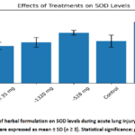 |
Figure 3: Effect of herbal formulation on SOD levels during acute lung injury induced by LPS (3 hours). |
In the groups given herbal formulations, SOD levels were significantly higher than the control group 3 hours after the administration, increasing by 16.5% at 528 mg/kg and by 20.8% at 1320 mg/kg (p < 0.05).
Histological examination results
In the LPS-induced acute lung injury model, tissue alterations were evaluated using the Acute Respiratory Distress Syndrome Network scoring system described by Matute-Bello et al. (2011). Microparticles were administered at doses of 528 mg/kg and 1320 mg/kg, and histological assessment revealed dose-dependent improvements in lung morphology compared with the pathological control group.
Alveolar wall thickening, no inflammatory cells were observed; the alveolar spaces were clear, and the blood vessels appeared normal. (Figure 4A) Lung tissue from the disease control group showed thickening of the alveolar walls, infiltration of inflammatory cells and macrophages in the alveolar spaces and interstitium, focal alveolar atelectasis, and hemorrhage. (Figure 4B) Histological analysis of lung tissue from the experimental group treated with 528 mg/kg of lung microparticles showed decreased hemorrhage (Figure 4C). Compared with the 528 mg/kg group, treatment with 1320 mg/kg of lung microparticles restored alveolar structure, thinned alveolar walls, and reduced both inflammatory cell infiltration and hemorrhage in the alveolar and interstitial areas (Figure 4D).
Table 5: Results of Histological Assessment of Lung Tissue
| № | Histopathological Features | Score | 1320 mg/kg (n=5) | 528 mg/kg (n=5) | LPS |
| 1 | Alveolar wall thickening | 3 | 2,6 | 2.2 | 2.7 |
| 2 | Intra-alveolar and interstitial neutrophils | 3 | 1 | 2 | 1.5 |
| 3 | Hemorrhage or alveolar edema | 3 | 1,8 | 1,8 | 3 |
| 4 | Alveolar destruction/ architectural disruption | 3 | 1,6 | 1,6 | 2 |
| 5 | Macrophage infiltration | 3 | 2 | 2 | 2 |
| Total | 15 | 9 | 9.6 | 11.2 |
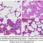 |
Figure 4: Changes in lung tissue after the experiment: 3(A) Health control, 3(B) Disease Control, 3(C) 528mg/kg, 3(D) 1320mg/kg. Legend: |
Molecular docking analysis results
The molecular interactions between the inflammatory proteins TNF-α, HMGB1, and IL-10 and the compounds in the herbal formulation, including glycyrrhizic acid, inulin, ascorbic acid, alantolactone, and isoalantolactone, were modelled using the molecular docking program DOCK 6.9v. The grid scores calculated using all possible bubbles for all protein interactions are shown in Table 6.
Table 6: Results of interactions between TNF-α, HMBG1, IL-10 proteins, and active ingredients molecules
| № | Active ingredients molecule | Protein binding affinity | ||
| TNF-α (ccal/mol) | HMGB1 (ccal/mol) | IL10 (ccal/mol) | ||
| 1 | Alantolactone | -38.97(LYS A:251, CYS A:208, PRO A:239) Hydrogen and carbon bonds | -30.03(ARG A:10, ASP A:5, LYS A:8) Hydrogen and alkyl bonds | -31.16(CYS A:45, ARG A:10, TYR A: 55)Charge attraction interactions; Carbon–hydrogen bonds; π–anion interactions |
| 2 | Isoalantolactone | -37.16(TRP A:253, CYS A:208)hydrogen bonding; Sulfur-based interactions (Sulfur-X); π–alkyl interactions | -30.83(ARG A:10, LYS A:8, PRO A:6 GLYA:4, GLY A:2) Hydrogen and alkyl bonds | -30.36(PHE G:94, PRO A:96, CYS G:45) AlkylAlkyl và π-alkyl π-π stacked |
| 3 | Glycyrrhizic acid | -79.61(LYS A:237, 89,103,251, GLU A:55, ARG A:94, TYR, A:253, 254, GLN A:93)Van der Waals, electrostatic and hydrogen interactions. | -95.38(LYS A:87,90,96, LYS88, PRO A:99, PHE A:103, ARG A:97)Hydrogen, electrostatic and hydrophobic interactions | -81.86(CYS A:45, TYR A:55, ARG A:10)Electrostatic (salt bridges, charge attraction), hydrogen (C–H) and π–anion interactions. |
| 4 | Inulin | -72.96(LYS A:251, CYS A:208, 92, PRO A:91, 239, GLU A:107, A:255, SER A:90, TYR A:254) Hydrogen and carbon bonds | -46.44(LYS A:167, 96, ARG A:163, A:97, ALA A:94, ASP A:91, PRO A:95, PHF A:89) Hydrogen and carbon bonds | -44.46(TYR A:55) hydrogen bond, π-donor hydrogen bond |
| 5 | Ascorbic acid | -53.6(GLY A:247, 207, GLU A:249, 246, CYS A:208, ARG A:242, THR A:244, LYS A:251) Hydrogen and carbon bonds | -30.47(LYS A:96, ASP A:158, LYS A:154, PRO A:98) Hydrogen and carbon bonds | -30.99(MET A:5, ASN A:80, ARG A:87) Hydrogen bonding |
Depending on the parameters and scoring function used, we evaluated the score by comparing the molecules under study rather than using absolute thresholds. As a result, glycyrrhizic acid and inulin showed the most negative scores for TNF-α, HMGB1, and IL-10, indicating relatively high binding affinity.
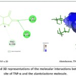 |
Figure 5(A): 2D and 3D representations of the molecular interactions between the active site of TNF-α and the alantolactone molecule. |
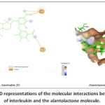 |
Figure 5(B): 2D and 3D representations of the molecular interactions between the active site of interleukin and the alantolactone molecule. |
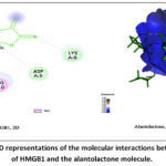 |
Figure 5(C).2D and 3D representations of the molecular interactions between the active site of HMGB1 and the alantolactone molecule. |
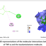 |
Figure 6(A): 2D and 3D representations of the molecular interactions between the active site of TNF-α and the isoalantolactone molecule. |
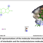 |
Figure 6(B): 2D and 3D representations of the molecular interactions between the active site of interleukin and the isoalantolactone molecule. |
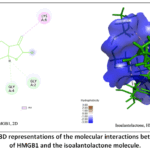 |
Figure 6(C): 2D and 3D representations of the molecular interactions between the active site of HMGB1 and the isoalantolactone molecule. |
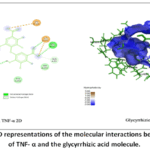 |
Figure 7(A): 2D and 3D representations of the molecular interactions between the active site of TNF- α and the glycyrrhizic acid molecule. |
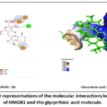 |
Figure 7(B): 2D and 3D representations of the molecular interactions between the active site of HMGB1 and the glycyrrhizic acid molecule. |
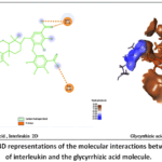 |
Figure 7(C): 2D and 3D representations of the molecular interactions between the active site of interleukin and the glycyrrhizic acid molecule. |
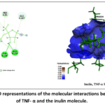 |
Figure 8(A): 2D and 3D representations of the molecular interactions between the active site of TNF- α and the inulin molecule. |
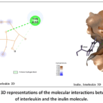 |
Figure 8(B): 2D and 3D representations of the molecular interactions between the active site of interleukin and the inulin molecule. |
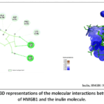 |
Figure 8(C): 2D and 3D representations of the molecular interactions between the active site of HMGB1 and the inulin molecule. |
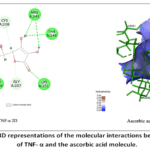 |
Figure 9(A): 2D and 3D representations of the molecular interactions between the active site of TNF- α and the ascorbic acid molecule. |
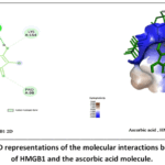 |
Figure 9(B). 2D and 3D representations of the molecular interactions between the active site of HMGB1 and the ascorbic acid molecule. |
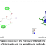 |
Figure 9(C): 2D and 3D representations of the molecular interactions between the active site of interleukin and the ascorbic acid molecule. |
Discussion
Analysis of nitric oxide (NO) production indicated that the herbal product had no significant effect on NO release in unstimulated RAW 264.7 cells. In contrast, upon LPS stimulation, the formulation significantly suppressed NO production compared with the LPS-treated groups. In the LPS-induced acute lung inflammation model, administration of the herbal product significantly increased SOD protein levels while markedly decreasing TNF-α and HMGB1 protein levels. Histopathological evaluation revealed that the formulation demonstrated a protective effect against LPS-induced acute lung inflammation, indicating its potential efficacy in mitigating inflammatory lung injury. Furthermore, molecular docking analysis demonstrated that glycyrrhizic acid and inulin form strong binding interactions with TNF-α and HMGB1 proteins, supporting their potential role in mediating the observed anti-inflammatory effects.
Glycyrrhizic acid also exhibited moderate binding affinity to flavonoids (isorhamnetin and kaempferol), as well as JUN–FOS–DNA, MAPK14, IL-6. Notably, glycyrrhizin demonstrated the highest binding affinity for COX-2 (C-score = 13.07). These interactions align with the inhibition of pro-inflammatory cytokine production and HMGB1 translocation observed in both in vitro and in vivo models, underscoring the potential of the herbal formulation to modulate key inflammatory pathways. Our results indicate that glycyrrhizic acid binds to TNF-α, interleukins, and HMGB1, supporting its effectiveness.
The activity of NO synthase in RAW 264.7 cells is an important marker for evaluating the effects of anti-inflammatory drugs.25 After the treatment of RAW 264.7 macrophage-like cells with various concentrations of the extract, nitric oxide (NO) levels were measured using the Griess assay. In this study, no statistically significant differences were observed between the extract-treated groups and the phosphate-buffered saline (PBS) control group at 12 and 24 hours. However, NO production was decreased at extract concentrations of 1–2 mg/mL compared to the control group. This suggests that it may have a potent anti-inflammatory effect. In a study by Park et al., high concentrations of marigold flower extracts which contain ISO were shown to inhibit the synthesis of NO and pro-inflammatory cytokines, including TNF-α and IL-1β, in LPS-stimulated RAW 264.7 cells.26 The effects of ISO on the release of TNF-α and IL-1β in bronchoalveolar lavage fluid (BALF) following LPS induction were assessed. However, treatment with isoalantolactone significantly reduced the level of these pro-inflammatory cytokines.27
Inulin has been shown to regulate the TNF-α by modifying the pathway proteins that are involved in the “cellular response to TNF-α in LPS-induced macrophages.28 Additionally, inulin reduces pro inflammatory cytokines by regulating gene and protein sensing pathways that modulate TNF-α responses 28, as well as in our study, the formulation significantly reduced the TNF-α level.
The study showed that glycyrrhizin prevented the translocation and release of HMGB1 from the nucleus to the cytoplasm in LPS-stimulated RAW 264.7 macrophages and significantly decreased the production of TNF-α and IL-6. Glycyrrhizic acid blocks the activation of the Toll-like receptor 4 (TLR4)/NF-κB signaling pathway, which in turn inhibits the cytoplasmic translocation and extracellular release of HMGB1 and lowers the levels of pro-inflammatory cytokines like TNF-α and IL-6. Glycyrrhizic acid also reduced HMGB1 expression in in vivo models and lowered lung inflammatory markers, including TNF-α and IL-630, aligning with the findings of our study.
Conclusion
Treatment of the herbal formulation significantly reduced TNF-α and HMGB1 levels compared with the control group. Histopathological evaluation of LPS-induced acute lung inflammation demonstrated preservation of lung architecture, by reduced alveolar wall thickening, decreased inflammatory cell infiltration, and lowered hemorrhage in alveolar and interstitial regions. These protective effects are supported by molecular docking results, which revealed strong binding interactions between glycyrrhizic acid and inulin with TNF-α and HMGB1, suggesting a potential mechanism for the observed anti-inflammatory activity.
Acknowledgement
The authors gratefully acknowledge the financial support from the Science and Technology Fund of the Mongolian National University of Medical Sciences (MNUMS). They also sincerely appreciate the support and collaboration of the School of Graduate Studies, the School of Pharmacy, the Institute of Physics and Technology, and the Institute of Traditional Medicine and Technology at MNUMS throughout this study.
Funding source
This project was carried out with support from the Mongolian National University of Medical Science, under contract number 02/2023.
Conflict of Interest
The authors declare no competing interests associated with the manuscript.
Data Availability Statement
This study did not involve any data.
Ethics statement
This study was approved by the Research Ethics Review Committee of the University of Ashgabat (Permit number №2023/3-07).
Informed Consent Statement
This study did not include human participants, so informed consent was not necessary.
Clinical Trial Registration
This study did not include any clinical trials.
Permission to reproduce material from other sources
Not applicable
Author Contributions
- Ariunzaya Lkhagvaa: Data collection, Analysis, review and & Editing, Writing- Original Draft
- Jambanninj Dambiinyam: Data collection, Analysis, Review& Editing
- Anujin Tseveenjav: Data collection, Analysis, Analysis,
- Anand Altankhuyag: Data collection, Analysis,
- Khongor Bat-Erdene: Data collection, Analysis, Analysis,
- Maralgua Avirmed: Data collection, Analysis
- Munkhzul Boldbaatar: Data collection, Analysis
- Turtushikh Damba: Data collection, Analysis, Review& Editing
- Tsengelmaa Jamiyan: Data collection, Analysis
- Ariunzaya Bat-Erdene:Data collection, Analysis, Review& Editing;
- Purevsuren Sodnomtseren: Data collection, Analysis, Review, & Editing;
- Shiirevnyamba Avirmed: Data collection, Analysis, Review& Editing
- Sarnai Tsagaankhuu: Data collection, Analysis, review and & Editing, and Project Administration
Reference
- World Health Organization. WHO Traditional Medicine Strategy 2014–2023. Geneva, Switzerland: World Health Organization; 2013.
- Fabricant DS, Farnsworth NR. The value of plants used in traditional medicine for drug discovery. Environ Health Perspect. 2001;109(Suppl 1):69-75. doi:10.1289/ehp.01109s169
CrossRef - Lkhagvaa A, Tsagaankhuu S, Sodnomtseren P et al. A technological study on developing granules from plants used in traditional Mongolian medicine. Cent Asian J Med Sci. 2025;11(1):34-45. doi:10.24079/cajms.2025.01.005
CrossRef - Health Development Center (HDC), Ministry of Health of Mongolia. Health Indicators 2024. Ulaanbaatar, Mongolia: HDC, Ministry of Health of Mongolia; 2024.
- Su X, Wu L, Hu M et al. Glycyrrhizic acid: a promising carrier material for anticancer therapy. Biomed Pharmacother. 2017;95:670-678. doi:10.1016/j.biopha.2017.08.123
CrossRef - Fouladi S, Masjedi M et al. The in vitro impact of glycyrrhizic acid on CD4+ T lymphocytes through OX40 receptor in patients with allergic rhinitis. Inflammation. 2018;41(5):1690-1701. doi:10.1007/s10753-018-0813-8
CrossRef - Qu L, Chen C, He W et al. Glycyrrhizic acid ameliorates LPS-induced acute lung injury by regulating autophagy through the PI3K/AKT/mTOR pathway. Am J Transl Res. 2019;11(4):2042-2055.
- Lee SA, Lee SH, Kim JY et al. Effects of glycyrrhizin on lipopolysaccharide-induced acute lung injury in a mouse model. J Thorac Dis. 2019;11(4):1287-1302. doi:10.21037/jtd.2019.04.14
CrossRef - Yang G, Yang L, Xu F. Isoalantolactone: a review on its pharmacological effects. Front Pharmacol. 2024;15:1453205. doi:10.3389/fphar.2024.1453205
CrossRef - Qu Y, Li JH, Zhang C et al. Content determination of twelve major components in Tibetan medicine Zuozhu Daxi by UPLC. China J Chin Mater Med. 2015;40:1825-1830.
- Qiburi Q, Ganbold T, Bao Q et al. Bioactive components of ethnomedicine Eerdun Wurile regulate the transcription of pro-inflammatory cytokines in microglia. J Ethnopharmacol. 2020;246:112241. doi:10.1016/j.jep.2019.112241
CrossRef - Li T, Xu H, Rezengcaidan, et al. Rapid identification of Radix inulae and its active component alantolactone in the Tibetan medicine Manuxitang. Biosci Trends. 2008;2:64-67.
- Liu X, Bian L, Duan X et al. Alantolactone: a sesquiterpene lactone with diverse pharmacological effects. Chem Biol Drug Des. 2021;98(6):1131-1145. doi:10.1111/cbdd.13972
CrossRef - Wang J, Zhang HH, Tian JM et al. Simultaneous determination of six components in commercial Roukou Wuwei pills using ultra-high-performance liquid chromatography with a diode-array detector. Biomed Chromatogr. 2019;33(12):e4677. doi:10.1002/bmc. 4677
CrossRef - Qu Z, Jiang D, Liu Y et al. Protects the gastric mucosa from gastric ulcer in rats by regulating the JAK2/STAT3 pathway. Tissue Cell. 2023;83:102145. doi:10.1016/j.tice.2023.102145
CrossRef - Olennikov DN, Chemposov VV, Chirikova NK et al. Metabolites of prickly rose: chemodiversity and digestive-enzyme-inhibiting potential of Rosa acicularis and the main ellagitannin rugosin D. Plants (Basel). 2021;10:Article number unavailable. doi:10.3390/plants
CrossRef - WHO Regional Office for the Western Pacific. Medicinal Plants in Mongolia. Manila, Philippines: World Health Organization Regional Office for the Western Pacific; 2013. ISBN:9789290616320
- Patel S. Rosehip as an underutilized functional food: an evidence-based review. Trends Food Sci Technol. 2017;63:29-38. doi:10.1016/j.tifs.2017.03.001
CrossRef - Matute-Bello G, Downey G, Moore BB, et al. An official American Thoracic Society workshop report: features and measurements of experimental acute lung injury in animals. Am J Respir Cell Mol Biol. 2011;44:725-738. doi:10.1165/rcmb.2009-0210ST
CrossRef - Mosmann T. Rapid colorimetric assay for cellular growth and survival: application to proliferation and cytotoxicity assays. J Immunol Methods. 1983;65:55-63. doi:10.1016/0022-1759(83)90303-4
CrossRef - Fotakis G, Timbrell JA. In vitro cytotoxicity assays: comparison of LDH, neutral red, MTT, and protein assay in hepatoma cell lines following exposure to cadmium chloride. Toxicol Lett. 2006;160(2):171-177. doi:10.1016/j.toxlet.2005.07.001
CrossRef - Green LC, Wagner DA, Glogowski J et al. Analysis of nitrate, nitrite, and [¹⁵N]nitrate in biological fluids. Anal Biochem. 1982;126:131-138. doi:10.1016/0003-2697(82)90118-X
CrossRef - Li G, Zhou CL, Zhou QS et al. Galantamine protects against lipopolysaccharide-induced acute lung injury in rats. Braz J Med Biol Res. 2016;49(2):e5008. doi:10.1590/1414-431X20155008
CrossRef - Ijaz M, Huang X, Buabeid M et al. Mechanistic investigation of Glycyrrhiza uralensis effects against respiratory ailments: application of network pharmacology and molecular docking approaches. Lett Drug Des Discov. 2021;18(5):397-412.
CrossRef - Ito K, Barnes PJ, Adcock IM. Glucocorticoid receptor recruitment of histone deacetylase 2 inhibits interleukin-1-induced histone H4 acetylation on lysines 8 and 12. Mol Cell Biol. 2000;20(18):6891-6903.
CrossRef - Chun J, Song K, Kim YS. Anti-inflammatory activity of standardized fraction from Inula helenium via suppression of NF-κB pathway in RAW 264.7 cells. Nat Prod Sci. 2019;25(1):16-22. doi:10.20307/nps.2019.25.1.16
CrossRef - Yuan CB, Tian L, Yang B et al. Isoalantolactone protects lipopolysaccharide-induced acute lung injury through Nrf2 activation. Microb Pathog. 2018;123:213-218. doi:10.1016/j.micpath.2018.07.010
CrossRef - Wu CX, He LX, Guo H et al. Inhibitory effect of glycyrrhizin on lipopolysaccharide-induced high-mobility group box 1 release and expression in RAW264.7 cells. Shock. 2015;43(4):412-421. doi:10.1097/SHK.0000000000000309
CrossRef - Wang G, Hiramoto K, Ma N et al. Glycyrrhizin attenuates carcinogenesis by inhibiting the inflammatory response in a murine model of colorectal cancer. Int J Mol Sci. 2021;22(5):2609. doi:10.3390/ijms22052609
CrossRef - Sun X, Zeng H, Wang Q et al. Glycyrrhizin ameliorates inflammatory pain by inhibiting microglial activation-mediated inflammatory response via blockage of the HMGB1–TLR4–NF-κB pathway. Exp Cell Res. 2018;369(1):112-119. doi:10.1016/j.yexcr.2018.05.012
CrossRef

