
Manuscript accepted on :26-09-2025
Published online on: 20-10-2025
Plagiarism Check: Yes
Reviewed by: Dr. Stephen Adepoju
Second Review by: Dr. Anjaneyulu Vinukonda
Final Approval by: Dr. Kamal Upreti
Abhimanu Singh1,2* and Smita Jain2
1Department of Applied Mathematics, Bhagwan Parshuram Institute of Technology, Delhi, India
2Department of Mathematics, JECRC University, Jaipur, India
Corresponding Author E-mail:asingh19669@gmail.com
Abstract
Brain tumors create life threatening consequences. Timely and accurate detection of brain tumors is critical for effective treatment planning and improving patient survival chances. In this study, we present a deep learning–hybrid model for automated brain tumor detection using MRI images. Our model consists of two phases. First phase extracts features and the second phase performs classification. EfficientNetB1 convolutional neural network is employed to extract features and Support Vector Machine (SVM) as a classifier. The EfficientNetB1 model, pre-trained on ImageNet, is employed as a fixed feature extractor to leverage its optimized architecture for capturing meaningful spatial representations from medical images. Without the use of data augmentation or dimensionality reduction techniques, the model extracts deep features directly from the MRI images, maintaining the integrity of anatomical information critical for accurate diagnosis. These extracted features are then used to train an SVM classifier with a radial basis function (RBF) kernel, enabling precise binary classification between tumor and non-tumor cases. The proposed method is evaluated on a publicly available brain MRI dataset and achieves a validation accuracy of 99.33%, along with high precision, recall, and F1-score. Compared to conventional CNN-based or transfer learning methods that rely on complex pipelines or augmented data, this architecture remains simple, fast, and highly effective. This framework offers a practical, low-overhead solution for assisting radiologists in brain tumor detection.
Keywords
Brain Tumor Detection; Deep Feature Extraction; Diagnostic Accuracy; Support Vector Machine; Transfer Learning
| Copy the following to cite this article: Singh A, Jain S. Improving Diagnostic Accuracy in Brain Tumor Detection using EfficientNetB3 Transfer Learning and Support Vector Machine. Biomed Pharmacol J 2025;18(October Spl Edition). |
| Copy the following to cite this URL: Singh A, Jain S. Improving Diagnostic Accuracy in Brain Tumor Detection using EfficientNetB3 Transfer Learning and Support Vector Machine. Biomed Pharmacol J 2025;18(October Spl Edition). Available from: https://bit.ly/47yYCNK |
Introduction
Brain tumors are among the most critical and life-threatening conditions affecting the central nervous system. Timely and accurate diagnosis is essential to formulate effective treatment strategy which may result in improved patient survival rates. Magnetic Resonance Imaging (MRI) is the widely used modality for non-invasive brain imaging due to its high spatial resolution and soft tissue contrast. However, manual interpretation of MRI scans by radiologists is time-consuming and highly error prone, consequently demands an automated and reliable diagnostic method. Recent advances in artificial intelligence, particularly deep learning, have shown great achievements in medical image analysis. Convolutional Neural Networks (CNNs) which are a subclass of deep learning, have performed excellently in image classification tasks, specifically brain tumor detection. However, training deep CNNs from scratch requires large labelled datasets and significant computational resources, which are often limited in medical applications. Transfer learning, which reuses pre-trained models, trained on large-scale datasets like ImageNet, provides an effective solution by enabling powerful feature extraction even with limited medical data.
In this study, we propose a deep learning–based framework for brain tumor detection using EfficientNetB3 as a feature extractor, followed by classification by Support Vector Machine (SVM). EfficientNetB3, known for its balance of accuracy and computational efficiency, extracts important features from MRI images. These features are then passed to an SVM classifier, which distinguishes between tumor and non-tumor cases with high precision. The proposed model is evaluated on a publicly available brain MRI dataset and achieves a classification accuracy of 99.33%, demonstrating its effectiveness and reliability. By combining the power of transfer learning with classical machine learning, this approach offers a lightweight and highly accurate solution for real-time brain tumor detection, potentially assisting clinicians in detecting brain tumor and finally making appropriate decision for the welfare of the suffering person. Following are the related works done by the researchers:
Agarwal, M., et al. proposed an automated, robust, and intelligent hybrid system consisting of two phases, for early diagnosis and classifying brain tumor. In the first phase Optimized Double Threshold Weighted Constraints Histogram Equalization technique is employed to enhance image contrast. In the second phase, the classifier using transfer learning having Inception V3 as base model, classifies the images. The model achieved an accuracy value of 98.89%.1 Appiah, R., et al. proposed a model consisting of Proper Orthogonal Decomposition (POD) paired with convolutional neural network, classifies brain MR images very fast with an accuracy value of 95.88%.2 Dutta, T.K., et al. proposed an attention based residual multiscale CNN referred to as ARM-Net and achieved a maximum accuracy of 97.11% on BRATS 2020 dataset.3 Khoramipour, S., et al. proposed a brain tumor classifier consisting of a convolutional neural network enhanced with support vector machine and achieved 99% accuracy.4 Priya, A., et al. proposed a hybrid AlexNet-GRU (Gated Recurrent Unit) architecture to detect and classify multiclass brain tumors and obtained an accuracy of 97%.5 Raghuwanshi, S., et al. proposed a methodology using VGG19 and Inception V3 for feature extraction and logistic regression and K-Nearest Neighbors as classifiers and found that Inception V3 along with augmentation gives the best accuracy value of 95.43%.6 Sandhiya, B., et al. developed an enhanced machine for brain tumor classification. They used Inception V3 model and DenseNet201 models to retrieve the basic features and Particle Swarm Optimized Kernel Extreme Learning Machine (PSO-KELM) for classification into four categories. They reported the accuracy value of 98.21% on the test dataset.7 Simo, A.M.D., et al. designed a sequential brain tumor detection and classification model based on deep learning technique. The proposed model works in two phases. First phase distinguishes between the non-neoplastic brain from neoplastic brain; the second phase determines the tumor type from the neoplastic brains. They experimented with four optimizers namely: Adam, Nesterov momentum, root-mean-square propagation, and adaptive gradient. They reported that Nesterov performed best with 95% on testing and validation datasets.8 Singh, T., et al. developed BrainNet to classify brain tumor categories. The proposed model achieved an accuracy value of 97.71% on test dataset.9 Sultanpure, K.A., et al. presented a technique for identifying brain cancer using Internet of Things (IoT), Particle Swarm Optimization (PSO), deep learning techniques. The proposed model achieved 98.00% accuracy.10 Tejashwini, P.S., et al. designed an Ensemble Brain Tumor Deep Net for brain tumor detection and obtained an accuracy value of 98.80 %.11 Agrawal, P., et al. employed 3D-UNet for volumetric segmentation of MRI images and convolutional neural network model for classification and obtained an accuracy value of 90% on test data.12 Ali, M. et al. proposed a framework for brain tumor detection and categorical classification. In their framework, they used Particle Swarm Optimization (PSO) algorithm for tumor segmentation and Convolutional Neural Network (CNN) algorithm for classification purpose. For classification purpose they used AlexNet and Inception-V3 pre-trained models using transfer learning. On BRATS-2018 and BRATS-2017 datasets, they reported accuracy value of 99.0%.13 Irfan, M., Shaf, A., used pre-trained models ResNet50, VGG16, VGG19, and U-Net and their combinations. They report that U-Net model itself gives highest accuracy value of 95%.14 Isaza, AA., et al. proposed a framework for brain tumor detection and classification. They performed experimentation with no method, transfer learning, data augmentation, and transfer learning and data augmentation both. They reported that combination of transfer learning and data augmentation enhance accuracy up to 97%.15 Islam, M M, et al. used transfer learning using Inception V3, VGG19, DenseNet121, and MobileNet as the base models on three databases of figshare, SARTAZ, and Br35H. They report that MobileNet outperforms giving an accuracy value of 98.40% on test data.16 Zulfikar F, et al. used transfer learning -based method for multi-class classification of brain tumors using variants of EfficientNets i.e. EfficientNetB0-EfficientNetB4, under different experimental settings. Their findings report a highest accuracy value of 98.86 given by EfficientNetB2.17 Lamba K, et al. presented an integrated deep learning and supervised learning approach for early detection of brain tumor using magnetic resonance imaging and achieved an accuracy value of 98.87%.18 Malakouti S M, et al. used transfer learning technique using GoogleNet as the base model to classify the sick and healthy brain MRI images and achieved an accuracy value of 99.3%.19 Bhardwaj N, et al. proposed a deep learning model for brain tumor detection and achieved accuracy value of 98.56%.20 Singh A and Jain S, designed a convolutional neural network and by leveraging the hyperparameters achieved an accuracy value of 99.31% on the test data.21
Materials and Methods
The purpose of this study is to enhance the accuracy of detection of brain tumor. Recent researches show that deep learning methods, especially the convolutional neural network models are outperforming in the field of medical image analysis. Training a deep learning model from scratch typically requires large annotated datasets and substantial computational resources. In the field of medical imaging, especially for brain tumor detection, acquiring such large-scale labeled data is often challenging. Privacy and ethical constraints, limited availability of annotated medical images, etc. are the major challenges in the path of getting large-scale labeled data. To address these limitations, transfer learning offers a practical and effective solution by leveraging deep neural networks that have been pretrained on large, general-purpose image datasets such as ImageNet. These pretrained models capture universal low- and mid-level features (e.g., edges, textures, and shapes) that are also relevant to medical images, despite differences in domains. In this paper we using transfer learning method. We employed EfficientNetB3 for feature extraction and support vector machine for classification.
Dataset
This study utilizes a brain MRI image dataset consisting 3000 MRI images labeled as “yes” (having tumor) and “no” (without tumor). This dataset is available with Kaggle’s platform titled as “Brain_Tumor_Detection_MRI”. All MRI images were resized to 300 × 300 pixels to match the input size requirements of the EfficientNetB3 architecture. Since EfficientNetB3 expects three-channel input, grayscale images were converted to RGB by duplicating the single channel No augmentation is applied. The dataset was partitioned to get training and validation subsets using an 80:20 split ratio. The computation work was conducted on Google Colab using T4 GPU with Keras library of TensorFlow API.
Transfer Learning with EfficientNetB3
Transfer learning allows the use of pretrained weights that already encapsulate rich feature representations. This significantly reduces the amount of data required for effective training, which is especially valuable in medical imaging where labeled data is scarce. Fine-tuning or using a frozen feature extractor from a pretrained model accelerates the training process and helps the model converge faster with better generalization. This leads to more stable performance even on small datasets. Models like EfficientNetB3 are designed for optimal performance-to-complexity ratio. When used with transfer learning, they achieve high accuracy without demanding high-end hardware, making them suitable for real-time and resource-constrained clinical environments. In small medical datasets, training a deep network from scratch often results in overfitting. Using transfer learning with frozen convolutional layers helps retain generalized features and minimizes the risk of fitting to noise in the limited dataset. Numerous studies have demonstrated the effectiveness of transfer learning in tasks such as lung disease detection, skin lesion classification, and retinal disease diagnosis. Its successful application in similar domains further justifies its adoption for brain tumor detection.
EfficientNetB3, a lightweight convolutional neural network pretrained on ImageNet, was employed as a feature extractor. The convolutional base was frozen to retain learned weights and prevent overfitting due to the limited size of the medical dataset. The feature maps generated from the final convolutional layers were fed to a support vector machine classifier.
Feature Extraction
The output of the final pooling layer of EfficientNetB3 was flattened to get a one-dimensional feature vector for each image. These vectors, representing high-level learned features, were used as input to a support vector machine classifier for the classification task.
Classification using Support Vector Machine (SVM)
A Support Vector Machine with a Radial Basis Function (RBF) kernel was employed for binary classification task. SVM was chosen for its robustness in high-dimensional feature spaces and its ability to handle non-linear decision boundaries effectively. The SVM classifier was trained on 80% of the dataset and validated on the remaining 20%.
Evaluation Metrics
The performance of the model was assessed using standard classification metrics including accuracy, precision, recall, and F1-score. A confusion matrix was also generated to visualize the distribution of true positives, false positives, true negatives, and false negatives. The model achieved an accuracy of 99.33% on the validation set, demonstrating high diagnostic reliability.
Results
The proposed EfficientNetB3 + SVM model was evaluated on a curated dataset of brain tumor MRI images. The dataset was partitioned to get training and validation subsets using an 80:20 split ratio. The input MRI images were resized to 300×300 pixels to match the EfficientNetB3 architecture requirements. Feature extraction was performed by EfficientNetB3 model with ImageNet weights, followed by SVM-based classification. The confusion matrix and the ROC curve obtained are presented in figure1 and figure 2, respectively, and are given below:
Class 0: Negative (No Tumor)
Class 1: Positive (Tumor)
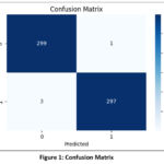 |
Figure 1: Confusion Matrix |
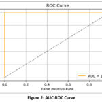 |
Figure 2: AUC-ROC Curve |
Using the confusion matrix, we get:
True Positives (TP) = 297
True Negatives (TN) = 299
False Positives (FP) = 1
False Negatives (FN) = 3
From these values, key performance metrics computed are as follows:
Accuracy = 99.33%
Precision = 99.66%
Recall (Sensitivity) = 99.00%
F1 Score = 99.32%
Table 1: Metrics obtained by the proposed model
| Accuracy | Precision | Recall | Specificity | F1 Score |
| 99.33 % | 99.66% | 99.00% | 99.66 | 99.32 |
Discussion
These results demonstrate the strong classification capability of the EfficientNetB3 + SVM framework in accurately identifying brain tumors from MRI images.
The confusion matrix reveals only a very small number of misclassifications, demonstrating the model’s ability to generalize well on unseen data. The model achieved an overall classification accuracy of 99.33% on the validation dataset. The precision, recall, specificity and F1-score for the proposed model are 99.66%, 99.00%, 99.66 and 99.32 respectively. All the metrics for all tumor classes were consistently above 99%, indicating both high sensitivity and specificity. The performance metrics are summarized in Table 1, given above. Figure 2 presents the ROC curve, which shows the perfect value of AUC being equal to 1.
Table 2. Accuracy comparison of various models
| S. No. | Ref. | Year | Method | Accuracy |
| 1 | Agarwal M. et al. | 2024 | Deep CNN | 98.89% |
| 2 | Appiah, R. et al. | 2024 | Deep CNN | 95.88% |
| 3 | Dutta, T. K. | 2024 | Attention-Guided CNN | 97.11% |
| 4 | Khoramipour, S. et al. | 2024 | CNN+SVM | 99.00% |
| 5 | Priya, A. et al. | 2024 | Hybrid | 97.00% |
| 6 | Raghuwanshi, S. et al. | 2024 | Machine Learning Techniques | 95.43% |
| 7 | Sandhiya, B. et al. | 2024 | Deep Learning with Optimized Learning | 98.21% |
| 8 | Simo, A. M. D. et al. | 2024 | Deep Learning | 95.00% |
| 9 | Singh, T. et al. | 2024 | Deep Learning | 97.71% |
| 10 | Sultanpure, K. A. et al. | 2024 | IoT+ Deep Learning | 98.00% |
| 11 | Tejashwini, P. S. et al. | 2024 | Ensemble | 98.80% |
| 12 | Agarwal P. et al | 2022 | CNN | 90.00% |
| 13 | Ali M. et al | 2022 | PSO+CNN+ Transfer Learning | 99.00% |
| 14 | Irfan M. et al. | 2023 | Ensemble | 95.00% |
| 15 | Isaza A A. et al. | 2023 | Transfer Learning | 97.00% |
| 16 | Islam M M. et al. | 2023 | Transfer Learning | 98.40% |
| 17 | Zulfiqar F. et al. | 2023 | Transfer Learning | 98.86% |
| 18 | Lamba K. et al. | 2024 | Deep CNN | 98.87% |
| 19 | Malakouti S M. et al. | 2024 | Transfer Learning | 99.30% |
| 20 | Bhardwaj N. et al. | 2024 | Deep Learning | 98.56% |
| 21 | Singh A. et al. | 2025 | CNN | 99.31% |
| 22 | Our proposed Model | 2025 | Hybrid Transfer Learning | 99.33% |
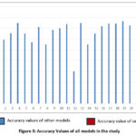 |
Figure 3. Accuracy Values of all models in the study |
The results presented in table 2 and figure 3, clearly indicate that the combination of EfficientNetB3 with SVM offers superior classification performance for brain tumor detection and surpasses all existing models reported in the literature.
The success of this hybrid approach can be attributed to the following factors:
Deep Feature Extraction
EfficientNetB3, being a deeper and more optimized convolutional neural network, is capable of learning rich hierarchical features that capture the complex patterns present in brain tumor MRI images.
SVM Classifier Strength
The use of SVM as the final classifier leverages its ability to find optimal separating hyperplanes in the high-dimensional feature space, particularly effective when dealing with small to medium-sized medical datasets where overfitting is a concern.
Transfer Learning Efficiency
By employing EfficientNetB3 model, the need for large-scale training data was mitigated, making the approach suitable even for limited datasets common in medical imaging.
It was also observed that the use of deeper EfficientNetB3 variants resulted in improved performance compared to smaller variants such as EfficientNetB0, B1 or B2. However, increasing model complexity must be balanced with computational cost and training time, which remains an area for future optimization.
Overall, this study demonstrates that a hybrid transfer learning-based framework using EfficientNetB3 and SVM is highly effective for accurate and robust brain tumor detection, offering potential for practical deployment in clinical settings.
Conclusion
In this study, we proposed an effective transfer learning-based approach to detect brain tumors using MRI images. By leveraging the deep feature extraction capabilities of EfficientNetB3 combined with the robust classification performance of a support vector machine (SVM), our model achieved a high classification accuracy of 99.33% on the validation dataset. The results demonstrate that using a deeper and more efficient CNN architecture such as EfficientNetB3 significantly enhances the ability to distinguish between tumor and non-tumor, compared to shallower models or traditional handcrafted feature approaches. Furthermore, the hybrid framework of deep learning with a classical classifier such as SVM proved to be a powerful combination, especially when dealing with limited medical imaging datasets. Future work will explore the incorporation of additional MRI modalities, automated tumor grading, and the development of more interpretable AI models to further support clinical decision-making.
Acknowledgement
The authors are thankful to their parent organizations for providing the facility to conduct this research.
Funding Sources
The author(s) received no financial support for the research, authorship, and/or publication of this article.
Conflict of Interest
The author(s) do not have any conflict of interest.
Data Availability Statement-
This statement does not apply to this article.
Ethics Statement
This research did not involve human participants, animal subjects, or any material that requires ethical approval.
Informed Consent Statement
This study did not involve human participants, and therefore, informed consent was not required.
Clinical Trial Registration
This research does not involve any clinical trials.
Permission to reproduce material from other sources
Not Applicable
Author Contribution
- Abhimanu Singh: Concept, drafting, design, data acquisition, editing, experimenting. Analysis.
- Smita Jain: concept, design, manuscript reviewing, analysis.
References
- Agarwal M, Rani G, Kumar A, Kumar P K, Manikandan R, Gandomi A H. Deep learning for enhanced brain Tumor Detection and classification. Results in Engineering. 2024; 22. https://doi.org/10.1016/j.rineng. 2024.102117
CrossRef - Appiah R, Pulletikurthi V, Esquivel-Puentes H A, et al. Brain tumor detection using proper orthogonal decomposition integrated with deep learning networks. Computer Methods and Programs in Biomedicine. 2024; 250. https://doi.org/10.1016/j.cmpb.2024.108167
CrossRef - Dutta T K, Nayak D R, Zhang Y. ARM-Net: Attention-guided residual multiscale CNN for multiclass brain tumor classification using MR images. Biomedical Signal Processing and Control. 2024; 87. https://doi.org/10.1016/j.bspc.2023.105421
CrossRef - Khoramipour S, Gandomkar M, Shakiba M. Enhancement of brain tumor classification from MRI images using multi-path convolutional neural network with SVM classifier. Biomedical Signal Processing and Control. 2024; 93. https://doi.org/10.1016/j.bspc.2024.106117
CrossRef - Priya A, Vasudevan V. Brain tumor classification and detection via hybrid alexnet-gru based on deep learning. Biomedical Signal Processing and Control. 2024; 89.https://doi.org/10.1016/j.bspc.2023.105716
CrossRef - Raghuwanshi S, Sukhad A, Rasool A, Meena V K, Jadhav A, Shivakarthik K. Early Detection of Brain Tumor from MRI Images Using Different Machine Learning Techniques. Procedia Computer Science. 2024; 235: 3094–3104. https://doi.org/10.1016/j.procs.2024.04.293
CrossRef - Sandhiya B, Raja S K S. Deep Learning and Optimized Learning Machine for Brain Tumor Classification. Biomedical Signal Processing and Control. 2024; 89. https://doi.org/10.1016/j.bspc.2023.105778
CrossRef - Simo A M D, Kouanou A T, Monthe V, Nana M K, Lonla B M. Introducing a deep learning method for brain tumor classification using MRI data towards better performance. Informatics in Medicine Unlocked. 2024; 44. https://doi.org/10.1016/j.imu.2023.101423
CrossRef - Singh T, Nair R R, Babu T, Wagh A, Bhosalena A, Duraisamy P. BrainNet: A Deep Learning Approach for Brain Tumor Classification. Procedia Computer Science. 2024; 235: 3283-3292. https://doi.org/10.1016/j.procs. 2024.04.310
CrossRef - Sultanpure K A, Bagade J, Bangare S L, Bangare M L, Bamane K D, Patnakar A J. Internet of things and deep learning based digital twins for diagnosis of brain tumor by analyzing MRI images. Measurement: Sensors. 2024; 33. https://doi.org/10.1016/j.measen.2024.101220
CrossRef - Tejashwini P S, Thriveni J, and Venugopal K R. EBT Deep Net: Ensemble brain tumor Deep Net for multi-classification of brain tumor in MR images. Biomedical Signal Processing and Control. 2024; 95. https://doi.org/10.1016/j.bspc.2024.106312
CrossRef - Agrawal P, Katal N. Segmentation and classification of brain tumor using 3D-UNet deep neural networks. International Journal of Cognitive Computing in Engineering. 2022; 3: 199-210. https://doi.org/10.1016/ j.ijcce.2022.11.001
CrossRef - Ali M, Shah J H, Khan M A, et al. Brain Tumor Detection and Classification Using PSO and Convolutional Neural Network. Computers, Materials & Continua. 2022; 73(3):4501-4518. DOI:10.32604/cmc.2022.030392
CrossRef - Irfan M, Shaf A, Ali T. Effectiveness of Deep Learning Models for Brain Tumor Classification and Segmentation. Computers, Materials & Continua. 2023; 76(1): 711-729.https://doi.org/10.32604/ cmc.2023.038176
CrossRef
- Isaza A A, Jimenez L M, Alejo L V, Sarasti L. Optimizing MRI-based brain tumor classification and detection using AI: A comparative analysis of neural networks, transfer learning, data augmentation, and the cross-transformer network. European Journal of Radiology. 2023; 10, 100484. https://doi.org/10.1016/ j.ejro.2023.100484
CrossRef - Islam M M, Barua P, Rahman M, Ahammed T, Akter L, Uddin J. Transfer learning architectures with fine-tuning for brain tumor classification using magnetic resonance imaging. Healthcare Analytics. 2023; 4, 100270. https://doi.org/10.1016/j.health.2023.100270
CrossRef - Zulfiqar F, Bajwa U I, Mehmood Y. Multi-class classification of brain tumor types from MR images using EfficientNets. Biomedical Signal Processing and Control. 2023; 84, 104777. https://doi.org/10.1016/j.bspc. 2023.104777 12
CrossRef - Lamba K, Rani S, Anand M, Maguluri L P. An integrated deep learning and supervised learning approach for early detection of brain tumor using magnetic resonance imaging. Healthcare Analytics. 2024; 5. https://doi.org/10.1016/j.health.2024.100336
CrossRef - Malakouti S M, Menhaj M B, Suratgar A A. Machine learning and transfer learning techniques for accurate brain tumor classification. Clinical eHealth. 2024; 7:106-119. https://doi.org/10.1016/j.ceh.2024.08.001
CrossRef - Bhardwaj N, Sood M, Gill S S. Design and Development of Hypertuned Deep learning Frameworks for Detection and Severity Grading of Brain Tumor using Medical Brain MR images. Current Medical Imaging. 2024; 24.https://doi.org/10.2174/0115734056288248240309044616
CrossRef - Singh A, Jain S. Enhanced brain tumor detection from brain MRI images using convolutional neural networks. AIMS Bioengineering. 2025; 12(2): 215-224.DOI: 10.3934/bioeng.2025010
CrossRef - Panigrahi A. Brain_Tumor_Detection_MRI. Kaggle. (2021). https://www.kaggle.com/abhranta
Abbreviations
Full Term Abbreviations
Magnetic Resonance Imaging MRI
Artificial Intelligence AI
Deep Learning DL
Machine Learning ML
Convolutional Neural Networks CNN
Support Vector Machine SVM
Receiver Operating Characteristic ROC
Area Under the Curve AUC

