Ashima Koundal , Sumit Budhiraja*
 and Sunil Agrawal
ECE, University Institute of Engineering and Technology, Panjab University, Chandigarh, India.
Corresponding Author E-mail: sumit@pu.ac.in
DOI : https://dx.doi.org/10.13005/bpj/3056
Abstract
Image segmentation is a way to simplify and analyze images by separating them into different segments. Fuzzy c-means (FCM) is the most widely used clustering algorithm, as it can handle data with blurry boundaries; where points belong to multiple clusters with varying strengths. The segmentation performance of this method is sensitive to the initial cluster centers. The fact that every feature in the image contributes equally and is given equal weight is another issue with this algorithm. In this paper, an image segmentation technique based on Fuzzy C-means (FCM) method is proposed. The proposed technique uses an extended feature set consisting of homogeneity, CIELAB, texture and edge is used for feature extraction in order to enhance segmentation quality. Further, weight optimization is done to help clustering process leverage the strengths of each feature, while downplaying less significant ones. The subjective and objective performance analysis of the proposed algorithm on medical images show improved performance as compared to existing standard image segmentation techniques.
Keywords
Clustering; Cluster weighting; Feature weighting; Medical image segmentation
Download this article as:| Copy the following to cite this article: Koundal A, Budhiraja S, Agrawal S. Medical Image Segmentation using Enhanced Feature Weight Learning Based FCM Clustering. Biomed Pharmacol J 2024;17(4). |
| Copy the following to cite this URL: Koundal A, Budhiraja S, Agrawal S. Medical Image Segmentation using Enhanced Feature Weight Learning Based FCM Clustering. Biomed Pharmacol J 2024;17(4). Available from: https://bit.ly/4hPQbkx |
Introduction
Image segmentation is the process of partitioning an image into different parts in the sets of pixels or superpixels. The segmentation is a crucial step for image analysis and its understanding. It aims to represent the image information in a form that is more suitable for different applications. Image segmentation finds a variety of applications such as face recognition1, object detection2, fingerprint recognition3, biomedical image processing4 and industrial applications5-8. Image segmentation may be broadly classified as threshold-based methods9, 10 region-extension methods11 and clustering based methods12. These methods have their different advantages and limitations. In recent years, cluster-based methods have become very popular because of its fast implementation and superior performance13.
The Fuzzy c-means (FCM) is a popular data clustering algorithm for colour images due to its easy and fast implementation14. In this method, pixels can be assigned to different clusters; providing improved information and better segmentation performance15 as compared to hard clustering techniques such as k-means16. In addition, the fuzzy membership set in this method helps discover complex evaluation between samples and clusters with more accuracy. The limitations of FCM algorithm are its sensitivity to the selection of initial cluster centres17, and the property that different features of the image contribute equally and hold same importance18, 19. As the number of features increase in source images; some features hold more importance than others. So assigning same weights to all features limits the performance of image segmentation20, 21.
The RGB components of an input image are shown in Fig 1. As shown in the figure, R channel should be better than the other channels within the feature of segmentation. This can also be true for other features and sub-features; so assigning same weights reduces the efficiency of image segmentation.
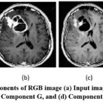 |
Figure 1: Color components of RGB image (a) Input image, (b) Component R, (c) Component G, and (d) Component B.
|
The paper is organized in the following sections. The current state of research in the area of image segmentation is presented in section 2. The proposed algorithm is presented in section 3. Section 4 describes the results and analysis. Conclusion is presented in section 5.
Related Work
In recent years, a variety of image segmentation algorithms based on clustering techniques have been proposed. An algorithm based on combination of modified k-means and Imperialistic Competitive Algorithm (ICA) used cluster centers to improve segmentation performance22. The CSFCM method improved the segmentation performance by combining three meta-heuristics of biogeography, genetic and firefly23. A fuzzy model based on unsupervised learning combined color and Gaussian density into fuzzy clustering algorithm and improved clustering by incorporating the neighbouring information into the learning step24. A technique named A-PSO-IT2IFCM used particle swarm optimization (PSO) to find suitable cluster center and the fuzzifiers for image segmentation25. Other similar proposed methods tried to obtain required set of clusters or primary centers26-28. G. Silva analysed improvements in dental X-ray using automated teeth segmentation29. The article provided insights into current trends and potential future directions in this field by introducing a new dataset and benchmarking different segmentation techniques improve dental diagnostics efficiency and accuracy.
In the past years, Multi-Objective Evolutionary Algorithms (MOEAs) have been frequently used for color image segmentation25, 30. The purpose of these algorithms is to find optimal cluster centers. But these methods also suffer from higher computational requirements. To address this limitation, Kriging-assisted reference vector evolutionary algorithm (KREVA) based methods were proposed30, 31. A new deep learning method combined attention mechanisms and JGate modules into the Residual U-Net architecture for brain tumor segmentation32. This technique successfully extracted contextual information and minute details from brain MRI images to improve segmentation accuracy.
A method combined level sets with Backchannel Filling Convolutional Neural Networks (CNNs) for segmentation of skin lesions in images33. Lesion boundaries were enhanced by delineation of by filling in missing or unclear regions in skin lesion images; leading to improvement in segmentation accuracy. A method combined YOLOv8 with SAM and HQ-SAM models to achieve thorough multimodal segmentation in medical imaging34. While SAM and HQ-SAM improve segmentation by utilizing multi-scale features and hierarchical contexts; YOLOv8 offers accurate object detection. The combined method improved accuracy and robustness in the segmentation of complex medical images.
An algorithm named entropy regularized weighted FCM (EWFCM) used an improved objective function using local-feature weighted entropy regularization method, to select the optimal weights for image features35. A clustering method proposed an objective function to use local feature weighting scheme for finding clusters in images36. A new FCM based cluster weighting and feature weighting method overcame limitations in existing methods37. In CGGFCM, a feature weighting technique was used to improve clustering accuracy and a strategy for automatic cluster weighting to lessen sensitivity to cluster initialization38.
Early detection and diagnosis of cancer leads to successful treatment and lower mortality rates. In recent years, deep learning techniques have been used to achieve improved performance in image segmentation algorithms. An algorithm based on improved fuzzy local information C means (IFF-FLICM) segmentation was successfully used for detection and classification of Dataset-255 brain tumor images39. A study used a variety of machine learning classification method for predicting cervical cancer using different risk factors. Based on this study and analysis of a dataset of around 850 patients, a deep-learning method was proposed for early diagnosis and detection of cervical cancer40. The flowchart of the proposed method incorporates the collection of patient’s information, pre-processing step, training of model, threshold prediction and setting, validation, and final diagnosis.
A new machine learning model and FCM based segmentation algorithm was used for classification and detection of breast cancer from mammogram images41. Firstly, fuzzy factor improved fast and robust fuzzy c means (FFI-FRFCM) segmentation segmented the input image by modifying the member partition matrix of the FRFCM technique. Then an improved particle swarm optimization (PSO) was used for weight optimization of the ensemble extreme learning machine (EELM) model. The proposed method showed improved performance for classification of breast cancer images. Another algorithm for classification of breast cancer images introduced a novel hybrid DenseNet121-based ELM Model42. The features collected after the pooling and flatten layers at the first stage of the classification were used as input to the proposed DenseNet121-ELM model. The AdaGrad optimization algorithm was used for updating the weights of ELM. The performance evaluation of proposed method for batch size up to 128 showed improved performance and robustness of algorithm.
Many techniques have been proposed for mitigating the limitations of sensitivity to equalization and equal importance of features issue in FCM method for image segmentation. In this paper, an extended set of image feature has been used to improve the representation of image information. An improved weight optimization method has been used to overcome the limitations of FCM; in the quest to improve color image segmentation for medical images.
Proposed Work
The proposed image segmentation technique is presented in Fig. 2. Initially, important features are extracted from the input image; which are used in the clustering process. ICA is used for finding the optimal parameters, which then are used to improve CGFFCM by finding weights of features for feature groups. The proposed algorithm has been described in the following sub-sections.
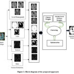 |
Figure 2: Block diagram of the proposed approach
|
Feature Extraction
The proposed method uses an effective combination of image features, including texture, edge, local homogeneity, and CIELAB20, 43. The features represent the important properties of the images and are described as follows:
Local homogeneity: In the context of image processing, it refers to the degree of uniformity in pixel values within a small neighbourhood surrounding a specific pixel. It quantifies the extent of constant value of image intensity or color in a localized area. By identifying regions with distinct homogeneity values (high for uniform areas, low for edges/textures); the segmentation algorithm groups distinct pixels into meaningful objects or regions.
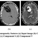 |
Figure 3: Local homogeneity features (a) Input image (b) Component H (c) Component S (d) Component V
|
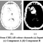 |
Figure 4: Color features from CIELAB colour channels (a) Input image (b) Component L (c) Component A (d) Component B
|
Color: It is represented in CIELAB pictures (Lab*) by L*, a*, and b* values. The brightness scale Lightness (L)*, goes from 0 (complete black) to 100 (white). Value of Green-Red (a)* changes from negative (green) to positive (red). The Yellow-Blue (b)* scale goes from positive (yellow) to negative (blue).
Grayscale or achromatic colors have values of a* = 0 and b* = 0. Distinct color zones within an image can be identified by analyzing these values for every pixel in the image.
Texture: This feature go beyond simple color gradation to depict recurring patterns in an image by describing how intensity or color varies throughout the image. This is essential for segmenting images, particularly when dividing things according to their surface textures rather than merely their colors. Texture enhances color in segmentation process. Color conveys basic information, while texture describes how color variations are arranged in space. When these features are combined, segmentation accuracy is better than, when color is used alone.
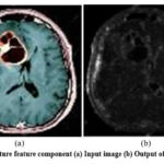 |
Figure 5: Texture feature component (a) Input image (b) Output of Gabor filter
|
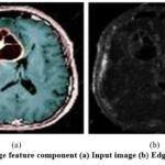 |
Figure 6: Edge feature component (a) Input image (b) Edge component
|
Edges: The boundaries between objects or regions in an image are crucial for image segmentation. These often arise from significant changes in pixel intensity, color, or texture. Identifying these edges is the key to separate objects and achieve accurate segmentation. Extracting edge features can be done through various techniques like gradient-based methods. Sobel and Prewitt filters calculate the rate of intensity change between pixels, highlighting areas with sharp transitions. The Laplacian operator emphasizes areas with substantial intensity variations, with edges corresponding to zero-crossing points. Finally, Canny edge detection combines gradient information to locate edges precisely while minimizing false detections. While edge features offer advantages like simplicity, efficiency, and clear interpretability; these can be sensitive to noise and might not capture blurry boundaries or provide enough detail for complex textures.
Clustering Method
The clustering-based algorithms may become sensitive to image artifacts if the spatial information is ignored. This will lead to shifting of the intensities values of pixels, leading to the situation where pixels from different clusters may possess similar features. Therefore cluster-based algorithms, look for means to suppress the adverse effect of noise.
Among the two strategies; one is group-local feature strategy, where weights are allocated to the group of features in a cluster. Other is local feature weighting strategy, in this weight is allocated to each feature in the group of feature. Both these methods are used to improve clustering accuracy. An algorithm based on automatic weighting of clusters reduced the sensitivity of the segmentation method and obtained improved result by using cluster initialization and group feature weighting method38.
In image segmentation, the different group of features can have different importance; so the advantage of group feature weighting is more obvious than local weighting which assigns one weight to all the features37. Within a group also, different features might be less or more important than the other. So assigning same weights to all the components will reduce the quality of segmented image. So, we use features and sub-features with more varying degree of importance in image clusters. The improved weighting of clusters is directly related to better segmentation performance. Further; to control the sensitivity to initial clusters, an effective cluster weighting technique is incorporated. The relative importance of features and sub-features is considered while selecting the weight of image clusters38.
Optimization
The clustering method is used in combination with the Imperialist Competitive Algorithm (ICA) for optimization of feature weighing step44. It finds optimized values of the weight coefficients in the segmentation method. Imperialist competitive algorithm (ICA) is a classic evolutionary optimization technique which incorporates the general political and social behaviour of imperialist countries in an attempt to dominate weaker countries44. In past few years, various algorithms based on ICA and its improved versions have been proposed; which have successful solved several practical optimization methods. Another advantage of ICA is its higher convergence rate as compared to other evolutionary optimization algorithms; delivering significant results in less time45. The Optimization procedure involving integration of ICA with FCM clustering can be described in following steps:
Initial Population and Weight Coefficient Ranges
Population Initialization: Generate a random initial population of solutions, where each country represents a weight vector [v1,v2,v3] for three feature groups.
Weight Constraints: Set weight range to [0.1, 0.8] to ensure participation of all groups, preventing any feature from being entirely excluded in initial clustering.
Objective Function Evaluation
Run FCM clustering for each country and calculate the objective function value based on clustering performance; reflects how well the weights align with optimal feature grouping.
Imperialist Selection
Selecting Imperialists: Rank countries based on objective function values and select the best-performing solutions as imperialists; the remaining become colonies.
Empire Configuration: Experiment with different numbers of initial imperialists Nimp = [10,20] to find the best structure for the clustering task.
Colony Allocation Based on Imperialist Power
Assigning Colonies: Allocate colonies to imperialists based on their power, calculated using cost differences and normalized to determine each imperialist’s control proportionally as per eq 1 and eq 2. The values are rounded for the final number of colonies per empire.

Assimilation – Moving Colonies Toward Imperialists
Cultural Assimilation: Shift colonies incrementally towards their imperialists, with movement influenced by a random variable 𝑎 and an angle 𝜃 for directional diversity.
Exploration Parameters: Set β = 2 for balanced convergence and γ = π/4 to encourage exploration in multiple directions.
Update Colony Costs and Improvement Check
Recompute colony costs after movement. If a colony’s cost surpasses its imperialist’s, they swap positions, enhancing the empire with potential improvements.
Calculate Total Empire Cost
Empire Cost Calculation: Compute each empire’s total cost TCn, combining the imperialist’s cost and an averaged colony cost.
Weight Coefficient: Set attenuation coefficient ξ = 0.1 to balance emphasis on imperialist vs. colony contributions, reinforcing high-quality solutions.
Inter-Empire Competition for Colonies
Competitive Redistribution: Weaker empires lose colonies to stronger ones based on normalized total costs and possession probabilities.
Roulette Selection: A roulette wheel mechanism selects winning empires, promoting dynamic growth of powerful empires and reallocation of resources.
Empire Elimination
Remove empires with no colonies left, and merge remaining imperialists into stronger empires, preserving potentially viable solutions.
Termination and Resulting Clusters
The algorithm stops when a single empire remains, marking the most dominant clustering solution.
Final Clustering Output: The remaining empire’s colonies represent the final clusters, with each colony position corresponding to feature weights optimized for FCM clustering. Evaluate clusters with benchmark datasets for segmentation quality.
Results and Discussion
To demonstrate the performance of proposed segmentation method, a set of medical images have been taken from a variety of environments. The images are taken from Berkley image dataset, The BRATS 2016 skin lesion challenge datasets, including color images having balanced and imbalanced regions. To evaluate the generalizability of the proposed method, test images Cystoid fluid image and Kvasir image have been taken respectively from OPTIMA 13 dataset and Kvasir SEG dataset. Cystoid fluid refers to fluid that accumulates within cyst-like spaces, often in the retina or other tissues. Kvasir is a medical image dataset, particularly useful for researchers working on computer-aided diagnosis systems in gastroenterology. The segmentation methods are implemented using MATLAB R2018b, and results of the proposed segmentation method are compared with existing standard methods JGate-AttResUNet32, New fuzzy c-means18, Automatic segmenting37, and CGFFCM38. The objectively evaluation of these methods is performed using performance metrics Normalized Mutual Information (NMI), Accuracy, and F-score. The performance metrics are described as follows:
Normalized Mutual Information (NMI)
Instead of focusing on individual pixels, NMI considers both the number of segments (quantity) and their arrangement (spatial distribution) in the image to observe segmentation performance.
Accuracy
It is defined as the number of clustered pixels divided by the total number of pixels. It measures the degree of similarity between the segmentation result and the given ground truth.
F-score
This metric calculates the harmonic mean of recall and precision values. This metric is effective in evaluation of imbalanced color regions in the image.
Subjective Performance Evaluation
For subjective evaluation, the results of proposed segmentation method and existing methods are shown in Fig. 7 and Fig 8. The results for images named MRI-1, MRI-2, Teeth, skin lesion, buffalo, and bird are different rows in Fig. 7. The segmentation results of Cystoid fluid and Kvasir images are shown in Fig 8. It is observed that results obtained by proposed method are sharper with clear cut boundaries. It can be attributed to enhanced feature set which has a direct impact on the segmentation results. It is observed that proposed algorithm performs better than the existing standard methods.
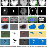 |
Figure 7: Segmentation results obtained using the CIELAB space for (a) Input image,(b) JGate-AttResUNet32 (c) Automatic segmenting37 (d) CGFFCM38 (e) Proposed method.
|
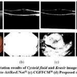 |
Figure 8: Segmentation results of Cystoid fluid and Kvasir images (a) Input image, (b) JGate-AttResUNet32 (c) CGFFCM38 (d) Proposed method.
|
Objective Performance Evaluation
For objective performance evaluation, the results of proposed method are compared with existing clustering-based segmentation techniques JGate-AttResUNet32, New fuzzy C-means18, and CGFFCM38 using metrics Accuracy, Normalized Mutual Information (NMI), and F1-Score. The results for different images are shown in Table 1.
Table 1: Objective performance evaluation of proposed method
|
Images |
Metric |
CNN& level sets33 |
JGate-AttResUNet32 |
New fuzzy C-means18 |
CGFFCM38 |
Proposed method |
|
MRI-1 |
Accuracy NMI F1-Score |
83.08 45.09 – |
83.72 45.29 71.14 |
95.91 77.57 96.90 |
99.59 88.62 99.78 |
98.478 95.524 93.9212 |
|
MRI-2 |
Accuracy NMI F1-Score |
93.51 76.74 |
93.85 76.02 70.16 |
94.65 78.86 90.97 |
96.52 83.21 95.89 |
96.70 95.98 89.42 |
|
Teeth |
Accuracy NMI F1-Score |
98.83 74.81 – |
– – – |
99.12 81.26 92.92 |
99.46 87.19 95.60 |
99.25 99.56 92.67 |
|
Skin lesion |
Accuracy NMI F1-Score |
– – – |
– – – |
73.85 77.48 – |
96.43 93.77 86.26 |
96.8 94.36 87.60 |
|
Buffalo |
Accuracy NMI F1-Score |
– – – |
92.92 77.02 83.80 |
98.71 74.23 88.63 |
99.68 91.66 97.37 |
99.70 99.858 97.07 |
|
Bird |
Accuracy NMI F1-Score |
99.18 81.22 – |
99.27 88.24 77.96 |
96.12 31.32 97.98 |
99.59 88.62 99.78 |
99.61 96.40 95.68 |
|
Cystoid fluid |
Accuracy NMI F1-Score |
– – – |
91.42 87.35 76.80 |
– – – |
97.34 93.21 96.11 |
98.14 96.15 95.66 |
|
Kvasir |
Accuracy NMI F1-Score |
– – – |
92.22 87.92 78.46 |
– – – |
97.81 89.71 98.08 |
97.13 95.32 96.22 |
From Table 1, it can be observed that proposed method provides best value of most of the metrics. The superior performance of proposed method can be attributed to enhanced feature set and use of optimal weights of groups and sub features in the proposed group feature weighting method. So it be concluded that for medical images, the proposed method delivers improved segmentation performance as compared to existing techniques.
The propose algorithm provides improved performance due to improved feature extraction and optimizations steps. Computational time of proposed segmentation is compared with other segmentation methods in Table 2. It is observed that computational time of proposed algorithm is around 15% higher than CGFFCM. As the processing capabilities are improving, the higher computational cost can be accepted for improved performance for most of the segmentation applications, although this can limit the real-time applications of proposed algorithm.
Table 2: Average Computational time (seconds) of segmentation methods
|
CNN& level sets33 |
JGate-AttResUNet32 |
New fuzzy C-means18 |
CGFFCM38 |
Proposed method |
|
25.23 |
29.19 |
24.67 |
24.93 |
28.71 |
Further, like other FCM-based methods, the proposed method’s dependence on local feature information and pixel intensities can make it difficult to maintain performance for noisy images; as noise can distort the clustering process and lead to inaccurate segmentation. These effects can be controlled to some extent by pre-processing the source images before segmentation process.
Conclusion
In this paper, a medical segmentation technique based on group feature weighting and cluster weighing scheme is proposed. Feature extraction is done using Homogeneity, CIELAB, texture and edge. The clustering step is used with Imperialist Competitive Algorithm for feature weight optimization. The segmentation performance of the proposed method is compared with existing algorithm in subjective and objective evaluations on medical images using a variety of parameters. Through performance evaluation, it is established that proposed algorithm provides superior segmentation performance as compared with existing techniques.
Acknowledgement
The authors would like to thank Department of ECE, University Institute of Engineering & Technology, Panjab University, Chandigarh for providing guidance and laboratory facilities for carrying out the research work.
Funding Sources
The author(s) received no financial support for the research, authorship, and/or publication of this article
Conflict of Interest
Authors have no conflict of interest to declare.
Conflict of Interest
The authors do not have any conflict of interest
Data Availability Statement
This statement does not apply to this article
Ethics Statement
This research did not involve human participants, animal subjects, or any material that requires ethical approval
Informed Consent Statement
This study did not involve human participants, and therefore, informed consent was not required
Clinical Trial Registration
This research does not involve any clinical trials
Authors Contribution
Ashima Koundal contributed in conceptualising the idea, implementing the algorithm, and writing the manuscript.
Dr. Sumit Budhiraja contributed in designing the analysis, implementing the algorithm, analysis of results, reviewing and editing the manuscript.
Dr. Sunil Agrawal contributed in analysis and validation of results. Each author mentioned has significantly and directly contributed intellectually to the project and has given their approval for its publication.
References
- Farajzadeh N, Hashemzadeh M. Exemplar-based facial expression recognition. Information Sciences. 2018;460-461:318-330.
CrossRef - Farajzadeh N, Karamiani A, Hashemzadeh M. A fast and accurate moving object tracker in active camera model. Multimed Tools Appl., 2018;77(6): 6775-6797.
CrossRef - Sharma R P, Dey S. Two-stage quality adaptive fingerprint image enhancement using Fuzzy C-means clustering based fingerprint quality analysis. Image Vis Comput. 2019;83–84:1–16.
CrossRef - Hashemzadeh M, Azar B A. Retinal blood vessel extraction employing effective image features and combination of supervised and unsupervised machine learning methods. Artif Intell Med. 2019;95:1-15.
CrossRef - Hashemzadeh M, Farajzadeh N. A Machine Vision System for Detecting Fertile Eggs in the Incubation Industry. International Journal of Computational Intelligence Systems. 2016;9: 850-862.
CrossRef - Hashemzadeh M. Hiding information in videos using motion clues of feature points. Computers & Electrical Engineering. 2018;68:14-25.
CrossRef - Hashemzadeh M, Asheghi B, Farajzadeh N. Content-aware image resizing: An improved and shadow-preserving seam carving method. Signal Processing. 2019;155:233-246.
CrossRef - Hashemzadeh M, Zademehdi A. Fire detection for video surveillance applications using ICA K-medoids-based color model and efficient spatio-temporal visual features. Expert Syst Appl. 2019; 130:60-78.
CrossRef - Lifang H, Songwei H. Modified firefly algorithm based multilevel thresholding for color image segmentation. Neurocomputing. 2017; 240: 152-174.
CrossRef - Pare S, Kumar A, Bajaj V, Singh G K. A multilevel color image segmentation technique based on cuckoo search algorithm and energy curve. Appl Soft Comput. 2016;47:76-102.
CrossRef - Feng L, Li H, Gao Y, Zhang Y. A Color Image Segmentation Method Based on Region Salient Color and Fuzzy C-Means Algorithm. Circuits Syst Signal Process. 2020;39:586-610.
CrossRef - Tan K S, Isa N A M, Lim W H. Color image segmentation using adaptive unsupervised clustering approach. Appl Soft Comput. 2013;13(4):2017-2036.
CrossRef - Farshi T R, Drake J H, Özcan E. A multimodal particle swarm optimization-based approach for image segmentation. Expert Syst Appl. 2020;149:113233.
CrossRef - Son L H, Tuan T M. Dental segmentation from X-ray images using semi-supervised fuzzy clustering with spatial constraints. Eng Appl Artif Intell. 2017;59:186-195.
CrossRef - Zhang X, Jian M, Sun Y, Wang H, Zhang C. Improving image segmentation based on patch-weighted distance and fuzzy clustering. Multimedia Tools and Applications. 2020;79:633-657.
CrossRef - MacQueen J. Some methods for classification and analysis of multivariate observations. Proceedings of the 5th Berkeley Symposium on Mathematical Statistics and Probability. 1967;1: 281−297.
- Choy SK, Yuen K, Yu C. Fuzzy bit-plane-dependence image segmentation R. Signal Processing. 2019;154:30–44.
CrossRef - Hashemzadeh M, Oskouei A G, Farajzadeh N. New fuzzy C-means clustering method based on feature-weight and cluster-weight learning. Appl Soft Comput. 2019;78:324-45.
CrossRef - Pimentel BA, de Souza RMCR. Multivariate Fuzzy C-Means algorithms with weighting. Neurocomputing. 2016;174B:946-65.
CrossRef - Zhou Z, Zhao X, Zhu S. K-harmonic means clustering algorithm using feature weighting for color image segmentation. Multimedia Tools and Applications 2018;77:15139-15160.
CrossRef - Xing HJ, Ha MH. Further improvements in Feature-Weighted Fuzzy C-Means. Information Sciences. 2014; 267:1-15.
CrossRef - Babrdelbonab M, Mohd Hashim SZ, Bazin NEN. Data analysis by combining the modified k-means and imperialist competitive algorithm. J Teknol. 2014;70(5):51-57.
CrossRef - Abdellahoum H, Mokhtari N, Brahimi A, Boukra A. CSFCM: An improved fuzzy C-Means image segmentation algorithm using a cooperative approach. Expert Syst Appl. 2021;166:114063.
CrossRef - Choy SK, Ng C, Yu C. Unsupervised fuzzy model-based image segmentation. Signal Processing. 2020;171:107483.
CrossRef - Zhao F, Chen Y, Liu H, Fan J. Alternate PSO-Based Adaptive Interval Type-2 Intuitionistic Fuzzy C-Means Clustering Algorithm for Color Image Segmentation. IEEE Access. 2019;7:64028-64039.
CrossRef - Mikaeil R, Haghshenas SS, Haghshenas SS, Ataei M. Performance prediction of circular saw machine using imperialist competitive algorithm and fuzzy clustering technique. Neural Comput Appl. 2018;29:283-292.
CrossRef - Aliniya Z, Mirroshandel SA. A novel combinatorial merge-split approach for automatic clustering using imperialist competitive algorithm. Expert Syst Appl. 2019;117:243-266.
CrossRef - Niknam T, Fard ET, Ehrampoosh S, Rousta A. A new hybrid imperialist competitive algorithm on data clustering. Sadhana – Academy Proceedings in Engineering Sciences. 2011; 36:293–315.
CrossRef - Silva G, Oliveira L, Pithon M. Automatic segmenting teeth in X-ray images: Trends, a novel data set, benchmarking and future perspectives. Expert Systems with Applications. 2018;107:15–31.
CrossRef - Zhao F, Zeng Z, Liu H, Lan R, Fan J. Semisupervised Approach to Surrogate-Assisted Multiobjective Kernel Intuitionistic Fuzzy Clustering Algorithm for Color Image Segmentation. IEEE Transactions on Fuzzy Systems. 2020;28(6):1023-1034.
CrossRef - Chugh T, Jin Y, Miettinen K, Hakanen J, Sindhya K. A Surrogate-Assisted Reference Vector Guided Evolutionary Algorithm for Computationally Expensive Many-Objective Optimization. IEEE Transactions on Evolutionary Computation. 2018;22(1):129-142.
CrossRef - Ruba T, Tamilselvi R, Beham MP. Brain tumor segmentation using JGate-AttResUNet – A novel deep learning approach. Biomed Signal Process Control. 2023;84: 104926.
CrossRef - Huang L, Zhao YG, Yang TJ. Skin lesion image segmentation by using backchannel filling CNN and level sets. Biomed Signal Process and Control. 2024;87A:105417.
CrossRef - Pandey S, Chen KF, Dam EB. Comprehensive Multimodal Segmentation in Medical Imaging: Combining YOLOv8 with SAM and HQ-SAM Models. IEEE/CVF International Conference on Computer Vision Workshops (ICCVW 2023). 2023;2584-2590.
CrossRef - Zhou J, Chen L, Chen CLP, Zhang Y, Li HX. Fuzzy clustering with the entropy of attribute weights. Neurocomputing. 2016;198:125-134.
CrossRef - Zhou Z, Zhu S. Kernel-based multiobjective clustering algorithm with automatic attribute weighting. Soft comput. 2018;22:3685-3709.
CrossRef - Hashemzadeh M, Oskouei AG, Farajzadeh N. New fuzzy C-means clustering method based on feature-weight and cluster-weight learning. Applied Soft Computing Journal. 2019;78:324-345.
CrossRef - Oskouei GA, Hashemzadeh M. CGFFCM: A color image segmentation method based on cluster-weight and feature-weight learning. Software Impacts. 2022;11:100228.
CrossRef - Dash S, Siddique M, Mishra S, Gelmecha DJ, Satapathy,S, Rathee DS, Singh RS. Brain Tumor Detection and Classification Using IFF-FLICM Segmentation and Optimized ELM Model. Journal of Engineering, 2024, Article ID 8419540, 2024:1-24.
CrossRef - Mohapatra S, Siddique M, Paikaray BK, Riyazbanu S, Automated Invasive Cervical Cancer Disease Detection at Early Stage Through Deep Learning, International Journal of Bioinformatics Research and Applications, 2023;19(4):306-326.
CrossRef - Pattnaik RK, Siddique M, Mishra S, Gelmecha DJ, Singh RS, Satapathy S. Breast cancer detection and classification using metaheuristic optimized ensemble extreme learning machine. International Journal of Information Technology, 2023;15(8):4551-4563.
CrossRef - Pattanaik RK, Mishra S, Siddique M, Gopikrishna T, Satapathy S. Breast Cancer Classification from Mammogram Images Using Extreme Learning Machine-Based DenseNet121 Model. Journal of Sensors. 2022, Article ID 2731364, 2022:1-12.
CrossRef - Gamino-Sánchez F, Hernández-Gutiérrez I V, Rosales-Silva AJ, Gallegos-Funes FJ, Mújica-Vargas D, Ramos-Díaz E, Carvajal-Gámez BE, Kinani JMV. Block-Matching Fuzzy C-Means clustering algorithm for segmentation of color images degraded with Gaussian noise. Engineering Applications of Artificial Intelligence. 2018;73:31–49.
CrossRef - Talatahari S, Farahmand Azar B, Sheikholeslami R, Gandomi AH. Imperialist competitive algorithm combined with chaos for global optimization. Commun in Nonlinear Sci Numer Simul. 2012;17(3):1312-1319.
CrossRef - Peri D. Hybridization of the imperialist competitive algorithm and local search with application to ship design optimization. Computers and Industrial Engineering. 2019;137:106069.
CrossRef


