
Manuscript accepted on :03-01-2024
Published online on: 09-02-2024
Plagiarism Check: Yes
Reviewed by: Dr. B. Kirthika and Dr Hisham Orban
Second Review by: Dr. Takkella Nagamma
Final Approval by: Dr. Patorn Promchai
Areej Jameel M. Alghabban
Biology Department, Faculty of science, University of Tabuk, KSA.
Corresponding Author E-mail: a_alghabban@ut.edu.sa
DOI : https://dx.doi.org/10.13005/bpj/2857
Abstract
The smallest known human nematode parasite is Trichinella spiralis (T. spiralis). A parasitic zoonosis that can be found anywhere in the world is trichinellosis. When a host consumes raw meat contaminated with Trichinella larvae, the larvae mature into adults in the host's stomach in 5 to 6 days. The females subsequently give birth to additional larvae, some of which eventually make it to muscle cells after travelling through blood channels. These cells allow infective larvae to survive for years because, after two to three weeks, the larvae become infectious and the host cell transforms into a nurse cell. The current study used parasitological assessment to count adult worms and encysted larvae at 5 weeks after infection (5WPI) to determine the effect of coriander seeds extract (CSE) against encysted larvae further to hematological and biochemical tests. Current results revealed that; Trichinella spiralis experimentally infection (5WPI) induced significant elevation in white blood cells (WBCs) counts, neutrophil, monocyte, eosinophil percentage, alanine transaminase (ALT), aspartate transaminase (AST), alkaline phosphatase (ALP), urea, creatinine and a significant depletion hemoglobin (Hb), red blood cell (RBC) count, hematocrit (HCT), mean corpuscular volume (MCV), mean corpuscular hemoglobin (MCH), platelet count (PLT), lymphocytes percentage, albumin, total proteins as compared to control and CSE groups. Meanwhile, treatments of 5WPI with CSE (5WPI+CSE) revealed a significant reduction in the number of Trichinella spiralis encysted larvae and improvements in all hematological parameters, liver and kidney functions as compared to 5WPI. We can conclude that; CSE could be an alternative method for treatment against trichinellosis.
Keywords
Coriander seeds; kidney functions; Liver; Mice; Trichinella spiralis
Download this article as:| Copy the following to cite this article: Alghabban A. J. M. Therapeutic Potential of Coriander (Coriandrum sativum) Seeds Extract Treatment on Hematological and Biochemical Parameters in Healthy and Trichinella spiralis Infected Mice. Biomed Pharmacol J 2024;17(1). |
| Copy the following to cite this URL: Alghabban A. J. M. Therapeutic Potential of Coriander (Coriandrum sativum) Seeds Extract Treatment on Hematological and Biochemical Parameters in Healthy and Trichinella spiralis Infected Mice. Biomed Pharmacol J 2024;17(1). Available from: https://bit.ly/4bxFnEj |
Introduction
Trichinellosis is a parasitic zoonosis that is spread through ingesting raw or undercooked meat that has been contaminated with Trichinella spiralis larvae.1-3 It has three clinical phases: intestinal, migratory, and muscular. Pork and its products are the main sources of infection.4-6 According to Saad et al.7 and Abou Rayia et al.8, T. spiralis has the unusual capacity to change the infected muscle cell into a new kind of cell known as a nurse cell in the host body. Trichinellasag spiralis has been frequently used as an experimental model to determine the effects of numerous anthelmintic agents because it can develop into adult, migratory, and encysted stages in the same host and infects a wide range of mammalian hosts.9 According to Gottstein et al.10, mebendazole is the standard therapy and the main anthelmintic medication for the treatment of trichinellosis in the majority of cautery and in KSA. However, according to Caner et al.11, they exhibit modest efficacy against encapsulated larvae, a high level of resistance, and restricted bioavailability. Medical plants create a wide range of chemical components that can treat a wide range of illnesses.12,13 The seeds of the fragrant, carminative, stomachic, and antispasmodic annual plant Coriandrum sativum L. (coriander, family Apiaceae) are used in medicine to treat abdominal symptoms such dyspepsia, and gastralgia. To avoid stomach aches, they are also a component in laxative remedies. Thanks to the identification of certain polyphenolic and antioxidant components present in the coriander plant, it has been shown to play a substantial role in the aetiology of a number of disorders.14 Coriander is used in folk medicine to treat rheumatism and joint discomfort as well as intestinal parasites.15 Because of this, the current investigation looked at how therapy with coriander affected hematological and biochemical variables in mice with Trichinella spiralis infection as well as healthy mice.
Materials and Methods
Making coriander seed extracts (CSE)
In accordance with Moustafa et al.16, coriander seeds (CS) were powdered, soaked in boiling water for 24 hours, extracted, and then stored at -30°C in the dark until use.
Animals and Ethical Considerations
A total of 40 male Swiss male albino mice were employed in this investigation, and they appeared to be in good health and free of parasites. 10 mice each were divided into 4 equal groups (Gps) of mice. The mice were bought in Giza, Egypt, from NRC. Prior to the trial, mice were kept at our faculty’s animal home for a week. They were kept in controlled lighting conditions with a conventional mouse feed and access to water at all times. Experimental design and animal groups
Gp1: Control group (Control), in which normal healthy non-infected mice.
Gp2: Coriandrum Gp (CSE) where mice treated orally (50 mg/kg body weight) daily for one week.
Gp3: Infected group (5WPI), in which mice were challenged with 300 larvae of trichinella spiralis for 5 weeks.
Gp4: Post treated group (5WPI+CSE), in which mice were challenged with 300 larvae of trichinella spiralis muscle larvae for 5 weeks then treated with CSE for 1 week post infection.
Sample collection
At the end of the experiment, overnight fasted rats will be anaesthetized with diethyl ether, dissection and blood will be collected in EDTA tubes for CBC determination and half of tubes were centrifuged at 3000 g for 20 min. plasma will be carefully separated, each of samples will label and kept at – 20 0C until parasitological, and biochemical analysis.
Isolation and infection of Trichinella spiralis muscle larvae
T. spiralis muscle larvae were obtained from laboratory bred infected rats in parasitology unit, Faculty of Medicine, Tanta University. Larval preparation and extraction of inoculums were made after Dunn and Wright.17 Five weeks after infection (5WPI),1% pepsin and 1% concentrated HCL are added to warm tap water to create an artificial gastric juice that is used to digest the muscles of infected mice. The mixture was incubated for 2 hours at 37°C while being continuously stirred by an electric stirrer. The digest was then filtered by sieve (50mesh/cm2), then by sieve (200mesh/cm2).18 After being collected, the larvae were rinsed in tap water two to three times before being suspended in a conical flask for 30 minutes to allow for sedimentation. Sediment larvae were counted microscopically while using a hemocytometer, and the supernatant fluid was discarded. Dead larvae were non-mobile and comma-shaped, whereas living larvae were coiled and motile. The Concentration of counted larvae in the fluid was adjusted to the appropriate dose for each rat, which is 0.25 ml of fluid containing 300 living larvae. Mice were starved for 12 hours before infection, then provided with 0.25ml of the infection orally by using a tuberculin syringe fitted with blunt, curved, 18-gauge needle to introduce infective larvae into mouse stomach.
Hematological studies
Complete blood picture (CBC) measurements were made in 2007 using a Nihon Kohden Corporation, Tokyo, Japan, automated hematology cell counter (serial number 11649, model Celltac, MEK-6410K). The following blood parameters were measured: mean corpuscular volume (MCV), mean corpuscular hemoglobin (MCH), mean corpuscular hemoglobin concentration (MCHC), white blood cell count (WBC) and its differential, red blood cell (RBC) count, hemoglobin (Hb) level, hematocrit (HCT), and platelet count (PLT).
Liver and Kidney functions estimation
Reitman and Frankel’s 19 approach was used to measure the activities of ALT and AST in serum, whereas Belfield and Goldberge’s 20 method was used to measure the activity of ALP. Total protein concentration was calculated using Simonian’s method 21, whereas albumin concentration was calculated using Abd Eldaim et al.’s approach 22. According to Patton and Crouch (1977) urea and creatinine were estimated.
Statistical Analysis
Results were analysed using an adapted SPSS programme. Data are presented as mean ± standard error of mean (SEM). A one-way ANOVA followed by Dunnett’s test was used to analyse the differences between groups. Unpaired T-test was used to assess the level of statistical significance between the groups, with the threshold set at p<0.01.
Results
Clinical signs
Only six out of twenty rats given an injection of T. spiralis larvae exhibited the peri-orbital edema, dyspnea, and respiratory issues that are indicative of trichinellosis during the infection period (Fig. 1). Figure 1 Photomicrograph revealed skeletal muscle fibers with marked fibrosis and marked inflammatory cellular infiltration with massive numbers of T. spiralis encysted encapsulated larvae. Table 1 revealed the mean number of Trichinella spiralis encysted larvae in mice diaphragms after 5 weeks of infections and in one week treatments with CSR after 5 week post infection with Trichinella spiralis (5WPI+CSE). 5WPI+CSE induced reduction in the number of Trichinella spiralis encysted larvae.
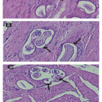 |
Figure 1: Photomicrograph of skeletal muscle sections stained with Haematoxylin & Eosin revealed muscle fibers with marked fibrosis and marked inflammatory cellular infiltration (arrow heads) with massive numbers of T. spiralis encysted encapsulated larvae (arrows). |
Table 1: Changes in complete blood picture (CBC) parameters in different groups.
|
|
5WPI |
5WPI+CSE |
|
Number mean ± SE |
1849.3 ± 380.0 |
746.5 ± 129.1 |
|
Range |
2250-1604 |
962-570 |
|
% of reduction |
0 ± 0 |
59.7% |
Table (2) revealed a significant decrease in the levels of Hb, RBCs count, HCT, MCV, MCH, PLT and lymphocytes percentage in 5 week post infection with Trichinella spiralis (5WPI). On the other hand; 5WPI induced significant increase in WBCs counts, neutrophil, monocyte and eosinophil percentage when compared to control and CSE. Treatments of 5WPI with CSE for 1 week (5WPI+CSE) induced significant increase in the levels of Hb, RBCs count, HCT, MCV, MCH, PLT, lymphocytes percentage and significant decrease in WBCs counts, neutrophil, monocyte and eosinophil percentage when compared to 5WPI group (Table 2).
Table 3 showed a significant elevation in the activities of ALT, ALP, AST, urea, creatinine and a significant depletion in the levels of albumin, total proteins in 5WPI group as compared to control and CSE groups. Meanwhile, treatments of 5WPI with CSE (5WPI+CSE) revealed a significant depletion in the activities of ALT, ALP, AST, urea, creatinine and a significant elevation in the levels of albumin, total proteins as compared to 5WPI group.
Discussion
It is still crucial to find novel ways to diagnose and manage zoonotic infections in developing nations, especially considering the possibility of human-animal contact there growing over time. With the ability to infect a wide range of mammals, including humans, T. spiralis is still regarded as one of the most dangerous and widely spread foodborne zoonotic nematodes.8 Drugs administered in trichinellosis patients include anthelmintics and steroids. Anthelmintics are the principal drugs for the treatment of Trichinellosis. A strong effort is currently being directed toward the development of an effective treatment against Trichinellosis.
Table 2: Changes in complete blood picture (CBC) parameters in different groups.
|
|
Control |
CSE |
5WPI |
5WPI+CSE |
|
Hb (g/dl) |
10.8# ± 0.69 |
11.1# ± 0.88 |
9.6* ± 0.40 |
10.4# ± 0. 70 |
|
RBC (million/ul) |
4.23# ±0.17 |
4.41# ±0.35 |
4.09*±0.25 |
4.16*# ± 0.39 |
|
Hct % |
35.6# ±2.20 |
36.3# ±2.07 |
31.7* ± 1.98 |
34.3# ± 1.33 |
|
MCV(fl) |
84.2±6.04 |
83.0±5.39 |
77.5* ± 4.55 |
82.5# ± 4. 05 |
|
MCH (pg) |
25.5# ± 1.60 |
25.2# ± 0.91 |
23.5* ± 1.14 |
4.74*# ± 0. 33 |
|
MCHC (g/dl) |
30.3 ±2.19 |
30.3 ±1.75 |
30.3±2.05 |
30.3 ± 2.17 |
|
Platelets (103 /ul) |
735.0# ±11.88 |
741.0# ±11.50 |
559.0* ± 9.35 |
618.0*# ± 10.23 |
|
WBC (103 /ul) |
6.2# ± 0.44 |
6.1# ± 0.42 |
11.45* ± 1.16 |
8.9*# ± 0. 60 |
|
Neutrophil % |
29.0# ±1.06 |
30.6# ±1.82 |
36.0*±1.75 |
33.9*# ± 2.08 |
|
Lymphocyte % |
61.2# ±3.81 |
62.0# ±3.25 |
50.0* ± 2.92 |
56.2*# ± 3.70 |
|
Monocyte % |
6.3*# ±0.51 |
5.0# ±0.40 |
8.0* ± 0.55 |
5.2# ± 0. 64 |
|
Eosinophil % |
3.5# ± 0.28 |
2.4# ± 0.17 |
6.0* ± 0.49 |
4.7*# ± 0. 29 |
*: Significant difference from the control group at p < 0.05, #: Significant difference from the Trichinella spiralis infection(5WPI) group at p < 0.05.
Table 3: Changes in the liver and kidney functions in different groups.
|
|
Control |
CSE |
5WPI |
5WPI+CSE |
|
ALT (U/I) |
37.9# ± 1.65 |
35.5#± 1.70 |
83.0* ± 3.25 |
61.3*#± 3.08 |
|
AST (U/I) |
81.0# ± 3.45 |
72.5# ± 4.15 |
109.2* ± 7.52 |
96.0*# ± 5.14 |
|
ALP (U/I) |
102.5# ± 6.75 |
93.9# ± 5.42 |
125.0* ± 8.76 |
116.2* ± 8.51 |
|
Albumin (mg/dl) |
4.11# ± 0.39 |
4.25# ± 0.28 |
3.05* ± 0.31 |
4.74*# ± 0. 33 |
|
Total protein (mg/dl) |
5.86# ±0.42 |
6.11# ±0.48 |
3.85*±0.29 |
4.54*# ± 0.35 |
|
Creatinine (mg/dl) |
0.65# ±0.05 |
0.63# ±0.08 |
0.95* ± 0.08 |
0.70# ± 0.05 |
|
Urea (mg/dl) |
31.9±2.14 |
28.5±2.65 |
43.5* ± 2.31 |
33.0# ± 2. 83 |
*: Significant difference from the control group at p < 0.05, #: Significant difference from the Trichinella spiralis infection(5WPI) group at p < 0.05.
These treatments have not proven their ability to fight and eliminate Trichinella spiralis effectively, so it was necessary to find new drug more effective for T. spiralis treatments. Consequently, the current research sought tothe impact of coriander treatment as new treatments on hematological and biochemical parameters in healthy and Trichinella spiralis infected mice.
30% of infected rats with Trichinella spiralis showed clinical signs as peri-orbital edema, dyspnea, and respiratory problems without changes in body temperature during the infection time. Current results agree with Ribicich et al. 24 who reported that; only two of nine pigs inoculated with T. spiralis larvae showed clinical signs consistent with trichinellosis. Treatments of 5WPI with CSE for one week showed a 59.6% reduction in number of larvae encysted in diaphragms of infected rats. Our findings are consistent with those of Abu El Ezz 25 and Soliman et al. 26, who found that untreated rats’ diaphragms had a large number of migratory larvae. Both in vitro and in vivo studies have shown that biological components found in CSE, including as polyphenols, tocopherols, and sterols, have potent anti-parasitic activities. 27,28
Current results showed that; infected rats with Trichinella spiralis for 5WPI induced significant decrease in the levels of Hb, RBC count, HCT, MCV, MCH, platelet count (PLT), lymphocytes percent and significant increase in WBCs, neutrophil, monocytes, eosinophil percent and the treatments with CSE improved these parameters. Low MCV and MCH means your hemoglobin production is less than normal. As a result, the number of healthy red blood cells also decreases, leading to anemia. Mean corpuscular volume and mean corpuscular hemoglobin are lower than the healthy range, it may indicate iron-deficiency anemia and microcytosis. Our findings are consistent with those of Ribicich et al. 24, who discovered that pigs implanted with 500 and 5000 larvae between 1 and 6 weeks after contracting Trichinella spiralis had lower haemoglobin values and higher white blood cell counts. Our results in the line of Sugane et al. 29 who reported that T. canis infection was induced elevation in WBCs count and eosinophil percent. Eosinophilia and infection severity are correlated. Additionally with Oto et al. 30, who investigated T. canis-caused eosinophilic meningo-encephalo-myelitis. On the first day after infection, a rise in eosinophils and basophils directly denotes the initiation of a primary-allergic reaction in the body. T. spiralis create a significant allergic reaction when they enter the body. 31 Allergy symptoms are brought on by the histamines that basophil cells produce. It is feasible to check the blood’s neutrophil count in addition to the clinical signs of invasion during T. spiralis infection and its early identification. This study shows that during the 5-week experiment, this marker is raised when compared to the control. According to Ovington and Behm 32, the presence of neutrophils and monocytes in the infiltrates of enclosing nurse cells indicates that an isolated rise in neutrophils is a sign of capsule development.
Our findings showed that, in comparison to control (healthy mice who were not infected with T. spiralis), T. spiralis infection (5WPI) caused a substantial increase in blood AST, ALT, ALP, urea, and creatinine levels and a significant decrease in serum total proteins and albumin. According to Gamble et al. 33 and Nada et al. 34, who confirmed that elevated AST and ALT is pointing to hepatic damage, while increased urea and creatinine is indicative of a kidney disease, these changes may be attributed to liver and kidney damages induced during larval migration. According to Saggu et al. 35, ALP is an enzyme that serves as a marker for the plasma membrane. Any of the two anomalies (an increase in normal levels or a decrease in normal levels) may result from damage to the biological membrane. This shows potential injury to the plasma membrane of the experimental rat tissues, and this conclusion is consistent with that of Adeyemi et al.36. Current study agreed with Mikhail 37 who find that T. spiralis infection induced in ALT and AST. Additionally, this investigation supported the findings of Basyoni and El-Sabah 38 and Soliman et al. 26 who reported that; After T. spiralis infection, levels of total proteins and albumin decreased. The decrease in total proteins and albumin levels may be caused by the migratory larvae damaging the liver parenchyma or by the metabolic byproducts of the parasites harming the liver.
Conclusion
Trichinella spiralis experimentallyinfection (5WPI) induced significant changes in hematological parameters, liver and kidney functions and the treatments of 5WPI with CSE (5WPI+CSE) induced a significant reduction in the number of Trichinella spiralis encysted larvae and improvements in all hematological parameters, liver and kidney functions.
Acknowledgement
None
Conflict of Interest
The authors declare no competing interests.
Funding Sources
There is no funding Sources
Data availability
All the data and material were available. The data of this article are included within the article and its additional files.
References
- Pozio, E., Rinaldi, L., Marucci, G., Musella, V., Galati, F., Cringoli, G., Boireau, P. and La Rosa, G. Hosts and habitats of Trichinella spiralis and Trichinella britovi in Europe. International journal for parasitology, 2009;39(1): 71-79.
CrossRef - Pozio, E., and Marucci, G. Trichinella-infected pork products: a dangerous gift. Trends in parasitology. 2003; 19(8): 338.
CrossRef - Ibrahim, S., Sarhan, M.H., Farag, T.I., and Mohamed, A.H. Apoptic and vascular changes in trichinella spiralis infected mice after parenteral artmether treatment. Journal of the Egyptian Society of Parasitology, 2019; 49(1): 17-27.
CrossRef - Wu, Z., Sofronic-Milosavljevic, L., Nagano, I., and Takahashi, Y. Trichinella spiralis: nurse cell formation with emphasis on analogy to muscle cell repair. Parasit Vectors 2008; 1: 27.
CrossRef - Ding, J., Liu, X., Bai, X., Wang, Y., Li, J., Wang, C., Li, S., Liu, M. and Wang, X., Trichinella spiralis: inflammation modulator. Journal of helminthology. 2020;94:e193.
CrossRef - ElGhannam, M., Dar, Y., ElMehlawy, M.H., Mokhtar, F.A. and Bakr, L., Eugenol; Effective Anthelmintic Compound against Foodborne Parasite Trichinella Spiralis Muscle Larvae and Adult. Pathogens 2023;12(1):127.
CrossRef - Saad, A.E., and Ghanem, H.B. Trichinella spiralis as a potential therapeutic agent: from a risky disease to a friend. Journal of the Egyptian Society of Parasit. 2020; 50(1):119-26.
CrossRef - Abou Rayia, D., Othman, A., Harras, S., Helal, D., Dawood, L. and Soliman, S. A new take on therapy of muscle phase of Trichinella spiralis infection. Acta tropica. 2022; 230: 106409.
CrossRef - Yadav, A.K, and Temjenmongla, A. Efficacy of Lasia spinosa leaf extract in treating mice infected with T. spiralis. Parasitol. Res., 2012; 110: 1-493.
CrossRef - Gottstein, B., Pozio, E., and Nöckler, K. Epidemiology, diagnosis, treatment, and control of trichinellosis. Clin Microbiol Rev. 2009; 22: 127-145.
CrossRef - Caner, A., Doskaya, M., Degirmenci, A., et al. Comparison of the effects of Artemisia vulgaris and Artemisia absinthium growing inwestern Anatolia against trichinellosis (Trichinella spiralis) in rats. Exp. Parasitol. 2008; 119:173-9.
CrossRef - Mutar, T.F., Tousson, E., Hafez, E., Abo Gazia, M. and Salem, S.B. Ameliorative effects of vitamin B17 on the kidney against Ehrlich ascites carcinoma induced renal toxicity in mice. Environmental Toxicol. 2020; 35(4): 528-537.
CrossRef - Essawy, A.E., El-Sayed, S.A., Tousson, E., Abd El-gawad, H.S., Alhasani, R.H. and Abd Elkader, H.T.A.E., Anti-kindling effect of Ginkgo biloba leaf extract and L-carnitine in the pentylenetetrazol model of epilepsy. Environmental Science and Pollution Res. 2022; 29: 48573–48587.
CrossRef - Alankooshi A.A., Hasan A. F., Tousson E., El-Atrsh A, Mohamed T.M. Impact of coriander seeds extract against thyroidectomy induced testicular damage and DNA replication in male rats. OnLine Journal of Biological Sciences 2023; 23 (2): 193.201. DOI: 10.3844/ojbsci.2023.193.201
CrossRef - Momin, A.H., Acharya, S.S., and Gajjar, A.V. Coriandrum sativum-review of advances in phytopharmacology. Int. J. Pharm. Sci. 2012; 3: 1233.
- Moustafa, A.H.A., Ali, E.M.M., Moselhey, S.S., Tousson, E. and El-Said, K.S., Effect of coriander on thioacetamide-induced hepatotoxicity in rats. Toxicology and industrial health, 2014; 30(7): 621-629.
CrossRef - Dunn, IJ, and Wright, KA. Cell injury caused by Trichinella spiralis in the mucosal epithelium in mice. J. Parasitol. 1985; 71:757-66.
CrossRef - Bocktor, N.Z., EL-Saied, M.O., and Imam. N.F. Effect of lactobacillus acidophilus on trichinella spiralis muscle larvae in experimentally infected mice compared to its effect when combined with albendazole and/or nitazoxanide. Journal of the Egyptian Society of Parasitol. 2022; 52(1):107-16.
CrossRef - Reitman, S. and Frankel, S. A colorimetric method for the determination of serum glutamic oxalacetic and glutamic pyruvic transaminases. American journal of clinical pathology, 1957; 28(1):56-63.
CrossRef - Belfield, A., and Goldberg, D.M. Revised assay for serum phenyl phosphatase activity using 4-amino-antipyrine. Enzyme. 1971; 12(5):561-73.
CrossRef - Simonian, M.H. Spectrophotometric determination of protein concentration. Current Protocols in Cell Biology 2002; 15(1):A-3B.
CrossRef - bd Eldaim, M.A., Tousson, E., Soliman, M.M., El Sayed, I.E.T., Abdel Aleem, A.A.H. and Elsharkawy, H.N. Grape seed extract ameliorated Ehrlich solid tumor-induced hepatic tissue and DNA damage with reduction of PCNA and P53 protein expression in mice. Environmental Science and Pollution Research 2021; 28(32):44226-38.
CrossRef - Patton, C., and Crouch, S. Determination of serum urea. Anal Chem 1977; 49:464–469.
CrossRef - Ribicich, M., Gamble, H.R., Rosa, A., Sommerfelt, I., Marquez, A., Mira, G., Cardillo, N., Cattaneo, M.L., Falzoni, E. and Franco, A., Clinical, haematological, biochemical and economic impacts of Trichinella spiralis infection in pigs. Veterinary parasitol. 2007; 147(3-4):265-270.
CrossRef - Abu El Ezz, N.M. Effects of Nigella sativa and Allium cepa oils on Trichinella spiralis inexperimentally infected rats. J. Egypt. Soc. Parasitol.2005; 35 (2):511-23.
- Soliman, G.A., Taher, E.S., and Mahmoud, M.A., Therapeutic effects of Dormectin, ivermectinand levamisole against different stages ofTrichinella spiralis in rats. Turk. Parasitol. Deg. 2011; 35:86-91.
CrossRef - Laribi, B., Kouki, K., M’Hamdi, M., and Bettaieb, T. Coriander (Coriandrum sativum L.) and its bioactive constituents. Fitoterapia 2015; 103: 9–26.
CrossRef - Al-Snafi, A.E. Antiparasitic, antiprotozoal, molluscicidal and insecticidal activity of medicinal plants (part 2)–plant based review. Sch. Acad. J. Pharm. 2016; 5: 194–207.
CrossRef - Sugane, K., Kusama, Y. Takamoto, M., Tominaga, A.,Takatsu, K.: Eosinophilia, IL-5 level and recovery of larvae in IL-5 transgenic mice infected with Toxocara canis. J. Helminthol. 1996; 2: 153-158.
CrossRef - Oto, S., Komiyama, A., Johkura, K., Hasegawa, O., Kondo, K.: Eosinophilic meningo-encephalo-myelitis due to Toxocara canis. Rinsho Shinkeigaku. 1994; 11: 1148-1152.
- Takamoto, M., Wang, Z.X., Watanate, N., Matsuzawa, A., Nariuchi, H., Sugane., K.: Eosinophilia, IgE production, and cytokine by lung T cells in surface CD4-deficient mutant mice infected with Toxocara canis. Immunology 1998; 95: 97-104.
CrossRef - Ovington, K.S., Behm, C.A.: The enigmatic eosinophil investigation of the biological role of eosinophils in parasitic helminth infection. Mem. Inst. Oswaldo Cruz. 1997; 2: 93-104.
CrossRef - Gamble, H.R., Wisnewski, N., and Wasson, D.L. Diagnosis of trichinellosis in swine by enzyme immunoassay, using a synthetic glycan antigen. Am J Vet Res R 1997; 58: 1417-1421.
CrossRef - Nada, S., Mohammad, S.M., Moad, H.S., El-shafey, M.A., Al-ghandour, A.M., and Ibrahim, N. Therapeutic effect of Nigella sativa and ivermectin versus albendazole on experimental trichinellosis in mice. Journal of the Egyptian Society of Parasitol. 2018; 48(1): 85-92.
CrossRef - Saggu, S., Sakeran, M.I., Zidan, N., Tousson, E., Mohan, A. and Rehman, H. Ameliorating effect of chicory (Chichorium intybus L.) fruit extract against 4-tertoctylphenol induced liver injury and oxidative stress in male rats. Food and Chem. Toxicol. 2014; 72(10): 138-1.
CrossRef - Adeyemi, O., Ajayi, J.O., Olajuyin, A.M., Oloyede, O.B., Oladiji, A.T., Oluba, O.M., Ololade, I.A. and Adebayo, E.A. Toxicological evaluation of the effect of water contaminated with lead, phenol and benzene on liver, kidney and colon of Albino rats. Food and Chemical Toxicol. 2009; 47(2): 885–887.
CrossRef - Mikhail, E.The occurrence of T. spiralis larvae in tissues other than skeletal muscles. J. Egypt. Soc. Parasitol. 1979; 9(1):269-72.
- Basyoni, M.M., and El-Sabah, A.A. Therapeutic potential of myrrh and ivermectin against experimental Trichinella spiralis infection in mice. Korean J. Parasitol., 2013; 51(3): 297-304.
CrossRef

