
Manuscript accepted on :21-08-2023
Published online on: 23-01-2024
Plagiarism Check: Yes
Reviewed by: Dr. Doaa Sayed Rashwan and Dr. S Shahi
Second Review by: Dr. Mazhar Ozkan
Final Approval by: Dr. H Fai Poon
Ajay Krishnan U and Anuradha Carani Venkataraman*
Department of Biochemistry and Biotechnology, Faculty of Science, Annamalai University, Annamalai Nagar, Tamil Nadu, India
Corresponding Author E-mail: cvaradha975@gmail.com
DOI : https://dx.doi.org/10.13005/bpj/2865
Abstract
The liver is a vital organ in the human body and is the primary site for lipid metabolism. Impaired lipid metabolism causes an accumulation of lipids in the liver, a discernible indication of non-alcoholic fatty liver disease (NAFLD). The condition is characterized by pathological alterations in the liver like steatosis, fibrosis and cirrhosis. 5′ Adenosine monophosphate-activated protein kinase (AMPK) maintains energy balance by regulating glucose and lipid metabolism. Dysregulation of AMPK is observed in NAFLD. The present work investigates the effect of an AMPK activator, 5-aminoimidazole-4-carboxamide-1-β-D-ribofuranoside (AICAR), on lipid levels, peroxisome proliferator-activated receptor (PPAR)-α, a nuclear receptor and cytoskeletal proteins α – smooth muscle actin (α-SMA) and cytokeratin 18 (CK18) and bilirubin levels in C57BL/6 mice fed high fat, high fructose diet (HFFD). The animals were divided into four groups (n=6, each group), and the feeding duration was ten weeks. The standard pellet was provided to groups 1 and 4 animals while HFFD was fed to animals of two groups (Groups 2 and 3) to induce fatty liver. AICAR injection (150 mg/kg bw/day, i.p.) was given to groups 3 and 4 animals on the 9th and 10th weeks. An equal volume of saline was injected into groups 1 and 2 animals. HFFD-fed mice showed increased levels of cholesterol, free fatty acids (FFAs) and CK18 with decreased bilirubin levels in plasma along with downregulated PPAR- α mRNA level and upregulated expression of α-SMA mRNA in the liver. Mice given HFFD and AICAR had significantly reduced cholesterol and FFA levels, increased bilirubin levels and reduced CK18 protein in plasma. The hepatic mRNA expression of PPAR-α was upregulated, while AICAR downregulated α-SMA expression. These findings suggest that AICAR regulates lipid metabolism, fibrogenesis and overall liver integrity. Thus, AICAR serves as a potential therapeutic measure for diet-induced fatty liver and the accompanying changes in the liver.
Keywords
AMPK; AICAR; Cytokeratin 18; Liver, Peroxisome proliferator-activated receptor-α; α-Smooth muscle actin
Download this article as:| Copy the following to cite this article: Krishnan U. A, Venkataraman A. C. 5-Aminoimidazole-4-carboxamide-1-β-D-ribofuranoside Attenuates High Fat, High Fructose Diet-induced Fatty Liver and Fibrosis in Mice. Biomed Pharmacol J 2024;17(1). |
| Copy the following to cite this URL: Krishnan U. A, Venkataraman A. C. 5-Aminoimidazole-4-carboxamide-1-β-D-ribofuranoside Attenuates High Fat, High Fructose Diet-induced Fatty Liver and Fibrosis in Mice. Biomed Pharmacol J 2024;17(1). Available from: https://bit.ly/48IXZPY |
Introduction
The liver is a vital organ performing several functions. It is the primary site for lipid metabolism pathways like lipolysis, fatty acid oxidation, de novo lipogenesis and very low-density lipoprotein (VLDL) secretion.1 Impaired lipid metabolism results in excessive deposition of lipid droplets in the liver. The condition is steatosis, which can develop into non-alcoholic fatty liver disease (NAFLD) if unattended. NAFLD is characterized by different stages of liver disorders starting from steatosis, which progresses to inflammation, fibrosis, cirrhosis and ultimately, liver failure.2 The prevalence of NAFLD is on the rise due to unhealthy diets and sedentary lifestyles and is often accompanied by obesity.3 Consumption of foods rich in fat and simple sugars promotes fatty liver formation. High fat, high fructose diet (HFFD) fed animals are commonly used as a model for NAFLD.4
Peroxisome proliferator-activated receptor-α (PPAR-α) is a nuclear receptor present abundantly in the liver. PPAR-α is a significant regulator of free fatty acid (FFA) oxidation, fatty acid uptake and glucose homeostasis.5 PPAR-α agonists are reported to reverse steatosis in alcohol-fed mice.6
Fibrosis of the liver refers to the overabundance of extracellular matrix (ECM) in the liver. Fibrosis is the underlying pathology of liver failure.7 Hepatic stellate cells (HSCs) are quiescent, vitamin A-storing cells that drive liver fibrosis. Activation and proliferation of HSCs result in the transformation of HSCs to fibrogenic myofibroblasts. The fibroblasts start overexpressing α-smooth muscle actin (α-SMA), an actin isoform that predominates in the fibrotic liver.8 Down-regulation of α-SMA in HSCs has been widely tested as a potential NAFLD therapeutic approach.9
Cytokeratin 18 (CK18) is a cytoskeletal protein of the cytokeratin acidic type I group (CK9-CK12).10 CK18 forms heteropolymers with CK8 to form keratin filaments. The keratin filaments are the major components of epithelial cells.11 CK18 is expressed in the liver and maintains the integrity and stability of hepatocytes. It plays crucial roles in apoptosis, cell cycle progression and cancer-related signalling pathways.12–15 During apoptosis, CK18 is cleaved by active caspases. These fragments resist proteases and are released into circulation due to plasma membrane disruption during the later stages of apoptosis.16 Elevated CK18 level is associated with liver cell death and is a marker for NAFLD.17
Bilirubin, the final product of heme degradation, is toxic to the brain and central nervous system. However, recently the cytoprotective actions of bilirubin have been shown. Some studies show that bilirubin contributes to total antioxidant capacity, is anti-inflammatory, and acts as a scavenger of reactive oxygen species.18 Increased bilirubin levels correlate negatively with the risk of NAFLD and type 2 diabetes mellitus.19,20
5′ Adenosine monophosphate-activated protein kinase (AMPK) plays a vital role in maintaining cellular energy balance by regulating glucose and lipid metabolism.21 Previously, we showed that AMPK protein is lowered during HFFD feeding and that administration of 5-aminoimidazole-4-carboxamide-1-β-D-ribofuranoside (AICAR), a synthetic AMPK activator, decreased fatty liver by lowering triglyceride (TG) and collagen content in HFFD fed mice.22 We hypothesize that AICAR could attenuate liver abnormalities in high-calorie diet-fed C57BL/6 mice. To test this hypothesis, the levels of cholesterol and FFAs in plasma and liver, bilirubin in plasma and the markers of fibrosis (α-SMA) in the liver, apoptosis (CK18) in plasma and the lipid modulator (PPAR- α) in mice with fatty liver were analyzed in animals with and without AICAR administration and then compared.
Materials and methods
Chemicals, kits and animal food components
AICAR, casein and fructose were purchased from Toronto Research Chemicals, Toronto, Ontario, Canada, Clarion Casein Pvt. Ltd., Kheda, India and SFA Food and Pharma Ingredients Pvt Ltd., Thane, India, respectively. Mouse Cytokeratin 18 ELISA kit was procured from Bioassay Technology Laboratory, Shanghai, China. Primers for polymerase chain reaction (PCR) analysis were obtained from Eurofins, Ebersberg, Germany. iTaq Universal SYBR Green Supermix and Trizol reagent were obtained from Bio-Rad, Hercules, CA, USA and Invitrogen, CA, USA, respectively. The remaining chemicals were purchased from either Himedia Laboratories, Mumbai, India or Sigma Aldrich Pvt. Ltd., St Louis, MO, USA, or SD Fine Chem Limited, Mumbai, India or Sisco Research Laboratories Pvt. Ltd, Mumbai, India.
Animal Maintenance
Male C57BL/6 mice of body weight 20-25 g were obtained from Biogen Laboratory Animal Facility, Bangalore, India and kept in the Central Animal House, Rajah Muthiah Medical College (RMMC), Tamil Nadu, India. Feed and water were provided ad libitum. The animal room had ambient temperature and humidity for the experimental mice. Approval from the local Institutional Animal Ethics Committee (IAEC), RMMC (AU-IAEC/1307/12/21) was obtained. All the animal procedures were conducted according to the guidelines of the Committee for the Purpose of Control and Supervision of Experiments on Animals (CPCSEA).
Induction of fatty liver and animal sacrifice
After acclimatization, the animals were randomly assigned to either of the four groups (Groups 1–4). Animals in Groups 1 and 4 were provided with standard pellets, and animals in Groups 2 and 3 were supplied with HFFD to induce fatty liver. The dietary regimen of animals was followed for ten weeks. HFFD was prepared fresh every day. The composition of HFFD (g/100g) is as follows: fructose (45), casein (22.5), wheat bran (5.5), peanut oil (10), beef fat (10), DL-methionine (0.3), vitamin mixture (1.2) and salt mixture (5.5). The standard diet contained 60% starch, 22.08% protein and 4.38% fat. Groups 3 and 4 animals were injected with AICAR dissolved in saline (150 mg/kg bw/ i.p. daily)23 and the other two groups (Groups 1 and 2) with equal amounts of saline in the 9th and 10th week. The animals were sacrificed after overnight fasting at the end of the 10th week. Blood and liver samples were collected from the animals.
Analysis of plasma and liver
Standard methods were employed for the extraction of lipids from plasma and liver24 and for the assay of total cholesterol25, FFA26 and total bilirubin.27 The levels of hepatic marker CK18 were measured in plasma using the kit protocol.
Quantitative real-time PCR analysis
Mouse liver RNA was extracted using Trizol reagent, and the concentrations were measured at 260 nm (Biophotometer plus, Eppendorf, Hamburg, Germany). The purity of RNA was checked by measuring the absorbance ratio at 260/280 nm. cDNA was prepared from RNA (2.0 μg) by reverse transcription using the real-time PCR system Mastercycler ep Realplex (Eppendorf, Hamburg, Germany) and then quantified (Biophotometer Plus, Eppendorf, Hamburg, Germany). cDNA amplification was done in a 10-μL reaction mixture containing cDNA (0.5 μg), 0.5 μL each of reverse and forward primers, 5 μL RT Easy mix and sterile water. The primer sequences are given in Table 1. Quantitative real-time PCR analysis was performed using iTAQ Universal SYBR Green Supermix (Bio-Rad, Hercules, CA) in Lightcycler 96 (Roche, Switzerland). The cycling conditions were as follows: 2 min at 950C, 40 cycles of 10 s at 950C, 60 s at 580C and 60 s at 720C. Quantitative data were expressed concerning control by the 2-ΔΔCT method.28The target gene ΔCT values were normalized for each group with an endogenous control glyceraldehyde-3-phosphate dehydrogenase (GAPDH). The relative expression of each gene was obtained by calculating the fold change compared to the control group.
Table 1: Nucleotide sequences of primers used inquantitative real-time PCR
|
Gene |
Accession Number |
Forward Primer 5’<-sequence->3’ |
Reverse Primer 5’<-sequence->3’ |
Product length |
|
PPARα |
NM_001113418.1 |
TGCATGTCCGTGGAGACCGTCAC |
ACTCGGTCTTCTTGATGACC |
523 |
|
αSMA |
NM_007392.3 |
GACGTACAACTGGTATTGTG |
TCAGGATCTTCATGAGGTAG |
144 |
|
GAPDH |
NM_008084.4 |
ACCCAGAAGACTGTGGATGG |
GTCATCATACTTGGCAGGTT |
222 |
Statistical analysis
Values are expressed as means ± SD (n=6) for biochemical studies and (n=3) for PCR studies. The data were analyzed for statistical significance by one–way analysis of variance (ANOVA) followed by the Tukey HSD test using SPSS software. A value of p < 0.05 was considered significant.
Results
Effect of AICAR on lipid levels
The total cholesterol levels in the plasma and liver are shown in Figures 1A and 1C, respectively. A significant increase in total cholesterol was noted in HFFD-fed mice compared to CON mice. AICAR administration to HFFD mice caused a reduction in cholesterol levels. AICAR alone treated animals showed normal cholesterol, and the values were near to the value of control animals.
Figures 1B and 1D present the levels of FFA in plasma and liver, respectively. HFFD-fed mice showed a significant increase in the level of FFA as compared to mice fed a regular diet in both plasma and liver. AICAR reduced FFA levels. The FFA levels in AICAR alone treated mice were found to be normal and near the value of CON mice.
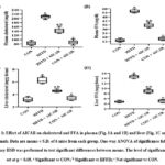 |
Figure 1: Effect of AICAR on cholesterol and FFA in plasma (Fig. 1A and 1B) and liver (Fig. 1C and 1D) of animals. Data are means ± S.D. of 6 mice from each group. |
Effect of AICAR on total bilirubin levels in plasma
The total bilirubin levels (Figure 2) were significantly reduced (59%) in animals fed HFFD compared to mice fed a regular diet. Significant increases in bilirubin values were observed in animals given HFFD and AICAR compared to AICAR untreated HFFD animals. AICAR alone treated animals had bilirubin levels near those of control group animals.
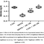 |
Figure 2: Effect of AICAR on plasma bilirubin levels of experimental animals. Data are means ± S.D. of 6 mice from each group. |
Effect of AICAR on plasma CK18 levels
The levels of CK18, a marker of liver cell integrity, were significantly higher (52%) in HFFD-fed mice compared to CON mice (Figure 3). CK18 levels were significantly reduced (32%) in animals that received HFFD and AICAR. There was no significant difference in the CK18 levels between AICAR alone treated and CON mice.
 |
Figure 3: Effect of AICAR on plasma CK18 levels of experimental animals. Data are means ± S.D. of 6 mice from each group. |
Expression of PPAR-α and α-SMA in liver
PPAR-α gene expression (Figure 4A) was downregulated in HFFD-fed mice compared to CON mice. However, in the HFFD + AICAR group, the gene expression was upregulated, whereas the AICAR alone treated group showed no significant change in expression compared to CON mice.
The gene expression of α-SMA is shown in Figure 4B. The levels of α-SMA were significantly higher in HFFD-fed mice (4-fold). AICAR treatment to HFFD-fed animals significantly reduced (2-fold) the expression of α-SMA. The gene expression in AICAR-alone treated mice was similar to that in the CON group.
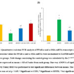 |
Figure 4: Quantitative real-time PCR analysis of PPAR-α and α-SMA mRNA transcripts of mice liver. Expression values for PPAR-α and α-SMA mRNA were normalized to GAPDH mRNA levels of respective groups. |
Discussion
The results of the present study show that HFFD feeding could cause hyperlipidemia characterized by a rise in cholesterol and FFAs in plasma and liver, elevate the levels of CK18 and reduce the level of bilirubin in plasma, increase the expression of α-SMA and decrease the expression of PPAR-α in the liver. AICAR treatment of HFFD-fed animals could reverse these changes.
Hyperlipidemia contributes to the development of NAFLD.29 Administration of a high fat diet can cause lipid accumulation and dysregulation of lipid metabolism in the liver. The rise in TG and lipid droplets have been reported earlier in the mice.22 NAFLD is often accompanied by insulin resistance, and fatty liver is one of the components of metabolic syndrome. Resistance to insulin action increases FFA flux, enhancing TG and VLDL synthesis in the liver, thereby triggering lipid accumulation.30 The presence of insulin resistance in HFFD mice has already been reported by many research groups31,32 and our previous study.22
Steatosis in HFFD mice liver can also be attributed to the downregulated expression of PPAR-α. PPAR-α, upon activation, triggers the expression of genes encoding enzymes of fatty acid oxidation in the peroxisomes and mitochondria.33 The anti-steatotic effect of PPAR-α has been attributed to the stimulation of fatty acid oxidation. The availability of FFA for TG synthesis is thereby decreased, and high-density lipoprotein (HDL) levels are increased.34
The rise in α-SMA expression in HFFD mice reflects the early fibrosis stage. Cytoskeletal proteins are upregulated during mitochondrial, endoplasmic reticulum, and oxidative stress, all of which occur in NAFLD.9 Feeding a high fat diet to minipigs caused hepatic fibrosis accompanied by the upregulation of α-SMA expression.35
CK18 is the major intermediate filament protein in the hepatocyte and acts as a mechanical stress absorber that maintains the entire cytoskeleton integrity.36 The changes in CK18 levels in serum are correlated to histologic changes in the liver, such as steatosis, lobular inflammation and hepatocellular ballooning in non-alcoholic steatohepatitis patients.16 Further, fragmented CK18 is positively correlated with changes in the levels of transaminases in NAFLD patients.37 Elevated levels of CK18 mRNA in the liver of HFFD mice might occur due to oxidative stress since cytoskeletal proteins are enhanced during cellular stresses.38
Serum bilirubin has a protective effect on various diseases.39 Bilirubin inhibits cholesterol synthesis, modulates the immune system,40 inhibits lipoprotein oxidation and prevents oxidative stress in the endothelial cells.41 Bilirubin in circulation is inversely correlated with HOMA-IR and may have a role in NAFLD since NAFLD is closely associated with insulin resistance.19 Biliverdin reductase (BVR) is an essential enzyme in the liver which catalyzes biliverdin to bilirubin during heme catabolism.18 Bilirubin gets oxidized to biliverdinduring oxidative stress. BVR also catalyzes the conversion of biliverdin back to bilirubin. Evidence of a link between impaired insulin signalling, tissue oxidative stress and BVR activity in regulating bilirubin metabolism has been emerging.19 Depletion of BVR increases oxidative stress. BVR also affects insulin signalling. Thus, reduced bilirubin in HFFD mice may be related to decreased BVR, oxidative stress and defective insulin signalling. This relationship needs to be confirmed in future studies.
In the cells, adenosine kinase converts AICAR to 5-aminoimidazole-4-carboxamide ribonucleotide (ZMP). ZMP is an analogue of AMP that binds and activates AMPK, activating various downstream targets.42 The benefits of AICAR through AMPK activation have been reported in the literature. AICAR attenuates lipid accumulation and cell death by inhibiting lipogenesis, activating lipolysis and enhancing fatty acid oxidation. AICAR inhibits apoptosis by inactivating pro-apoptotic factors and promotes the expression of anti-apoptotic factors in rat myoblasts.43 By reducing apoptosis, AICAR lowers CK18 and preserves liver function, slowing disease progression. Thus the effects of AICAR observed in this study are attributed to AMPK activation in the liver of HFFD mice.
Activation of AMPK stimulates the PPAR-α signalling resulting in the transcriptional activation of mitochondrial fatty acid oxidation resulting in lipolysis and utilization of existing lipid stores as a source of energy.44 Bilirubin also has been shown to activate PPAR-α and suppress lipid accumulation in 3T3-L1 adipocytes and in PPAR-α knock out mice.40,45
Studies have confirmed the role of AMPK activation in reducing fibrogenesis. For example, the inactivation of cellular AMPK by high phosphate level activates transforming growth factor-β1 (TGF-β1), which potentiates α-SMA expression and fibrosis in human mesangial cells.46 AICAR reduces TGF-β1-induced elevation of fibronectin and α-SMA in human renal proximal tubular epithelial cells.47
AICAR lowered the levels of TG and collagen and limited oxidative stress in the liver of HFFD-fed mice in our previous study.22 Reducing oxidative stress and preventing insulin resistance by AICAR may cause repletion of bilirubin levels. The present findings suggest that AICAR regulates lipid metabolism, fibrogenesis and overall liver integrity.
Conclusion
This study found that AICAR corrected hyperlipidemia, bilirubin and CK18 in plasma to near normal. Further, the hepatic mRNA expression of PPAR-α was upregulated, and α-SMA was downregulated in AICAR treated HFFD fed mice. AICAR can be a potential treatment for NAFLD in humans. Further research on dosage, treatment duration and side effects in various cell lines and animal models is required to determine its efficacy before entering clinical trials.
Acknowledgements
The authors thank DST-FIST and UGC-SAP for the facilities provided by the Department of Biochemistry and Biotechnology, Annamalai University, Tamil Nadu, India. The authors also thank the Center for Research on Molecular and Applied Sciences, Thiruvananthapuram, Kerala, India, for their help in performing PCR analysis.
Conflict of interest
The authors declare that they have no conflict of interest.
Funding Sources
This work was financially supported by the Indian Council of Medical Research, New Delhi, India, in the form of Senior Research Fellowship to the first author, Ajay Krishnan U (3/1/2(19)/OBS/2022-NCD-II).
References
- Gaggini M, Morelli M, Buzzigoli E, DeFronzo RA, Bugianesi E, Gastaldelli A. Non-alcoholic fatty liver disease (NAFLD) and its connection with insulin resistance, dyslipidemia, atherosclerosis and coronary heart disease. Nutrients. 2013;5(5):1544-1560. doi:https://doi.org/10.3390/nu5051544
CrossRef - Sayiner M, Koenig A, Henry L, Younossi ZM. Epidemiology of nonalcoholic fatty liver disease and nonalcoholic steatohepatitis in the United States and the rest of the World. Clin Liver Dis. 2016;20(2):205-214. doi:http://dx.doi.org/10.1016/j.cld.2015.10.001
CrossRef - Nseir W, Hellou E, Assy N. Role of diet and lifestyle changes in nonalcoholic fatty liver disease. World J Gastroenterol. 2014;20(28):9338-9344. doi:https://doi.org/10.3748/wjg.v20.i28.9338
- Im YR, Hunter H, de Gracia Hahn D, et al. A systematic review of animal models of NAFLD finds high-fat, high-fructose diets most closely resemble human NAFLD. Hepatology. 2021;74(4):1884-1901. doi:https://doi.org/10.1002/hep.31897
CrossRef - Todisco S, Santarsiero A, Convertini P, et al. PPAR alpha as a metabolic modulator of the liver: Role in the pathogenesis of nonalcoholic steatohepatitis (NASH). Biol (Basel). 2022;11(5):792. doi:https://doi.org/10.3390/biology11050792
CrossRef - Fischer M, You M, Matsumoto M, Crabb DW. Peroxisome proliferator-activated receptor α (PPARα) agonist treatment reverses PPARα dysfunction and abnormalities in hepatic lipid metabolism in ethanol-fed mice. J Biol Chem. 2003;278(30):27997-28004. doi:https://doi.org/10.1074/jbc.M302140200
CrossRef - Bataller R, Brenner DA. Liver fibrosis. J Clin Invest. 2005;115(2):209-218. doi:https://doi.org/10.1172/JCI24282
CrossRef - Xu J, Liu X, Koyama Y, et al. The types of hepatic myofibroblasts contributing to liver fibrosis of different etiologies. Front Pharmacol. 2014;5:167. doi:https://doi.org/10.3389/fphar.2014.00167
CrossRef - Pessoa J, Teixeira J. Cytoskeleton alterations in non-alcoholic fatty liver disease. Metabolism. 2022;128:155115. doi:https://doi.org/10.1016/j.metabol.2021.155115
CrossRef - Waseem A, Alexander CM, Steel JB, Lane EB. Embryonic simple epithelial keratins 8 and 18: chromosomal location emphasizes difference from other keratin pairs. New Biol. 1990;2(5):464-478.
- Menz A, Weitbrecht T, Gorbokon N, et al. Diagnostic and prognostic impact of cytokeratin 18 expression in human tumors: a tissue microarray study on 11,952 tumors. Mol Med. 2021;27:16. doi:https://doi.org/10.1186/s10020-021-00274-7
CrossRef - Cajaiba MM, Neves JI, Casarotti FF, et al. Hepatoblastomas and liver development: a study of cytokeratin immunoexpression in twenty-nine hepatoblastomas. Pediatr Dev Pathol. 2006;9(3):196-202. doi:https://doi.org/10.2350/05-12-0002.1
CrossRef - Gilbert S, Loranger A, Daigle N, Marceau N. Simple epithelium keratins 8 and 18 provide resistance to Fas-mediated apoptosis. The protection occurs through a receptor-targeting modulation. J Cell Biol. 2001;154(4):763-773. doi:https://doi.org/10.1083/jcb.200102130
CrossRef - Galarneau L, Loranger A, Gilbert S, Marceau N. Keratins modulate hepatic cell adhesion, size and G1/S transition. Exp Cell Res. 2007;313(1):179-194. doi:https://doi.org/10.1016/j.yexcr.2006.10.007
CrossRef - Faridi N, Bathaie SZ, Abroun S, et al. Isolation and characterization of the primary epithelial breast cancer cells and the adjacent normal epithelial cells from Iranian women’s breast cancer tumors. Cytotechnology. 2018;70(2):625-639. doi:https://doi.org/10.1007/s10616-017-0159-3
CrossRef - Vuppalanchi R, Jain AK, Deppe R, et al. Relationship between changes in serum levels of keratin 18 and changes in liver histology in children and adults with nonalcoholic fatty liver disease. Clin Gastroenterol Hepatol. 2014;12(12):2121-30. doi:https://doi.org/10.1016/j.cgh.2014.05.010.
CrossRef - Vos MB, Barve S, Joshi-Barve S, Carew JD, Whitington PF, McClain CJ. Cytokeratin 18, a marker of cell death, is increased in children with suspected nonalcoholic fatty liver disease. J Pediatr Gastroenterol Nutr. 2008;47(4):481-485. doi:https://doi.org/10.1097/mpg.0b013e31817e2bfb
CrossRef - Jang BK. Elevated serum bilirubin levels are inversely associated with nonalcoholic fatty liver disease. Clin Mol Hepatol. 2012;18(4):357-359. doi:https://doi.org/10.3350/cmh.2012.18.4.357
CrossRef - Lin LY, Kuo HK, Hwang JJ, et al. Serum bilirubin is inversely associated with insulin resistance and metabolic syndrome among children and adolescents. Atherosclerosis. 2009;203(2):563-568. doi:https://doi.org/10.1016/j.atherosclerosis.2008.07.021
CrossRef - Giral P, Ratziu V, Couvert P, et al. Plasma bilirubin and gamma-glutamyltransferase activity are inversely related in dyslipidemic patients with metabolic syndrome: Relevance to oxidative stress. Atherosclerosis. 2010;210(2):607-613. doi:https://doi.org/ 10.1016/ j.atherosclerosis.2009.12.026
CrossRef - Foretz M, Even PC, Viollet B. AMPK activation reduces hepatic lipid content by increasing fat oxidation in vivo. Int J Mol Sci. 2018;19(9):2826. doi:https://doi.org/10.3390/ijms19092826
CrossRef - Krishnan U A, Viswanathan P, Venkataraman AC. AMPK activation by AICAR reduces diet induced fatty liver in C57BL/6 mice. Tissue Cell. 2023;82:102054. doi:https://doi.org/10.1016/j.tice.2023.102054
CrossRef - Yang Z, Wang X, He Y, et al. The full capacity of AICAR to reduce obesity-induced inflammation and insulin resistance requires myeloid SIRT1. PLoS One. 2012;7(11):1-11. doi:http://dx.doi.org/10.1371/journal.pone.0049935
CrossRef - Folch J, Lees M, Stanley GHS. A simple method for the isolation and purification of total lipides from animal tissues. J Biol Chem. 1957;226(1):497-509. doi:http://dx.doi.org/10.1016/s0021-9258(18)64849-5
CrossRef - Zlatkis A, Zak B, Boyle AJ. A new method for the direct determination of serum cholesterol. J Lab Clin Med. 1953;41(3):486-492.
- Falholt K, Lund B, Falholt W. An easy colorimetric micromethod for routine determination of free fatty acids in plasma. Clin Chim Acta. 1973;46(2):105-111. doi:https://doi.org/10.1016/0009-8981(73)90016-8
CrossRef - Malloy HT, Evelyn KA. The determination of bilirubin with the photoelectric colorimeter. J Biol Chem. 1937;119(2):481-490. doi:https://doi.org/10.1016/S0021-9258(18)74392-5
CrossRef - Livak KJ, Schmittgen TD. Analysis of relative gene expression data using real-time quantitative PCR and the 2-ΔΔCT method. Methods. 2001;25(4):402-408. doi:https://doi.org/10.1006/meth.2001.1262
CrossRef - Perla FM, Prelati M, Lavorato M, Visicchio D, Anania C. The role of lipid and lipoprotein metabolism in non-alcoholic fatty liver disease. Child (Basel). 2017;4(6):46. doi:https://doi.org/10.3390/children4060046
CrossRef - Bjornstad P, Eckel RH. Pathogenesis of lipid disorders in insulin resistance: A brief review. Curr Diab Rep. 2018;18(12):127. doi:https://doi.org/10.1007/s11892-018-1101-6
CrossRef - Wada T, Kenmochi H, Miyashita Y, et al. Spironolactone improves glucose and lipid metabolism by ameliorating hepatic steatosis and inflammation and suppressing enhanced gluconeogenesis induced by high-fat and high-fructose diet. Endocrinology. 2010;151(5):2040-2049. doi:https://doi.org/10.1210/en.2009-0869
CrossRef - Zhuhua Z, Zhiquan W, Zhen Y, et al. A novel mice model of metabolic syndrome: the high-fat-high-fructose diet-fed ICR mice. Exp Anim. 2015;64(4):435-442. doi:https://doi.org/10.1538/ expanim.14-0086
CrossRef - Bougarne N, Weyers B, Desmet SJ, et al. Molecular actions of PPARα in lipid metabolism and inflammation. Endocr Rev. 2018;39(5):760-802. doi:https://doi.org/10.1210/er.2018-00064
CrossRef - Kersten S. Peroxisome proliferator activated receptors and lipoprotein metabolism. PPAR Res. 2008;2008:132960. doi:https://doi.org/10.1155/2008/132960
CrossRef - Wang H, Huang M, Bei W, Yang Y, Song L. FTZ attenuates liver steatosis and fibrosis in the minipigs with type 2 diabetes by regulating the AMPK signaling pathway. Biomed Pharmacother. 2021;138:111532. doi:https://doi.org/10.1016/j.biopha.2021.111532
CrossRef - Moll R, Divo M, Langbein L. The human keratins : biology and pathology. Histochem Cell Biol. 2008;129(6):705-733. doi:https://doi.org/10.1007/s00418-008-0435-6
CrossRef - Diab DL, Yerian L, Schauer P, et al. Cytokeratin 18 fragment levels as a noninvasive biomarker for nonalcoholic steatohepatitis in bariatric surgery patients. Clin Gastroenterol Hepatol.
2008;6(11):1249-1254. doi:https://doi.org/10.1016/j.cgh.2008.07.016
CrossRef - Gonsebatt ME, Razo LM Del, Cerbon MA, Zúñiga O, Sanchez-Peña LC, Ramírez P. Arsenite induced oxidative damage in mouse liver is associated with increased cytokeratin 18 expression. Arch Toxicol. 2007;81(9):619-626. doi:https://doi.org/10.1007/s00204-007-0192-7
CrossRef - Kwak M sun, Kim D, Chung GE, et al. Serum bilirubin levels are inversely associated with nonalcoholic fatty liver disease. Clin Mol Hepatol. 2012;18(4):383-390. doi:https://doi.org/10.3350/cmh.2012.18.4.383
CrossRef - Wen G, Yao L, Hao Y, Wang J, Liu J. Bilirubin ameliorates murine atherosclerosis through inhibiting cholesterol synthesis and reshaping the immune system. JJ Transl Med. 2022;20(1):1-18. doi:https://doi.org/10.1186/s12967-021-03207-4
CrossRef - Ziberna L, Martelanc M, Franko M, Passamonti S. Bilirubin is an endogenous antioxidant in human vascular endothelial cells. Sci Rep. 2016;6:29240. doi:http://dx.doi.org/10.1038/srep29240
CrossRef - Sengupta TK, Leclerc GM, Hsieh-Kinser TT, Leclerc GJ, Singh I, Barredo JC. Cytotoxic effect of 5-aminoimidazole-4-carboxamide-1-β-4-ribofuranoside (AICAR) on childhood acute lymphoblastic leukemia (ALL) cells: Implication for targeted therapy. Mol Cancer. 2007;6:46. doi:https://doi.org/10.1186/1476-4598-6-46
CrossRef - Vilchinskaya NA, Rozhkov S V., Turtikova O V., Mirzoev TM, Shenkman BS. AMPK phosphorylation impacts apoptosis in differentiating myoblasts isolated from atrophied rat soleus muscle. Cells. 2023;12(6):920. doi:https://doi.org/10.3390/cells12060920
CrossRef - Lee WH, Kim SG. AMPK-dependent metabolic regulation by PPAR agonists. PPAR Res. 2010;2010:549101. doi:https://doi.org/10.1155/2010/549101
CrossRef - Stec DE, John K, Trabbic CJ, et al. Bilirubin binding to PPARα inhibits lipid accumulation. PLoS One. 2016;11(4):e0153427. doi:https://doi.org/10.1371/journal.pone.0153427
CrossRef - Papadimitriou A, Peixoto EBMI, Silva KC, Faria JML de FJBL de. Inactivation of AMPK mediates high phosphate-induced extracellular matrix accumulation via NOX4/TGFß-1 signaling in human mesangial cells. Cell Physiol Biochem. 2014;34(4):1260-1272. doi:https://doi.org/10.1159/000366336
CrossRef - Thakur S, Viswanadhapalli S, Kopp JB, et al. Activation of AMP-activated protein kinase prevents TGF-B1- induced epithelial-mesenchymal transition and myofibroblast activation. Am J Pathol. 2015;185(8):2168-2180. doi:http://dx.doi.org/10.1016/ j.ajpath. 2015.04.014
CrossRef

