
Manuscript accepted on :15-11-2023
Published online on: 02-01-2024
Plagiarism Check: Yes
Reviewed by: Dr. Ayan Chatterjee and Maysaa Kadhim Al-Malkey
Second Review by: Tetty A
Final Approval by: Dr. Patorn Piromchai
Namariq Al-Saadi1 , Ohood A. Radhi2 and Ali Adnan Hashim1
1Al-Manara, College for Medical Sciences, Misan, Iraq.
2Department of Basic Science, College of Nursing, University of Kufa, Iraq.
Corresponding Author E-mail: nha15@scarletmail.rutgers.edu
DOI : https://dx.doi.org/10.13005/bpj/2792
Abstract
Klebsiella pneumoniae is a type of gram-negative bacterium that was initially discovered and isolated by Carl Friedlander in the year 1882. Klebsiella pneumoniae is responsible for a range of nosocomial and community-acquired illnesses, including urinary tract infections (UTIs), pneumonia, surgical site infections, and bloodstream infections. According to a number of studies, gram-negative sepsis ranks as the second most prevalent cause. A comprehensive analysis of the entire genome of Klebsiella pneumoniae was conducted using Illumina sequencing by synthesis (SBS) technology, specifically the Illumina HiSeq platform. The genetic material of the organism was comprised of a single circular chromosome of three thousand base pairs in length, exhibiting a GC content of 55.90%. In total, the genome exhibited a collective count of 164 genes associated with transfer RNA, 9 genes associated with ribosomal RNA, and 11,132 protein-coding sequences. Moreover, the analysis of genetic diversity entailed the application of sequence data, which was then compared to the reference genome that shared the highest degree of similarity. The genome annotation service offered by PATRIC and the RAST annotation system have successfully detected many antimicrobial resistance elements, clusters of genes associated with antibiotic resistance, as well as efflux pumps. This study represents the first known case of conducting a comprehensive genome sequencing of Klebsiella pneumoniae within the geographic area of Iraq, based on our current knowledge. The current investigation holds potential for advancing our comprehension of the antibiotic resistance traits demonstrated by Klebsiella pneumoniae, therefore enabling the healthcare facility to efficiently handle and alleviate outbreaks.
Keywords
Klebsiella Pneumoniae; Skin Burn; Whole Genome Sequencing
Download this article as:| Copy the following to cite this article: Al-Saadi N, Radhi O. A, Hashim A. A. Whole Genome Sequence for Klebsiella pneumoniae isolate from Burn Skin. Biomed Pharmacol J 2023;16(4). |
| Copy the following to cite this URL: Al-Saadi N, Radhi O. A, Hashim A. A. Whole Genome Sequence for Klebsiella pneumoniae isolate from Burn Skin. Biomed Pharmacol J 2023;16(4). Available from: https://bit.ly/41LhncA |
Introduction
Carl Friedlander isolated Klebsiella pneumoniae, a gram-negative bacterium, in 1882. Klebsiella pneumoniae causes a variety of nosocomial and community-acquired illnesses, including urinary tract infections (UTIs), pneumonia, surgical site infections, and bloodstream infections. Multiple investigations 1-3 have found that it is the second main cause of gram-negative sepsis.
Klebsiella pneumoniae is gram negative bacteria first isolate in 1882 by Carl Friedlander. It is the second leading cause of gram-negative sepsis. Capsular polysaccharide (CPS) and a critical virulence component that is a substantial contributor in developing sepsis are both present in K. pneumoniae 4. Two pathotypes, or clusters, of Klebsiella pneumoniae clinical isolates have been identified. Patients with compromised immune systems are the most common sources of classical strains (cKp), and these strains are often carbapenem-resistant (CR). On the other hand, carbapenem is effective against hypervirulent strains (hvKp), which are linked to invasive infections in the community 5,6. The HMV phenotype may be determined and hypervirulent K. pneumoniae can be distinguished from classical K. pneumoniae with the help of a simple microbiological test called the string test. Hyper-capsule generated by hypervirulent K. pneumoniae (hvKP) 7 confers the hyper-mucoviscous phenotype. Human defense mechanism expression and neutrophil and macrophage phagocytosis are both suppressed by CPS8. Additionally, the hyper-capsule enhances defenses against a wide variety of humoral defense mechanisms. This means that hvKP is protected against complement killing to a greater extent than cKP 9. Siderophores are low-molecular-weight secondary metabolites that can chelate iron. To transport ferric ions across the cell membrane, these compounds use small peptide molecules with side chains and functional groups that have high affinity ligands 10. Microbial siderophores are able to chelate iron and increase its uptake even at low concentrations thanks to the formation of a ferric-siderophore complex11. For K. pneumoniae to cause disease, siderophores are also required 5. To thrive during an infection, K. pneumoniae need a small amount of iron from the surrounding environment. As part of its nonspecific immune response, the host sequesters this metal during infection in an effort to curb the spread of multiple possible pathogens. Iron in the host plasma is usually bound to transferrin and other iron transport molecules, so there isn’t much free iron floating around 14. During a bacterial infection, mammals switch iron binding to lactoferrin, a naturally occurring defense protein found in bodily fluids 12,13. The creation of siderophores, molecules with a higher affinity for iron than host transport proteins, is the primary mechanism by which many infections, including K. pneumoniae, acquire iron. Siderophores are able to scavenge iron from the environment or the host’s iron-chelating proteins 14. Enterobactin, yersiniabactin, salmochelin, and aerobactin are just few of the siderophores that Klebsiella pneumoniae expresses 15. Only about 2–4% of nosocomial K. pneumoniae strains contain the salmochelin siderophore, which is expressed by the iroB gene. Since there are very few evidence explain the mechanism for Klebsiella pneumoniae, in current project try to study the mastery for multi antibiotics resistance by employ the NEXT GENERATION SEQUENCE NGS and bioinformatics tools to annotated the Klebsiella pneumoniae genome.
Materials and methods:
Bacterial isolate
Standard microbiological techniques were used for isolation and identification. The bacteria grow on nutritive and selective medium after being taken from swab samples taken from individuals who suffer from burned skin. After that, the colony that displayed multidrug resistance was isolated, and DNA was extracted from this solitary colony by utilizing a GENEAID, bacterial DNA extraction kit/Korea, extracted genomic DNA the included instructions, which were followed very strictly, albeit with minor adjustments made to cut down on the amount of contamination and boost the amount of pure DNA obtained. This research aimed to get insight into the whole genome of multidrug-resistant microorganisms because so little attention has been paid to doing so in the past.
Genome sequence
Illumina genome sequencing was performed on the DNA samples (Psomagen/USA, reference order number HN00194138). After quality control (QC), samples’ DNA was randomly fragmented before 5′ and 3′ adapter ligation for library creation. Create and sequence the library with NGS. After sequencing, raw results were evaluated for GC (percentage), total bases, and total reads. Quality filtering and Fast QC (https://www.bioinformatics.babraham.ac.uk/projects/fastqc/) reduced analytical biases. We assessed data quality at the end of each cycle using the phred quality score (Q20(%) and Q30(%)). De novo contigs were built from raw readings using SPAdes v.3.5 8.
Genome analysis:
The assembled genome of K. pneumoniae was obtained by the PATRS comprehensive genome analysis facility 9. The sequencing data were mapped using the reference genome of Klebsiella pneumoniae. The genome was fully annotated to identify functional genes within subsystem categories. Comparative mapping was conducted to analyze the findings, highlighting conserved and distinctive sequencing properties. Additionally, high-quality maps were constructed to validate and visually represent the annotated features. The removal of duplicates, identification of variations, and mapping of filtered data reads to the reference genome were accomplished by the utilization of several bioinformatics tools 10,11,12,13,14.
Results
Genome quality
The genome quality was good and the coarse consistency was 96.5 and fine consistency was 55.3. as it displays in figure (1).
Table 1: and give the table legend should be Genome quality of Klebsiella pneumoniae
|
Genome Quality |
Percentage |
|
coarse consistency |
96.5% |
|
fine consistency |
55.3% |
|
genome quality |
Good |
Antibiotics resistance profile
Antibiotic resistance was observed in the isolate. Furthermore, as shown in table 2, the isolate showed sensitivity to just nine antibiotics: ertapenem, ciprofloxacin, meropenem, imipenem, aztreonam, levofloxacin, cefoxitin, and piperacillin-tazobactam.
Table 2: showing the resistance and sensitivity for Klebsiella pneumoniae isolate.
|
Taxon ID |
Genome ID |
Genome Name |
Antibiotic |
Resistant Phenotype |
|
573 |
573.37830 |
Klebsiella pneumoniae |
tetracycline |
Resistant |
|
573 |
573.37830 |
Klebsiella pneumoniae |
ertapenem |
Susceptible |
|
573 |
573.37830 |
Klebsiella pneumoniae |
tobramycin |
Resistant |
|
573 |
573.37830 |
Klebsiella pneumoniae |
amikacin |
Resistant |
|
573 |
573.37830 |
Klebsiella pneumoniae |
ciprofloxacin |
Susceptible |
|
573 |
573.37830 |
Klebsiella pneumoniae |
meropenem |
Susceptible |
|
573 |
573.37830 |
Klebsiella pneumoniae |
imipenem |
Susceptible |
|
573 |
573.37830 |
Klebsiella pneumoniae |
aztreonam |
Susceptible |
|
573 |
573.37830 |
Klebsiella pneumoniae |
cefepime |
Susceptible |
|
573 |
573.37830 |
Klebsiella pneumoniae |
levofloxacin |
Susceptible |
|
573 |
573.37830 |
Klebsiella pneumoniae |
trimethoprim sulfamethoxazole |
Resistant |
|
573 |
573.37830 |
Klebsiella pneumoniae |
gentamicin |
Resistant |
|
573 |
573.37830 |
Klebsiella pneumoniae |
cefoxitin |
Susceptible |
|
573 |
573.37830 |
Klebsiella pneumoniae |
piperacillin tazobactam |
Susceptible |
Based on annotation statistics and PATRIC genomes of the same species, this genome looks to be high-quality. After readings filtering, phred quality scores of bases above Q20 and Q30 were 97.8% and 94.0 %. The Comprehensive Genome Analysis revealed 167 contigs, 11,068,038 bp, and 2,454 coding proteins in this assembled genome (Table-4). GC averages 55.90%. Figure-1 shows GC content and GC skew analysis schematically. A protein subsystem implements a biological activity or structural complex. 278 genome-specific subsystems were identified during annotation. Figure-2 provides a genome subsystem overview.
Table 3: Summary for the genome assembly and annotated features details.
|
Genome Annotation Pipeline (PGAP) |
Results |
|
Total length: |
11,068,038 bp |
|
GC Content % |
55.91 |
|
Number of Contigs: |
167 |
|
Number of Subsystems |
278 |
|
Genes (total) |
2,530 |
|
CDSs (total) |
5,074 |
|
Genes (coding) proteins |
11,132 |
|
rRNA |
9 |
|
tRNAs |
134 |
|
Contig L50 |
13 |
|
Contig N50 |
264,921 |
|
Plasmids |
0 |
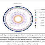 |
Figure 1: – K. pneumoniae circular genome. The red ring indicates genomic backbone (contigs) while the innermost ring reflects chromosomal position. |
This genome has too many contigs to be rendered clearly. The circular display has been limited to the 90 longest contigs of the 352 contigs in the genome.
Table 4: Summary proteins features details.
|
Proteins features |
PATRICS |
|
Hypothetical proteins |
3053 |
|
Proteins with functional assignments |
8079 |
|
Proteins with EC number assignments |
0 |
|
Proteins with GO assignments |
1928 |
|
Proteins with pathway assignments |
0 |
|
Proteins with Subsystem assignments |
0 |
|
Proteins with PATRIC genus-specific family (PLfam) assignments |
5374 |
|
Proteins with PATRIC cross-genus family (PGfam) assignments |
10435 |
Subsystem Analysis
A subsystem is a set of proteins that together implement a specific biological process or structural complexand PATRIC annotation includes an analysis of the subsystems unique to each genome. An overview of the subsystems for this genome is provided in Figure 2.
 |
Figure 2: Subsystem distribution of Serratia marcescens. RAST annotated the genome. The pie chart and SEED viewer showed subsystem feature counts and coverage. |
Phylogenetic Analysis
PATRIC offered reference and representative genomes for phylogenetic research. Figure-3 shows the closest reference and typical genotypes.
 |
Figure 3: Phylogenic relationship representation of the K. pneumoniae |
Discussion
Gram-negative bacilli infections are the most dangerous and potentially fatal infectious illness causes in hospitalized patients[1, 2]. K. pneumoniae is an opportunistic pathogen that causes pneumonia, abscess, bacteremia, and urinary tract infections in both community-acquired and nosocomial infections[3]. K. pneumoniae has become a global problem due to its propensity to rapidly acquire antibiotic resistance, requiring action to avoid the spread of multidrug-resistant bacteria[4]. Whole-genome sequencing (WGS) has grown more accessible and economical in recent years, leading in increased application in a variety of domains, including clinical microbiology[5-7]. One of the primary advantages of employing WGS is the ability to characterize the genetic content of clinically relevant bacteria and tie it to virulence-associated phenotypes, allowing for a better understanding of their transmission within the hospital and the use of timely medicines. The current investigation intended to describe K. pneumoniae isolates from burns skin patients hospitalized in Baghdad City. This study consider as is the first study in Iraq to use WGS to characterize an opportunistic pathogen in depth.
Conclusion
In conclusion, our study is the first in Iraq to analyze the genome of K. pneumoniae. Furthermore, the K. pneumoniae genome was compared to those of clinical reference strains, and its display of antibiotic resistance and virulence genes was identified. More whole genome sequencing and comparative genomics research is needed to better understand the background and assessment of multidrug-resistant isolates from different hospital rooms with special attention on delivery room.
Acknowledgments
The authors acknowledge Medical and science colleges/Misan University and Science college/ Kirkuk for their support.
Funding Sources
These authors declare that above-submitted work was not funded by any governmental or private funding source nor sup- ported by any financial projects.
Conflict of Interest
The authors declare that they have no conflict of interest.
References
- Podschun, R. and U. Ullmann, Klebsiella spp. as nosocomial pathogens: epidemiology, taxonomy, typing methods, and pathogenicity factors. Clin Microbiol Rev, 1998. 11(4): p. 589-603.
CrossRef - Kang, C.I., et al., Risk factors and pathogenic significance of severe sepsis and septic shock in 2286 patients with gram-negative bacteremia. J Infect, 2011. 62(1): p. 26-33.
CrossRef - Multistate Point-Prevalence Survey of Health Care-Associated Infections. N Engl J Med, 2022. 386(24): p. 2348.
CrossRef - Cortés, G., et al., Molecular analysis of the contribution of the capsular polysaccharide and the lipopolysaccharide O side chain to the virulence of Klebsiella pneumoniae in a murine model of pneumonia. Infect Immun, 2002. 70(5): p. 2583-90.
CrossRef - Paczosa, M.K. and J. Mecsas, Klebsiella pneumoniae: Going on the Offense with a Strong Defense. Microbiol Mol Biol Rev, 2016. 80(3): p. 629-61.
CrossRef - Sellick, J.A. and T.A. Russo, Getting hypervirulent Klebsiella pneumoniae on the radar screen. Curr Opin Infect Dis, 2018. 31(4): p. 341-346.
CrossRef - Fang, C.T., et al., A novel virulence gene in Klebsiella pneumoniae strains causing primary liver abscess and septic metastatic complications. J Exp Med, 2004. 199(5): p. 697-705.
CrossRef - Ohama, Y., et al., Accurate Identification of Klebsiella variicola by MALDI-TOF Mass Spectrometry in Clinical Microbiology Laboratories. Microbiol Spectr, 2022. 10(5): p. e0284422.
CrossRef - Pomakova, D.K., et al., Clinical and phenotypic differences between classic and hypervirulent Klebsiella pneumonia: an emerging and under-recognized pathogenic variant. Eur J Clin Microbiol Infect Dis, 2012. 31(6): p. 981-9.
CrossRef - Raymond, K.N., B.E. Allred, and A.K. Sia, Coordination Chemistry of Microbial Iron Transport. Acc Chem Res, 2015. 48(9): p. 2496-505.
CrossRef - Boiteau, R.M., et al., Siderophore-based microbial adaptations to iron scarcity across the eastern Pacific Ocean. Proc Natl Acad Sci U S A, 2016. 113(50): p. 14237-14242.
CrossRef - Bullen, J.J., Iron-binding proteins in milk and resistance to Escherichia coli infection in infants. Proc R Soc Med, 1972. 65(12): p. 1086.
CrossRef - Schubert, S., et al., Prevalence of the “high-pathogenicity island” of Yersinia species among Escherichia coli strains that are pathogenic to humans. Infect Immun, 1998. 66(2): p. 480-5.
CrossRef - Miethke, M. and M.A. Marahiel, Siderophore-based iron acquisition and pathogen control. Microbiol Mol Biol Rev, 2007. 71(3): p. 413-51.
CrossRef - Brock, J.H., et al., Relative availability of transferrin-bound iron and cell-derived iron to aerobactin-producing and enterochelin-producing strains of Escherichia coli and to other microorganisms. Infect Immun, 1991. 59(9): p. 3185-90.
CrossRef - Ares, M., et al., Antibiotic resistance of gram-negative bacilli isolated from pediatric patients with nosocomial bloodstream infections in a Mexican tertiary care hospital. Chemotherapy, 2013. 59(5): p. 361-8.
CrossRef - Hanna, M., et al., Infant isolation and cohorting for preventing or reducing transmission of healthcare-associated infections in neonatal units. Cochrane Database Syst Rev, 2023. 6(6): p. CD012458.
CrossRef - Gassama-Sow, A., et al., Genetic determinants of antibiotic resistance in diarrheagenic Klebsiella Pneumoniae subspecies ozaenae: an emerging enteropathogen in Senegal. Clin Infect Dis, 2010. 50(3): p. 453-5.
CrossRef - Rodriguez, L. and E.A. Berliner, Outbreak management of multidrug-resistant. J Feline Med Surg, 2023. 25(2): p. 1098612X231153051.
CrossRef - Huang, J., et al., Outbreak of KPC-producing. J Med Microbiol, 2022. 71(2).
CrossRef - Balushi, M.A., et al., Genomic analysis of the emerging carbapenem-resistant Klebsiella pneumoniae sequence type 11 harbouring Klebsiella pneumoniae carbapenemase (KPC) in Oman. J Infect Public Health, 2022. 15(10): p. 1089-1096.
CrossRef - Shen, S., et al., A Nationwide Genomic Study of Clinical Klebsiella pneumoniae Carrying. Microbiol Spectr, 2023. 11(3): p. e0386322.
CrossRef

