
Manuscript accepted on :27-04-2023
Published online on: 30-10-2023
Plagiarism Check: Yes
Reviewed by: Dr. Abidin Çalışkan
Second Review by: Dr. Menon,Kannan M
Final Approval by: Dr. Ian James Martin
Mansi Lather* and Parvinder Singh
Department of Computer Science and Engineering, Deenbandhu Chhotu Ram University of Science and Technology, Murthal, Sonepat, India.
Corresponding Author E-mail: mansi.schcse@dcrustm.org
DOI : https://dx.doi.org/10.13005/bpj/2770
Abstract
Brain tumor is one of the most prevalent and life-threatening illness these days. A tumor is an aberrant mass of tissue caused by unrestrained cell proliferation and multiplication. It is important to detect and diagnose brain tumors at the early stages. For disease diagnosis at an initial stage, there is a high demand for accurate analysis of healthcare data. But tumors vary greatly in size, shape, and existence, making it extremely difficult to collect precise measurements in order to properly diagnose them. Digital image processing enacts a crucial role in the analysis of medical images for timely and efficient planning of treatment. This paper provides an insight into brain tumors, the mechanism involved in their detection along with the different image processing steps that can be applied to medical images for automating the brain tumor detection process. This paper reviews a significant number of recently proposed brain tumor detection techniques related to the current study along with their tabulated comparison. This work can help in designing a solution that provides different applications such as detection, localization, or identifying the type of tumor under a single model.
Keywords
Brain Tumor; Image Processing; Medical Image Analysis; Tumor Detection
Download this article as:| Copy the following to cite this article: Lather M, Singh P. A Comprehensive Review on Strategies to Detect, Diagnose and Classify Brain Tumors. Biomed Pharmacol J 2023;16(4). |
| Copy the following to cite this URL: Lather M, Singh P. A Comprehensive Review on Strategies to Detect, Diagnose and Classify Brain Tumors. Biomed Pharmacol J 2023;16(4). Available from: https://bit.ly/3Ml7TOH |
Introduction
A tumor is an aberrant mass of tissue caused by unrestrained cell proliferation and multiplication 1. There are hundreds of different types of tumors. They can start in any of the trillion cells in our body. The name of tumor is reflected by the type of tissue they stem from such as brain tumor, lung tumor, breast cancer, ovarian tumor, etc. 2. Human brain is the most important organ in the body. It is often recognized as the human body’s regulating point. The brain is in charge of almost every critical activity inside the human body. Feelings, motion, intellect, speech, cognition, senses, reasoning, physical activity, taste, and creativity are all controlled by the brain 3. As a result, any mishap or impairment to this crucial organ will disrupt the human body’s usual working and result in an aberrant routine. It is therefore crucial to take the best possible care of this priceless organ.
The most prevalent and life-threatening illness that affects the brain these days is a brain tumor. It impacts both children and adults. Tumors can be malignant (cancerous) or benign (noncancerous). Tumors can also be classified as primary or secondary brain tumors. The characteristics of different types of tumors are shown in Fig. 1.
All the tumors are classified on the basis of a standard created by the World Health Organization (WHO) into four kinds: Gliomas, meningiomas, pituitary adenomas, and nerve sheath tumors 4. Tumors are named on the basis of the cells where they stem. Each of these categories is classified using a grading system, having a grading scale ranging from Grade I to IV. This grading standard is divided into benign and malignant tumors. Low-level tumors are classified as Grade I and II, whereas high-level tumors are classified as Grade III and IV. Grade I and II tumors comprise slow-growing cells that are generally not cancerous and are typically followed by long-lived survival of the affected individual 4. Grade III and Grade IV tumors comprise rapidly growing cells that are generally cancerous and termed as malignant. These tumors can invade and disrupt the nearby healthy tissues of the brain 4. The features of different grades of tumors are shown in Fig. 2. Because of the complexity of the brain anatomy, the overall influence of brain tumors on an individual can vary a lot as well as the repercussions may not be the same. Thus, it is much more vital to prepare a plan for treatment and forecast the patient’s reaction when treating a brain tumor at a preliminary stage 5.
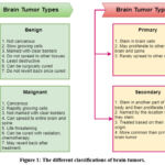 |
Figure 1: The different classifications of brain tumors. |
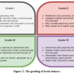 |
Figure 2: The grading of brain tumors. |
Cancer and its associated repercussions have a significant impact on public health. Years of life lost owing to premature deaths, economic expenditures connected with disease and treatments, and the long-term impact of cancer and its treatment on the quality of life of survivors, all take a toll on the population 6. Cancer patients encounter distinct physical and mental health, family functioning, and maintaining healthy lifestyle issues in the short and long term. Thus, it is very important to pinpoint and diagnose these tumors as early as possible.
This paper provides an insight into what a brain tumor is, its grading, and its classification. The different methods available to detect tumors along with different imaging scans have been highlighted. This article also discusses the different image processing steps that could be used to automate the brain tumor detection process. After studying and analyzing brain tumors and the role of computer-aided methods in their detection, a significant number of recently proposed brain tumor detection techniques related to our work have been reviewed along with their comparison. This work can help in designing a solution that is adaptive in nature, having capabilities to provide different applications such as the detection of tumor, localization of the tumor, or identifying the type of tumor under a single model.
This paper is divided into four sections: Section II describes the different methods available to detect brain tumors; Section III describes the different image processing steps that can be applied to a medical image for the purpose of detection of tumors; thus helping to automate the brain tumor detection process. Section IV gives an in-depth look into the work of numerous researchers in linked and related fields that are relevant to the current study, with a focus on segmentation and classification approaches, and finally, Section V concludes the paper.
Brain Tumor Detection Methods
Early recognition and diagnosis of brain tumors can significantly improve the patient’s chances of survival. A diagnosis of a brain tumor necessitates the following steps after assessing the physical signs of a patient suspected of a brain tumor: neurological examination, brain scan, and biopsy.
Neurological Examination
A neurological exam is generally the first step to figure out the cause of symptoms. A neurological assessment is a testing process used to determine the sensory system’s capabilities as well as the patient’s physiological condition. The functionality of nervous system is tested by this test. Mental status, motor function and balance, evaluation of nerves of the brain, responses, sensory exam, muscular strength, and coordination exam are the common tests 7.
Brain Scan Techniques
Brain scanning also known as neuroimaging makes use of different techniques to image the brain’s internal structure or function. The internal functioning of brain can be observed and monitored by sequencing scanned sections of brain. Brain scans are useful for detecting the presence, location, and size of a tumor. Advanced imaging techniques such as Computed Tomography (CT) scan, Magnetic Resonance Imaging (MRI), Functional MRI (fMRI), angiography, Magnetic Resonance Spectroscopy (MRS), Positron Emission Tomography (PET) scan, etc. are available to recognize tumors. CT scans and MRIs are the most commonly used diagnostic tools 8 9. The brain images captured using different medical imaging scanning techniques are shown in Fig.3 10 11 12 13 14 15.
CT Scan: It is the most commonly used scan to detect the presence of a tumor.
MRI: It provides more detailed information about the tumor’s location and size
fMRI: MRI images generated under this scan help in identifying brain areas responsible for important functions like speech or movement.
Angiography: These scans are used to locate blood vessels in the brain.
MRS: Examination of chemical profile of tumor and determination of type of lesions detected in MRI can be done using MRS.
PET Scan: This scan shows the metabolic functioning of tissues/organs. Persisting tumors can be detected using PET scans 8 9.
 |
Figure 3: The brain images obtained using different imaging modalities. |
Biopsy
A biopsy is a procedure during which a sample of tissue from a tumor location is removed and examined under a microscope. This process involves drilling a hole in the skull and removing a piece of tumor tissue which is then examined by the pathologist under a microscope to determine the tumor type and grade 9.
Stages Involved in Brain Tumor Recognition
Digital image processing is the process of converting digital data into pixels in an image. Many applications employ medical image processing to process medical data. In order to organize therapy in a timely and effective manner, digital image processing plays a critical role in medical image analysis. Any image processing application’s primary goal is to extract the required attributes from an image so that a machine can make appropriate assessment 16. The different image processing steps that can be applied to a medical image for the purpose of detection of tumors includes pre-processing, segmentation, feature extraction, and classification.
Pre-Processing
Pre-processing stage in medical image processing is the simplest phase. The main objective of this phase is to improve the MR image quality such that relevant information may be extracted from these images using either manual or automated approaches. Pre-processing boosts the image quality to make it more appropriate for subsequent processing, like classification or segmentation. Pre-processing is generally used to reduce noise and increase image resolution. Undesirable image pixels inside an image significantly impacting its resolution and clarity are referred to as noise. Due to the random nature of the noise formation process, it is really hard to anticipate the exact value of image distortion. The environmental conditions at the time of image acquisition, and the quality of sensing elements used to obtain medical images are the primary sources of noise in these images. Thus, it is critical to come up with a denoising technique that decreases the effect of noise and at the same time preserves the anatomical data that is important for clinical assessment. The pre-processing phase usually includes the following steps: removal and minimization of unnecessary noise, brightness preservation and contrast enhancement, transformation to grayscale, skull extraction, image registration or subsampling, among others 17.
Segmentation
Following the pre-processing phase, a segmentation process is typically performed. A medical image is divided into various segments during the segmentation process. These segments comprise similar qualities of texture, color, brightness, contrast, and gray level. Therefore, the major objective of this process is the division of objects existing in an image that are linked to one another in some way 18. Segmentation is the process of separating the specific area of interest from the rest of the image. For brain tumor detection, segmentation refers to extracting the tumor region from the brain MRI image. Segmentation techniques can be divided into thresholding, edge-based, clustering-based, and region-growing techniques.
Segmentation of a brain tumor is a key step in developing a Computer-Aided Diagnosis (CAD) system for MRI brain image processing because it allows clinicians to more precisely locate the tumor region. This type of process can be done manually, automatically, or semi-automatically.
Manual segmentation. A professional or radiologist visually locates and delineates the tumor region from the MRI slice in which the probable tumor emerges. Manual segmentation is costly, time-consuming, as well as prone to errors due to lack of constant updates, consistency, and repeatability.
Semi-automatic segmentation. In these types of segmentation methods, human interaction is incorporated to manually check and rectify the segmentation results produced by an automatic machine so as to boost the accuracy of brain tumor detection.
Fully-automatic segmentation. In these types of segmentation methods, segment boundaries are provided automatically by a program. There is no need for human interaction because the computer does all the work. Soft computing methods and other intelligent techniques can be used to create an approach to this goal.
Feature Extraction
The feature extraction technique extracts relevant information or features from medical images, such as statistics, shape, color, and texture. Feature extraction is a crucial part of medical image processing since it reveals the statistical characteristics of individual regions. The main goal of this phase is to minimize the amount of data used in the computation as it reduces the feature set by removing the redundant features. The extracted features contain relevant visual features that may be extended for future use such as detection and segmentation. Thus, the essential step in achieving a better outcome is to pick the best features derived from the image. Feature’s distinctiveness and integrity are important factors for selecting the best features. The use of feature extraction techniques in machine learning models leads to improved accuracy, reduction in overfitting risk, speed up in training, and improved data visualization 19. Feature extraction can be carried out using Stationary Wavelet Transform (SWT), Discrete Wavelet Transform (DWT), Gray Level Co-occurrence Matrix (GLCM), Gray Level Difference Method (GLDM), Local Binary Pattern (LBP), among others 20.
Classification
Classification is the most important stage in the CAD system. Classification helps in categorizing the medical images into appropriate classes depending on the characteristics collected from source images or images obtained from previous phases. For brain tumor detection, the classification phase can help in identifying whether a tumor is present in the brain MRI image or not and then identifying the type of tumor if a tumor is present. The different methods available for image classification are Artificial Neural Network (ANN), Back Propagation Neural Network (BPNN), Convolutional Neural Network (CNN), Decision Tree (DT), Support Vector Machine (SVM), Self-Organizing Map (SOM), etc. While selecting a particular classifier for classification, criteria such as reliability, efficiency, and processing capacity need to be considered 21 22.
Related Work
Timely detection of cancer plays an important role in beneficial therapy and revival of the patient. After studying and analyzing brain tumors and the role of computer-aided methods in their detection, a significant number of recently proposed brain tumor detection techniques have been reviewed. This section gives an in-depth look into previous brain tumor detection approaches that are relevant to the current study. It showcases the work of numerous researchers in linked and related fields, with a focus on segmentation and classification approaches.
Kalaivani et al. 23 presented machine learning-based software for the segmentation and classification of tumor types by utilizing brain MRIs. The MRI images were initially enhanced by employing the contrast optimization technique. Then double thresholding was performed through morphological processes, and finally, the skull stripping method was employed to eliminate undesirable non-cerebral tissues from the MR image. Features were also obtained using GLCM. Oversized aberrant cells were detected and tumor regions were sliced and segmented using classification algorithms such as k-NN, FCM, and K-means.
Jayadevappa and Kesava 24 proposed a Contourlet transform and the Chan-Vese active contour framework to automatically segment brain tumors. The Contourlet transform gathers edges and clean contours in any configuration and enhances the noise filtration process. The Contourlet transform, which was adding the property of directionality, produces high-resolution images. Chan-Vese active contour models were region-based segmentation algorithms that employ the best piecewise linear approximating algorithm. The proposed model generated precise results as compared to typical segmentation methods.
Islam et al. 25 suggested a modified brain tumor detection model based on the template-based K-means algorithm combining superpixels as well as Principal Component Analysis (PCA) within the given study. Initially, both the superpixels and PCA were used to retrieve key features that aid in the accurate detection of brain cancers. The image was then enhanced using a filter that aids in improving accuracy. Ultimately, to identify the brain tumor, segmentation was done using the template-based K-means clustering technique. The experiments indicated that the suggested strategy for brain tumor detection in MR images resulted in higher accuracy of 95%, and sensitivity of 97.36%, thus outperforming the other current recent schemes.
Padlia and Sharma 26 presented a method for detecting and segmenting brain tumors using T1-weighted and FLAIR brain MRI images. The fractional Sobel filter was used to reduce noise and improve the appearance of the brain MRI images. The fractional Sobel filter’s fractional-order provided more flexibility in enhancing segmentation performance. Bhattacharya factors and information gain were used to detect asymmetry across hemispheres. The statistical characteristics of a given window were generated and classified employing SVM for separating the tumor region from the tumor hemisphere. The images from the BRATS-2013 database were simulated, and quality factors like accuracy, sensitivity, and specificity were determined. The simulation results showed that the suggested scheme’s effectiveness was identical to that of the closest systems.
Lather and Singh 27 proposed an automated approachto detect the tumor from brain MR images based on segmentation technique. The brain MRI image dataset was initially pre-processed in order to handle the different types of noise and improve the quality of the medical images so that the components of the image can be effectively processed and assist in the segmentation phase for detecting the tumor from MRI images. As part of pre-processing, image quality was enhanced using Minimum Mean Brightness Error Bi-Histogram Equalization (MMBEBHE) technique and noise was removed from the images using a combination of Wiener and bilateral filters. Next, segmentation was done to extract the tumor region from these pre-processed MRI images. The Sine Tree Seed Algorithm (STSA) tuned K-means clustering approach was used for accurately segmenting the tumor region from the MRI images. In addition to this, the proposed segmentation approach was analyzed for its effectiveness by considering the impact of Gaussian and speckle noise on the original image. The results demonstrated that the proposed segmentation scheme outperforms the existing techniques in terms of various performance parameters for all the three cases of no noise, speckle, and Gaussian noise.
Kesav and Jibukumar 28 developed a region based Convolutional Neural Network (RCNN) approach for brain tumor classification and tumor type object recognition. The proposed approach was tested using two publicly available datasets from Figshare and Kaggle. Initially, a two-channel CNN was employed to distinguish between Glioma and a healthy tumor. The features for the RCNN were also detected using two-channel CNN. The RCNN was then used to recognize the tumor regions of the previously classified Glioma MRI data. With just an average level of confidence of 98.83 %, the technique was sufficient to attain a very low execution time when compared to other existing designs.
Abd El Kader et al. 29 presented a Differential Deep Convolutional Neural Network model (differential deep-CNN) in order to categorize various forms of brain tumors. The supplementary differentiated local features inside the original CNN feature maps were produced using differential operators in the differential deep-CNN architecture. The differential deep-CNN model was able to identify a large collection of images with high precision and without technological difficulties, as well as evaluate pixel directional patterns of images using contrast computations. The findings showed that the suggested differential deep-CNN model can be utilized to automatically categorize brain tumors.
Das et al. 30 focused on creating a CNN model for diagnosing brain tumors in T1-weighted contrast-enhanced MRI images. There were two major phases in the developed framework. Initially, various image processing algorithms were used to modify the images, and then classification was done using CNN. The study used a collection of 3064 medical images that had three different forms of brain tumors (glioma, meningioma, and pituitary). The proposed CNN model resulted in high testing accuracy of 94.39%, precision of 93.33%, and recall of 93%. The proposed work outscored a number of well-known current approaches.
Xuan and Liao 31 suggested a tumor segmentation technique that is based on statistical structure analysis. Initially, structural elements were analyzed for three types of features: intensity, symmetrical, and textural. The structural elements were then classified into normal and diseased tissues using an AdaBoost classification algorithm which trained itself by picking the most discriminant features. The proposed scheme resulted in a segmentation accuracy of 96.82%
Cherguif et al. 32 suggested a Deep Learning approach based on U-Net model. The proposed technique was tested on the BRATS 2017 dataset having both HGG and LGG cases. In comparison to traditionally defined ground truth, the proposed method was producing a segmentation that was efficient and resilient and resulted in Dice Similarity Coefficient (DSC) values of 0.81805, and 0.8103.
Garg and Garg 33 proposed a dynamic ensemble technique centered on the majority voting methodology that utilizes a combined model of Random Forest (RF), k-NN, and Decision Tree (DT) methods. Their goal was to calculate the tumor’s diameter and identify benign and malignant brain tumors. At first, Otsu’s threshold approach was used to segment the data. Then the SWT, PCA, and GLCM were used to extract features, yielding thirteen relevant features for the classification phase. Based on the majority voting approach, a hybrid ensemble classifier (kNN-RFDT) was used to classify the data. The suggested method resulted in an accuracy of 97.305%.
Lu et al. 34 presented an advanced CAD model called PBTNet for the detection of primary brain tumors from MRI images. In the proposed PBTNet model, they used a pre-trained ResNet-18 as a backbone framework fine-tuning it just for feature extraction. Furthermore, in the PBTNet, three randomized NNs named Schmidt, random vector functional-link, and extreme learning engine acted as detectors. The combination of the outputs from the classifiers formed the PBTNet’s finalized forecasts. The performance of the PBTNet classifier was evaluated using 5-fold cross-validation, and the results demonstrated the effectiveness of the PBTNet model for the detection of primary brain tumors.
Rehman et al. 35 presented an innovative deep learning-based methodology for brain tumor identification and tumor type prediction. Initially, a 3D CNN framework was created to extract brain tumors which were then passed to a pre-trained model for extracting the features. The retrieved features were then fed to a correlation-based selection technique, resulting in the best features selection as an output. Finally, the selected features were confirmed using a feed-forward neural network for the final prediction. For investigations and evaluation, three datasets were used, that is, BraTS 2015, BraTS 2017, and BraTS 2018.
Sajjad et al. 36 presented a novel multi-grade brain tumor classification scheme working on the principle of CNN based network. In the initial stage, deep learning was used to segregate tumor locations from the MRI images. Next, considerable data augmentation was performed to properly train the created methodology and eliminate the lack of data problems that can occur when employing MRI for multi-grade brain tumor classification. Finally, supplemented data was used to fine-tune a pre-trained CNN model for determining brain tumor grade. The proposed system was realistically evaluated using both enhanced and original data, demonstrating that it outperforms previous techniques.
Ismael and Abdel-Qader 37 presented a classification framework based on statistical features and NN models in order to classify the brain tumors in MRI images. Features were selected by using a hybrid approach of 2D DWT, and 2D Gabor filter. To analyze the influence of feature selection, a backpropagation NN classifier was chosen for classification. The proposed approach resulted in an accuracy of 91.9%.
Lather and Singh 38 presented an automated classification frameworkto identify tumor presence and tumor type from BraTS 2015 dataset. The brain MRI image dataset was initially pre-processed to enhance contrast using MMBEBHE method and remove unwanted noise from the images using a combination of Wiener and bilateral filters. Next, classification was done for detecting the presence of the tumor in MRI images as well as identifying the class of the tumor as High Grade Glioma (HGG) or Low Grade Glioma (LGG). The Dual Decision Voting Mechanism (DDVM) model employing dual classifiers – CNN, and Bi-directional Long Short-Term Memory (Bi-LSTM) was proposed to identify whether a tumor is present or not. Next, the Local Binary Pattern and Phase Quantization (LBP2Q) featured SVM model is proposed to identify the patterns from the tumor images to classify the type of tumor as HGG or LGG. In addition to this, the proposed classification framework was analyzed for its effectiveness by considering the impact of Gaussian and speckle noise on the original image. The results demonstrated that the proposed framework outperforms the existing techniques in terms of various performance parameters for all the three cases of no noise, speckle, and Gaussian noise.
A thorough comparative analysis of the research work of different researchers reviewed above is summarized in Table 1.
Table 1: Comparison of the reviewed work
|
S. No |
Authors |
Total images/ |
Pre-Processing |
Feature Extraction |
Segmentation |
Classification |
Result |
|
1 |
Kalaivani et al. 23 |
Chettinadu hospitals and research centers |
Median filtering double erosion, |
GLCM |
k-NN, FCM, |
NA |
Accuracy
|
|
2 |
Jayadevappa and Kesava 24 |
brain web database |
Gaussian |
Contourlet |
Chan-Vese |
NA |
NA |
|
3 |
Islam et al. 25 |
Kaggle |
Mean, and |
superpixels |
template-based |
NA |
Accuracy Sensitivity |
|
4 |
Padlia, and Sharma 26 |
BraTS 2013 |
fractional |
Statistical features |
SVM |
NA |
Accuracy Sensitivity |
|
5 |
Lather, and Singh 27 |
BraTS 2012 |
MMBEBHE, |
SWT |
STSA tuned |
NA |
Accuracy Precision Recall |
|
6 |
Kesav, and Jibukumar 28 |
Figshare and Kaggle |
NA |
two-channel CNN |
NA |
two-channel CNN, RCNN |
Accuracy |
|
7 |
Abd El Kader et al. 29 |
TUCMD database |
NA |
differential operators |
NA |
differential deep-CNN |
Accuracy Sensitivity Precision |
|
8 |
Das et al. 30 |
3064 images |
Gaussian filtering, Histogram Equalization |
NA |
NA |
CNN |
Accuracy Precision Recall |
|
9 |
Xuan, and Liao 31 |
140 images |
NA |
structural features: intensity, symmetrical, textural |
NA |
AdaBoost |
Accuracy |
|
10 |
Cherguif et al. 32 |
BraTS 2017 |
NA |
NA |
NA |
U-Net |
DSC HGG DSC LGG |
|
11 |
Garg, and Garg 33 |
2556 images |
NA |
SWT, PCA, and GLCM |
Otsu’s |
Hybrid ensemble classifier (kNN-RFDT) |
Accuracy |
|
12 |
Lu et al. 34 |
Kaggle |
NA |
pre-trained ResNet-18 |
NA |
PBTNet classifier |
Accuracy Sensitivity Precision |
|
13 |
Rehman et al. 35 |
BraTS 2015, 2017, 2018 |
NA |
3D CNN |
NA |
Feed forward NN |
Accuracy |
|
14 |
Sajjad et al. 36 |
Brain tumor dataset |
NA |
NA |
Input |
pre-trained CNN |
Accuracy Sensitivity Specificity =96.12% |
|
15 |
Ismael, and Abdel-Qader 37 |
3064 images |
NA |
hybrid approach of 2D DWT, 2D Gabor filter |
NA |
BPNN |
Accuracy= 91.9% |
|
16 |
Lather, and Singh 38 |
BraTS 2015 |
MMBEBHE, Wiener and bilateral filters |
LBP2Q |
NA |
DDVM model using dual classifiers – CNN, Bi-LSTM to identify tumor presence SVM to classify tumor type |
Tumor Presence Accuracy= 98.91% Tumor type Accuracy= 96.06% |
Conclusion
A tumor is an aberrant mass of tissues caused by unrestrained cell proliferation and multiplication inside the brain or around the brain. A brain tumor not only affects the cells in its immediate vicinity, but it can also harm other cells by generating inflammation. Brain tumor is the most prevalent and life-threatening illness these days. It is important to detect and diagnose brain tumors at the early stages. Early recognition and diagnosis of brain tumors can significantly improve the patient’s chances of survival. In this paper, we discussed about brain tumors, their grading, and classification. We also discussed about the mechanism followed to detect the brain tumor along with the different image processing steps that could be used to automate the brain tumor detection process. After studying and analyzing brain tumors and the role of computer-aided methods in their detection, we reviewed a significant number of recently proposed brain tumor detection techniques related to our work along with their tabulated comparison.
This work can be further extended to design an adaptive solution, having capabilities to provide different applications such as detection of tumor, localization of tumor, or identifying type of tumor under a single model.
Conflicts of Interest
The authors declare that they have no conflicts of interest.
Funding sources
This research received no external funding.
References
- What is a tumor? https://www.healio.com/news/hematology-oncology/20120331/what-is-a-tumor. Accessed May 21, 2022.
- Medical Definition of Tumor. MedicineNet. https://www. medicinenet.com/ tumor/definition.htm. Accessed May 21, 2022.
- Brain Tumor – Introduction. Cancer.Net. https://www.cancer.net/ cancer-types/brain-tumor/introduction. Published June 25, 2012. Accessed May 21, 2022.
- frankly-speaking-about-cancer-brain-tumors.pdf. http://blog. braintumor.org/files/public-docs/frankly-speaking-about-cancer-brain-tumors.pdf. Accessed May 21, 2022.
- Ghorpade N, Bhapkar H. Brain MRI Segmentation and Tumor Detection: Challenges, Techniques and Applications. 2021 5th International Conference on Intelligent Computing and Control Systems (ICICCS). 2021:1657-1664. doi:10.1109/ICICCS51141.2021.9432346
CrossRef - Research Areas: Cancer and Public Health – NCI. https://www.cancer. gov/research/areas/public-health. Published May 7, 2015. Accessed May 21, 2022.
- Neurological Exam. https://www.hopkinsmedicine.org/ health/conditions-and-diseases/neurological-exam. Published November 19, 2019. Accessed May 21, 2022.
- Brain_scanning_techniques.pdf. https://psicoterapiabilbao.es/wp-content/uploads/2015/12/Brain_scanning_techniques.pdf. Accessed May 21, 2022.
- Diagnosis – Learn More or Donate Today! | ABTA. https://www.abta.org/about-brain-tumors/brain-tumor-diagnosis/. Accessed May 21, 2022.
- Kamil M. Brain Tumor Area Calculation in CT-scan image using Morphological Operations. IOSR Journal of Computer Engineering. 2015;17:128-131.
- Perumal TS, Palanisamy V, Purusothaman T. Performance Analysis of Clustering Algorithms in Brain Tumor Detection of MR Images. European Journal of Scientific Research. 2011;62:321-330.
- Functional Magnetic Resonance Imaging | Radiology | University of Nebraska Medical Center. https://www.unmc.edu/ radiology/research/clinical/results/fmri.html. Accessed January 4, 2023.
- Castillo M. History and Evolution of Brain Tumor Imaging: Insights through Radiology. Radiology. 2014;273(2S):S111-S125. doi:10.1148/radiol.14140130
CrossRef - In vivo magnetic resonance spectroscopy. In: Wikipedia. ; 2022. https://en.wikipedia.org/w/index.php?title=In_vivo_magnetic_ resonance_ spectroscopy&oldid=1090268293. Accessed January 4, 2023.
- Verger A, Langen KJ. PET Imaging in Glioblastoma: Use in Clinical Practice. Exon Publications. September 2017:155-174. doi:10.15586/codon.glioblastoma.2017.ch9
CrossRef - Lather M, Singh P. Investigating Brain Tumor Segmentation and Detection Techniques. Procedia Computer Science. 2020;167:121-130. doi:10.1016/j.procs.2020.03.189
CrossRef - Kapoor L, Thakur S. A Survey on Brain Tumor Detection using Image Processing Techniques. In: 7th International Conference on Cloud Computing, Data Science & Engineering – Confluence. Noida, India: IEEE; 2017:582-585. doi:10.1109/CONFLUENCE.2017.7943218
CrossRef - Hassan E, Aboshgifa A. Detecting Brain Tumour from MRI Image Using Matlab GUI Programme. IJCSES. 2015;6(6):47-60. doi:10.5121/ijcses.2015.6604
CrossRef - Ippolito PP. Feature Extraction Techniques. Medium. https://towardsdatascience.com/feature-extraction-techniques-d619b56e31be. Published October 11, 2019. Accessed May 22, 2022.
CrossRef - Çalışkan A. A New Ensemble Approach for Congestive Heart Failure and Arrhythmia Classification Using Shifted One-Dimensional Local Binary Patterns with Long Short-Term Memory. The Computer Journal. 2022;65(9):2535-2546. doi:10.1093/comjnl/bxac087
- Kharat KD, Pawar V, Pardeshi SR. Feature Extraction and Selection from MRI Images for the Brain Tumor Classification. In: International Conference on Communication and Electronics Systems (ICCES). ; 2016:1-5. doi:10.1109/CESYS.2016.7889969
CrossRef - Çalışkan A. Detecting Human Activity Types from 3D Posture Data Using Deep Learning Models. Biomedical Signal Processing and Control. 2022;81:104479. doi:10.1016/j.bspc.2022.104479
CrossRef - Kalaivani I, Oliver AS, Pugalenthi R, Jeipratha PN, Jeena AAS, Saranya G. Brain Tumor Segmentation Using Machine Learning Classifier. In: 2019 Fifth International Conference on Science Technology Engineering and Mathematics (ICONSTEM). Vol 1. ; 2019:85-90. doi:10.1109/ICONSTEM.2019.8918918
CrossRef - Jayadevappa D, Kesava TC. Segmentation of Brain Tumor using Contourlet Transform and Chan-Vese Active Contour Model Approach. European Journal of Molecular & Clinical Medicine. 2021;8(2):2381-2389. https://ejmcm.com/article_8239.html. Accessed May 23, 2022.
- Islam MK, Ali MS, Miah MS, Rahman MM, Alam MS, Hossain MA. Brain Tumor Detection in MR Image Using Superpixels, Principal Component Analysis and Template Based K-means Clustering Algorithm. Machine Learning with Applications. 2021;5:100044. doi:10.1016/j.mlwa.2021.100044
CrossRef - Padlia M, Sharma J. Fractional Sobel Filter Based Brain Tumor Detection and Segmentation Using Statistical Features and SVM. In: Nath V, Mandal JK, eds. Nanoelectronics, Circuits and Communication Systems. Lecture Notes in Electrical Engineering. Singapore: Springer; 2019:161-175. doi:10.1007/978-981-13-0776-8_15
CrossRef - Lather M, Singh P. Tumor Segmentation From Brain MR Images Using STSA Based Modified K-means Clustering Approach. Journal of Intelligent & Fuzzy Systems. 2022;43:2579-2595. doi:10.3233/JIFS-212709
CrossRef - Kesav N, Jibukumar MG. Efficient and low Complex Architecture for Detection and Classification of Brain Tumor Using RCNN with Two Channel CNN. Journal of King Saud University – Computer and Information Sciences. May 2021. doi:10.1016/j.jksuci.2021.05.008
CrossRef - Abd El Kader I, Xu G, Shuai Z, Saminu S, Javaid I, Salim Ahmad I. Differential Deep Convolutional Neural Network Model for Brain Tumor Classification. Brain Science. 2021;11(3):352. doi:10.3390/brainsci11030352
CrossRef - Das S, Aranya OFMRR, Labiba NN. Brain Tumor Classification Using Convolutional Neural Network. In: 2019 1st International Conference on Advances in Science, Engineering and Robotics Technology (ICASERT). ; 2019:1-5. doi:10.1109/ICASERT.2019.8934603
CrossRef - Xuan X, Liao Q. Statistical Structure Analysis in MRI Brain Tumor Segmentation. In: Fourth International Conference on Image and Graphics (ICIG 2007). ; 2007:421-426. doi:10.1109/ICIG.2007.181
CrossRef - Cherguif H, Riffi J, Mahraz MA, Yahyaouy A, Tairi H. Brain Tumor Segmentation Based on Deep Learning. In: 2019 International Conference on Intelligent Systems and Advanced Computing Sciences (ISACS). ; 2019:1-8. doi:10.1109/ISACS48493.2019.9068878
CrossRef - Garg G, Garg R. Brain Tumor Detection and Classification Based on Hybrid Ensemble Classifier. arXiv; 2021. doi:10.48550/arXiv.2101.00216
CrossRef - Lu SY, Satapathy SC, Wang SH, Zhang YD. PBTNet: A New Computer-Aided Diagnosis System for Detecting Primary Brain Tumors. Front Cell Dev Biol. 2021;9:765654. doi:10.3389/fcell.2021.765654
CrossRef - Rehman A, Khan MA, Saba T, Mehmood Z, Tariq U, Ayesha N. Microscopic Brain Tumor Detection and Classification Using 3D CNN and Feature Selection Architecture. Microscopy Research and Technique. 2021;84(1):133-149. doi:10.1002/jemt.23597
CrossRef - Sajjad M, Khan S, Muhammad K, Wu W, Ullah A, Baik SW. Multi-grade Brain Tumor Classification Using Deep CNN with Extensive Data Augmentation. Journal of Computational Science. 2019;30:174-182. doi:10.1016/j.jocs.2018.12.003
CrossRef - Ismael MR, Abdel-Qader I. Brain Tumor Classification via Statistical Features and Back-Propagation Neural Network. In: 2018 IEEE International Conference on Electro/Information Technology (EIT). ; 2018:0252-0257. doi:10.1109/EIT.2018.8500308
CrossRef - Lather M, Singh P. DDVM: Dual Decision Voting Mechanism for Brain Tumour Identification with LBP2Q-SVM Type Classifier. International Journal of Computational Vision and Robotics. 2023;13(1):52-72. doi:10.1504/IJCVR.2023.127304
CrossRef

