
Manuscript accepted on :16-05-2023
Published online on: 24-08-2023
Plagiarism Check: Yes
Reviewed by: Dr. Farah Ramzi Nouri and Dr. Durgeshranjan Kar
Second Review by: Dr. Akhtar Ali
Final Approval by: Dr. Patorn Promchai
Ida Ayu Raka Astiti Asih* , I Ketut Giri Harta Subawa and I Made Oka Adi Parwata
Department of Chemistry, Faculty of Mathematics and Natural Sciences, Udayana University, Denpasar, Bali, Indonesia
Corresponding Author E-mail:astiti_asih@unud.ac.id
DOI : https://dx.doi.org/10.13005/bpj/2734
Abstract
Secang (Caesalpinia sappan L.) has a good effect on the health of the human body traditionally. It is a plant that has antioxidant activity because containing phenol and flavonoid compounds. The focus of this study was to evaluate the antioxidant activity of Secang wood water extract in vitro and in vivo. The Ferric Reducing Antioxidant Power method is used to examine antioxidant activity in vitro, while Wistar rats are used to test it in vivo. Test animals are given orally 50 mg/kgBW of the Secang wood water extract. Malondialdehyde levels and Superoxide Dismutase activity in rat heart and liver tissue with maximum physical activity were measured after the five-day intervention. In vitro, tests revealed that Secang wood water extract, which contained 393.7374 mg AAE/g of the sample, had the highest antioxidant capacity. In vivo, a test revealed that consumption of Secang wood water extract significantly (p < 0.05) boosted Superoxide Dismutase activity and lowered Malondialdehyde levels in the Wistar rats compared to the control.
Keywords
Malondialdehyde; Secang Wood; Superoxide Dismutase; Wistar rats
Download this article as:| Copy the following to cite this article: Asih I. A. R. A, Subawa I. K. G. H, Parwata I. M. O. A. Evaluation of Antioxidant Activity of Secang Wood (Caesalpinia Sappan L) Water Extract in Wistar Rats. Biomed Pharmacol J 2023;16(3). |
| Copy the following to cite this URL: Asih I. A. R. A, Subawa I. K. G. H, Parwata I. M. O. A. Evaluation of Antioxidant Activity of Secang Wood (Caesalpinia Sappan L) Water Extract in Wistar Rats. Biomed Pharmacol J 2023;16(3). Available from: https://bit.ly/3YM0kFA |
Introduction
Free radicals are unstable and very reactive atoms or molecules because there are unpaired electrons that makes this radicals can take electrons from other molecules 1,2. High levels of free radicals in the body caused by maximum physical activity that can increase the metabolism to form superoxide radicals (O2•) 3. Free radicals in the body can be detected by the presence of Malondialdehyde (MDA). Malondialdehyde is formed from the peroxidation reaction of unsaturated fats, namely the breaking of the fatty acid chains that make up the phospholipid cell membrane by free radicals 4,5.
Antioxidant compounds are able to neutralize free radicals by completing the electron deficiency 6. The human body produces endogenous antioxidants, one of them is Superoxide Dismutase (SOD) which can catalyze the reduction of O2• radicals to form H2O2 and O2 7. High levels of free radicals in the body cause endogenous antioxidants to be unable to neutralize these free radicals, which is called oxidative stress. This situation can be overcome by the presence of exogenous antioxidants to help the counteract process of free radicals. One of the active plants as a source of exogenous antioxidants is Secang wood (Caesalpinia sappan L.). The Secang wood is traditionally used to make an herbal drink by brewing it with hot water that is believed to have a good effect on the body’s health. A major specific active compound of Secang wood is brazilin, which can easily oxidize into brazilein in red color 8. Brazilin is a homo isoflavonoid compound known to has various bioactivity including antioxidant, antibacterial, anti-inflammatory, anti-photoaging, hypoglycemic (lowering fat levels), hepatoprotective (protecting the liver), and anti-acne9.
Tests for the antioxidant activity of Secang wood have been mainly done in ethanol extract. But, many people still consume Secang wood by brewing it in hot water, so it is necessary to evaluate the antioxidant activity of Secang wood in aqueous extract. In this study, the antioxidant activity of Secang wood aqueous extract was tested by using FRAP (Ferric Reducing Antioxidant Power) because it is cheap, simple, fast, and can be used in water solvents. This method is based on the ability of an antioxidant compound to reduce Fe3+ to Fe2+ ions into Prussian blue 10. Further evaluation was carried out with pre-clinical trials, such as in vivo. The test animals used in this study is rats because it has similar characteristics to human in physiology, anatomy, nutrition, pathology, and metabolism. Rats that are given maximum physical activity will increase the levels of free radicals, especially superoxide radicals (O2•), that can damage body tissues, such as the liver and heart. Maximum physical activity causes an increase the work of sympathetic nerves in heart tissue, resulting the increase in heart rate, heart contractions, and blood pressure, thus can increase the free radical levels in heart tissue 11. Liver tissue has microsomal membranes that are very susceptible to lipid peroxidation because it contains a lot of unsaturated fatty acids. The higher the levels of lipid peroxidation, the higher the levels of free radicals in liver tissue 12. Based on the description above, it is necessary to do research on the antioxidant activity test of Secang wood in water extract, conducted in vitro by determining its antioxidant capacity using the FRAP method and in vivo by measuring levels of MDA and SOD in the liver and heart tissue of Wistar rats, also the identification of their active compounds.
Materials and Methods
This study uses a descriptive exploratory and experimental design. This research was conducted from November 2021 to May 2022.
Materials
The materials used include Secang wood (Caesalpinia sappan L.) distilled water, n-hexane, chloroform, ethyl acetate, K₃[Fe(CN)₆] 0.2 M phosphate buffer pH 6.6, FeCl3, trichloroacetic (TCA), ascorbic acid, ethanol, vitamin E, Superoxide Dismutase Kit, thiobarbituric acid (TBA), phosphate-buffered saline (PBS), water-soluble tetrazolium (WST), 0.1 M Tris-HCl pH 7.4.
Methods
Sample Preparation
The Secang wood samples were sorted and washed under running water. Samples were chopped and then air-dried for 7 days. The dried samples were crushed with a chopper to become simplicia powder.
Extraction and partitioning of Secang wood
1 kg of simplicia powder was macerated with distilled water (50oC – 60oC) for 24 hours. The filtrate was concentrated with a vacuum rotary evaporator until crude extract was obtained. The crude extract was partitioned with n-hexane, chloroform and ethyl acetate to obtain the n-hexane, chloroform, ethyl acetate, and water fraction.
Ferric reducing antioxidant power (FRAP) assay
The FRAP method is used to determine antioxidant activity based on the ability of a compound to reduce Fe3+ ions to Fe2+ ions by providing one electron (single electron transfer). The FRAP assay was performed based on the methods of Benzie in Rabeta 13 with slight modification. An amount of 2 ml of sample, followed by 2 ml of 0.2 M phosphate buffer (pH 6.6) and 2 ml of K3[Fe(CN)6] 1%, then vortex and incubate for 20 minutes at 50°C. The mixture was then added to 2 mL of a 10% TCA solution, and then centrifuged for 10 minutes at 3000 rpm. Pipette 2 mL of the supernatant, followed by 2 mL of aqua DM and 0.4 mL of 0.1% FeCl3. A UV-visible spectrophotometer was used to measure the absorbance. Series of ascorbic acid stock solution 0,5,10,15,20,25 ppm (r2=0.9953) acted as standard curve. The antioxidant capacity is expressed as the equivalent weight of the sample per mg of ascorbic acid (mg AAE/g)
Phytochemical Test
Phytochemical tests were carried out on extracts with the highest antioxidant capacity including phenol, flavonoid, alkaloid, steroid, terpenoids, and saponin tests.
Identification of Active Compounds through Liquid Chromatography-Mass Spectrometer (LC- MS/MS)
Water extract of Secang wood which has the highest antioxidant capacity was analyzed by LC-MS/MS XEVO G2-S QTOF. All Mass Spectrometry spectra were analyzed using Masslynx v4.1 software. The chromatogram at each retention time was obtained, and then the molecular formula at each retention time was checked for its structure in the ChemSpider database (www.chemspider.com).
In Vivo Antioxidant Activity Test
The test animals using Wistar rats were divided into 4 treatment groups, each containing 6 rats. The increase in free radical levels was carried out by giving maximum physical activity, such as swimming until almost drowning (120 minutes/day). Group P0 (control 0) was not given any treatment, group P1 (negative control) was swimming, group P2 (positive control) was given vitamin E at a dose of 50 mg/kg BW and swimming, and group P3 (test group) was given Secang wood water extract at a dose of 50 mg/kg BW and swimming. The Intervention was carried out for 5 days. After that, they were taken as samples and dissected to obtain their heart and liver tissues. The Colorimetric method with Superoxide Dismutase Kit (BioVision, K335-100) was used for the determination of SOD activity in rat heart and liver tissue, and the TBARS technique was used to evaluate MDA levels. All test animals used have received ethical clearance from Animal Ethics Commitees the Faculty of Veterinary Medicine, Udayana University No. B/16/UN14.2.9/PT.01.04/2022
Statistical Analysis
Statistical analysis for all in vivo test data was analyzed using the Statistical Package for the Social Sciences (SPSS) application version 23.0. The method used was an analysis of variance (ANOVA) in the form of one-way ANOVA. Differences in MDA levels and SOD activity in heart and liver tissues in each group were further analyzed using the Least Significance Difference (LSD).
Results and Discussion
Results
Extraction and Partition
The Secang wood obtained through maceration was obtained around 54.32 grams of crude water extract in dark red. From the partition of 30 grams of aqueous extract, it was obtained 12.40 grams of dark red in ethyl fraction, while 15.74 grams of a reddish-brown in water fraction was obtained.
Ferric reducing antioxidant power (FRAP) assay
A linear regression equation, y = 0.0297x – 0.0138, was developed based on the calibration curve of the ascorbic acid standard solution. The antioxidant capacity of extract and fraction from the result of the partitioning of Secang wood was calculated using the linear regression equation, and the antioxidant capacity is expressed as the equivalent weight of the sample per mg of ascorbic acid. The results are displayed in Table 1.
Table 1: Antioxidant Capacity of Secang Wood Water Extract
|
Sample |
Antioxidant capacity (mg AAE/g sample) |
|
Water extract |
393.7374 |
|
Ethyl acetate fraction |
347.3401 |
|
water fraction |
214.2761 |
Phytochemical Test Results
The phytochemical test was carried out on the aqueous extract of Secang wood which had the highest antioxidant activity. The aqueous extract of Secang wood contains bioactive compounds, such as phenols, flavonoids, and saponins as shown in Table 2.
Table 2: Phytochemical Test of Secang Wood Water Extract
|
Phytochemical Test |
Reagent |
Colour Change |
Result |
|
Phenol |
FeCl3 |
Blackish blue |
Positive Phenol |
|
Flavonoids |
Wilsatter |
Brick red |
Positive Flavonoid |
|
Alkaloids |
Mayer Wagner |
None None |
None- Alkaloids |
|
Steroids |
Liebermann-Buchard |
None |
None- Steroids |
|
Terpenoids |
Liebermann-Buchard |
None |
None- terpenoids |
|
Saponins |
Aquades-HCl (shaken) |
Stable foam is formed |
Positive Saponins |
LC MS/MS results of Active Compounds of secang wood water extract
Identification of aqueous extract of Secang wood using LC-MS/MS spectrometry was carried out to determine the chromatographic and fragmentation patterns of each compound. Identification of the chromatogram and fragmentation of LC-MS/MS for aqueous extract of Secang wood shown in Table 3.
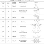 |
Table 3: Results of LCMS/MS analysis and suspected compounds |
In Vivo Antioxidant Activity Test Results
In Vivo test was conducted using the Randomize Posttest only control group design. The data obtained was analyzed statistically with IBM SPSS software version 23.0. The results of the statistical analysis of the average variable, the variance homogeneity of each variable and normal distribution of each group in rat heart and liver tissue are shown in Table 4.
Table 4: Average, normality and homogeneity of variants
|
Group P0 |
Group P1 |
Group P2 |
Group P3 |
(p*) |
|
|
SOD (%) liver tissue (p**) |
84.34 ± 3.91 0.945 |
84.34 ± 3.91 0.313 |
64.14 ± 4.46 0.805 |
49.46 ± 3.91 0.946 |
0.509 |
|
MDA (nmol/g) liver tissue (p**) |
2.10 ± 0.16 0.771 |
10.14 ± 0.19 0.836 |
4.02 ± 0.32 0.821 |
5.01 ± 0.20 0.991 |
0.424 |
|
SOD (%) heart tissue
|
78.78 ± 3.94 0.824 |
30.05 ± 4.93 0.406 |
58.01 ± 3.27 0.963 |
46.97 ± 3.95 0.818 |
0.751 |
|
MDA (nmol/g) heart tissue (p**) |
2,28 ± 0.17 0.560 |
11.35 ± 0.26 0.735 |
5.29 ± 0.34 0.846 |
6.15 ± 0.20 0.989 |
0.726 |
P0 = Control
P1 = Negative control
P2 = Positive control
P3 = Treatment Group
P* = homogeneity data (p>0.05)
P** = normality data (p>0.05)
The p-value of the average SOD activity and MDA levels was 0.0001 which indicates that the four treatments statistically showed a significant difference (p<0.05). The results of further analysis using the Least Significance Difference (LSD) method on SOD activity and MDA levels are presented in Table 5 and Table 6.
Table 5: LSD analysis of SOD activity and MDA levels in liver tissue.
|
Treatment Group |
Average of SOD activity (%) ± SD |
Treatment Group |
Average of MDA levels ± SD |
|
P0 |
84.34 ± 3.91 b,c,d |
P0 |
2.10 ± 0.16 b,c,d |
|
P1 |
31.81 ± 5.16 a,c,d |
P1 |
10.14 ± 0.19 a,c,d |
|
P2 |
64.14 ± 4.46 a,b,d |
P2 |
4.02 ± 0.32 a,b,d |
|
P3 |
49.46 ± 3.91 a,b,c |
P3 |
5.01 ± 0.20 a,b,c |
Table 6: LSD analysis of SOD Activity and MDA levels in heart tissue
|
Treatment Group |
Average of SOD activity (%) ± SD |
Treatment Group |
Average of MDA levels ± SD |
|
P0 |
78.78 ± 3.94 b,c,d |
P0 |
2.28 ± 0.17 b,c,d |
|
P1 |
30.05 ± 4.93 a,c,d |
P1 |
11.35 ± 0.26 a,c,d |
|
P2 |
58.08 ± 3.27 a,b,d |
P2 |
5.29 ± 0.34 a,b,d |
|
P3 |
46.97 ± 3.95 a,b,c |
P3 |
6.15 ± 0.20 a,b,c |
Information
SD = standard deviation;
a = There is a significant difference with the control group 0 (P0) (p <0.05)
b = There is a significant difference with the negative control group (P1) (p<0.05)
c = There is a significant difference with the positive control group (P2) (p<0.05)
d = There is a significant difference with the test group (P3) (p<0.05)
Discussion
Water extract of Secang wood has the highest antioxidant capacity, obtained 393.7374 mg AAE/g sample. This is because the antioxidant compounds contained in the water extract of Secang wood are synergistic, so they have a higher antioxidant capacity compared to the partition results from the water extract of Secang wood. Secang wood contains water-soluble flavonoids, namely brazilin, which is the dominant flavonoid compound contained in Secang wood 14. Brazilin was isolated from Secang wood which is known to have antioxidant activity higher than commercial antioxidants (BHT and BHA), so it is more potential as a free radical barrier 14,15.
Water extract of Secang wood contains bioactive compounds such as phenolic, flavonoid, and saponin groups (Table 2). Flavonoids and phenols have a hydroxyl group (-OH) attached to benzene, that can donate its hydrogen atom to free radicals 4. The ethanol extract of Secang wood contains phenolic compounds and flavonoid compounds which have antioxidant activity 16. The eight compounds contained in the aqueous extract of Secang wood have antioxidant activity because of the similarity of those structures with phenol and flavonoid groups.
The dominant compounds found were brazilin and brazilein which were also the dominant compounds contained in Secang wood. Brazilin is a flavonoid compound that structurally belongs to the iso-flavonoid group which has antioxidant activity 14,15,17. Brazilin can be an antioxidant because it has a catechol group, that can donate electrons to free radicals. In addition, the protosappanin compound is also a typical compound contained in the Secang wood which belongs to the class of phenolic compounds. Similar to brazilin, the protosappanin compound also has catechol groups that can donate electrons and neutralize free radicals. The glycoside quercetin compounds and chalcone compounds found are also a class of flavonoids that have a phenol group. Flavonoids can neutralize free radicals by donating hydrogen atoms so that they can neutralize the effects of free radicals and can act as chelate metals that play a role in the formation of ROS 4.
Maximum physical activity can trigger an increase in metabolism and oxygen consumption which increases up to 100-200 times to meet the energy required during physical activity. The increase in the amount of energy produced is proportional to the amount of ROS produced 18. When physical activity is excessive, oxygen is pumped to the muscles more, so other organs experienced hypoxia. The increased oxygen will trigger the conversion of ATP to ADP and AMP. Furthermore, AMP will be converted into hypoxanthine. The xanthine oxidase will break down hypoxanthine to form uric acid and xanthine. This process uses oxygen as an electron acceptor to form the radical •O2–. In addition, the increased oxygen causes an increase in electron leakage during electron transfer in mitochondria which will become superoxide anions 18,19,20. Superoxide radicals (•O2–) causes lipid peroxidation in breaking fatty acid chains to produce MDA through oxidation by free radicals. The •O2– radicals can be neutralized by an endogenous antioxidant, such as SOD, by catalyzing the reduction reaction of the O2• radical to produce H2O2 and O2 5,8. A comparison of MDA levels and SOD activity between groups are shown in Table 4.
The influence on each treatment group was examined using the average SOD activity and MDA levels across groups. The average SOD activity and MDA levels had a p-value in the one-way ANOVA test of 0.0001. This value showed that there was a statistically significant difference between the four treatments given to the rat (p<0.05). In both rat heart and liver tissue, the P0 group exhibited the lowest MDA concentrations and the highest SOD activity. This was because the P0 group (control 0) was not given maximum physical activity, such as swimming which caused the formation of free radicals to be very low, so the lipid peroxidation produced by MDA was lower. The P1 group (negative control) had the highest MDA levels and most insufficient SOD activity, because the P1 group was given maximum physical activity, so the formation of free radicals in the P1 group was high. Free radicals that are formed will cause lipid peroxidation reactions to produce Malondialdehyde (MDA). Cell membranes of body tissues are composed of lipids in the form of unsaturated fatty acids (LH). The peroxidation begins with the reaction between fatty acids (LH) and free radicals (oxidants) to form carbon radicals (L•) in the form of free fats (initiation). The free fat then reacts with oxygen to form peroxyl radicals (LOO•) which reacted again with other unsaturated fatty acids (LH) to form lipid hydro peroxides (LOOH) which are cytotoxic and free fatty acids (L•) which cause a chain reaction (propagation). This chain reaction ends when the radicals formed (at the initiation stage or at the propagation stage) react again with other radicals to become non-radical products (termination stage). At termination stage, it will produce endoperoxide which will further decompose into MDA 5,21,22,23.
The P2 group had lower MDA levels in heart and liver tissue and had higher SOD activity in rat heart and liver tissue than the P1 group. Giving vitamin E to the P2 group can neutralize free radicals that are formed as a result of the given maximum physical activity 24. By scavenging superoxide radical anion and lipid peroxyl free radicals, alpha-tocopherol prevents against lipid peroxidation in cell membranes 25. In its mechanism, vitamin E (α-tocopherol) reduces free radicals by donating their hydrogen atoms, where vitamin E reduced into tocopheryl radicals and becomes tocopheryl quinone 25,26.
The P3 group had lower levels of MDA in heart and liver tissue and had higher SOD activity in rat heart and liver tissue than the P1 group. This is because the intake of Secang wood extract can neutralize free radicals that are formed as a result of giving maximum physical activity, so it can reduce the formation of MDA and can increase SOD activity in the heart and liver tissues of rats. The decrease in MDA levels and the increase in SOD activity were caused by the presence of compounds contained in the water extract of Secang wood which has antioxidant activity. The compounds contained in the aqueous extract of Secang wood which is thought to act as antioxidants are phenolic compounds and flavonoids according to the results of the phytochemical tests obtained. Phenol compounds can donate their hydrogen atoms to free radicals, so that the lipid peroxidation reaction that produces MDA becomes lower. Likewise, flavonoid compounds that have -OH groups can also neutralize free radicals by donating their hydrogen atoms, as well as chelating metals which contribute to the production of ROS. 4,27. Flavonoids can also activate the nuclear factor erythroid-2 related factor 2 (Nrf2) which will increase the expression of endogenous antioxidants, that can also increases SOD activity 28.
The LSD test showed that there were significant differences in MDA levels and SOD activity in rat heart and liver tissues in each treatment group. All treatment groups were significantly different. Giving Secang wood extract intake can reduce MDA levels in group P3 with a decrease in MDA levels in heart tissue by 45.81% and in liver tissue by 50.59% compared to group P1 (negative control). Intake of Secang wood extract can also increase superoxide activity in P3 (test group) with an increased percentage of SOD activity in heart tissue by 56.30% and in liver tissue by 55.49% compared to group P1 (negative control).
Conclusion
Based on the study’s findings, a water extract from Secang wood has the highest antioxidant activity compared to its other extract. Intake of Secang wood water extract at a dose of 50 mg/kg BW significantly led to higher SOD activity and lower MDA levels in the liver and heart tissue of Wistar rats that were given maximum physical activity compared to SOD activity and MDA levels in the liver and heart tissue of Wistar rats that were not given Secang wood water extract.
Acknowledgment
Thanks to the Graduate Program of Chemistry at Udayana University and all parties who have assisted in this research.
Conflict of Interest
There is no conflict of interest.
References
- Selawa, W., M. R. J. Runtuwene, dan G. Cit raningtyas . Flavonoid content and total antioxidant capacity of the ethanol extract of binahong leaves (Anredera cordifolia (Ten.) Steenis.). J. Ilmiah Farmasi. 2013;2 (1): 18-22. https://ejournal.unsrat.ac.id/v3/index.php/ pharmacon/article/view/1018
- Senet, M.R.M., Parwata, I.M.O.A., and Sudiarta, I.W. Total Phenol and Flavonoid Content of Kersen Fruit (Muntingia calabura) and Antioxidant Activity. Chemistry Journal. 2017;11 (2) : 187-193. https://ojs.unud.ac.id/index.php/jchem/article/view/32539
CrossRef - Parwata, I. M. O. A., Laksmiwati., Sudiarta., Dina, M. N., and Sutirta Yasa I.W. The Potency of Flavonoid Compounds in water Extract Gyrinops Versteegii Leaves as Natural Antioxidants Sources. 2018; 11 (3) : 1501-1511. https://biomedpharmajournal.org/vol11no3/the-potency-of-flavonoid-compounds-in-water-extract-gyrinops-versteegii-leaves-as-natural-antioxidants-sources/
CrossRef - Asih, I.A.R.A., Manuaba, I.B.P., Berata, K., Satriyasa, B.K., dan Tunas, I.K. Intake Flavonoid Glycosides of Fruit Solanum Betaceum in Its Activity as a Candidate of Anti-Stress Oxidative. International Journal of Pharmaceutical and Phytopharmacological Research (eIJPPR). 2018; 8 (6) : 1-7. https://eijppr.com/yX1t1yo
- Maurya, R.P., Prajapat, M.K., Singh, V.P., Roy, M., Todi, R., Bosak, S., Singh, S.K,, Chaudhary, S., Kumar, A., andMorekar, S.R. Serum Malondialdehyde as a Biomarker of Oxidative Stress in Patients with Primary Ocular Carcinoma: Impact on Response to Chemotherapy. Clin Ophthalmol. 2021; 15 : 871-879 https://www.ncbi.nlm.nih.gov/pmc/ articles/PMC7924123/
CrossRef - Widyawati S, P., Wibawa B.T.D., Wahyu W.Y.D., and Olivia H.M. Antioxidant Activity of Black Tea Beluntas Leaf Drink (Pluchea indica Less-Camelia sinensis. Agritech .2018.;38(2) : 200 – 207 https://jurnal.ugm.ac.id/agritech/article/view/25699
CrossRef - Akyuz, O. Effect of Different-derm Cute Swimming Exercises on Oxidative Stres Parameters in Rat Cerebellum Tissue. US-China Education Review. 2016 ;6 (10) : 600- 608. https://davidpublisher.com/ Public/uploads/Contribute/5863781c4492b.pdf
CrossRef - Ngamwonglumlert L, Devahastin S, Chiewchan N, Raghavan GSV. Color and molecular structure alterations of brazilein extracted from Caesalpinia sappan L. under different pH and heating conditions. Sci Rep. 2020; 10 (1) : 12386 https://www.ncbi.nlm.nih.gov/pmc/ articles/ PMC7382456/
CrossRef - Pertamawati, Sriningsih, Fahrudin, F., and Efendi, J. Consumption of Secang Extract (Caesalpinia sappan L.) on Urine Volume of Spraque Dawley Male White Rats. Journal of Indonesian Jamu. 2017;2(3) : 121-126. https://www.academia.edu/50618802/Konsumsi_Ekstrak_Secang_ Caesalpinia_sappan_L_Terhadap_Volume_Urin_Tikus_Putih_Jantan_Galur_Spraque_Dawley
CrossRef - Maesaroh,K. Kurnia, D. Al-Anshori, J. Comparison of DPPH, FRAP and FIC Antioxidant Activity Test Methods Against Ascorbic Acid, Gallic Acid and Quercetin, Chimica et Natura Acta, 2018; 6(2) :93-100. http://jurnal.unpad.ac.id/jcena/article/view/19049/8961
CrossRef - Sherwood & Lauralee. Human physiology from cells to systems. 9th ed. United States: Brooks/Cole, Cengage Learning, 2016; Chapter 19, Peripheral Endocrine Glands; 682-684.
- Chiu, Y.J., Chou, S.C., Chiu, C.S., Kao, C.P., Wu, K.C., Chen, C.J., Tsai, JC., and Peng, W.H. Hepatoprotective effect of the ethanol extract of Polygonum orientale on carbon tetrachloride-induced acute liver injury in mice. J Food Drug Anal. 2018; 26(1):369-379.https://pubmed.ncbi.nlm.nih.gov/29389576/
CrossRef - Rabeta, M.S., dan Faraniza N. Total Phenolic Content and Ferric Reducing Antioxidant Power of The Leaves and Fruits of Garcinia atrovirdis and Cynometra cauliflora. International Food Research Journal 2013;20(4): 1691-1696 http://www.ifrj.upm.edu.my/ 20%20 (04)%202013/26%20IFRJ%2020%20(04)%202013%20Rabeta%20(018).pdf
- Nirmal, N.P., Rarput, M.S., Prasad, R.G.S.V., and Ahmad, M. Brazilin from Caesalpinia sappan heartwood and its pharmacological activities: A review. Asian Pasific Journal of Tropical Medicine. 2015; 8 (6) : 421-430.https://www.sciencedirect.com/science/article/pii/S1995764515000541
CrossRef - Farhana, H., Maulana, I.T., and Kodir, R.A. Comparison of Temperature and Time Rescue for Brazilian Contention in the wood.(Caesalpinia sappan L.). Prosiding Farmasi. 2015; 1 (2) : 19-25. https://karyailmiah. unisba.ac.id/index.php/farmasi/article/view/1564
- Prabawa, I.D.G.P., Khairiah, N., and Ihsan, H. Study Of Bioactivity And Secondary Metabolites From Secang Wood Extract (Caesalpinia Sappan L.) As Active Ingredient. Proceedings of the 2nd National Seminar Year 2019 Samarinda Industrial Research and Standardization Center. Samarinda. 2019. https://baristandsamarinda.kemenperin.go.id/ download/proceeding/2019_semnas2/Hal-B_1- 12_Dewa.pdf
- Hwang, H.S. & Shim, J.H.. Brazilin and Caesalpinia sappan L. extract protect epidermal keratinocytes from oxidative stress by inducing the expression of GPX7. Chinese Journal of Natural Medicines, 2018;16(3): 203-209. https://www.sciencedirect.com/science/article/pii/ S1875536418300487
CrossRef - Debevec, T., Millet, G.P. and Pialoux, V. Hypoxia-Induced Oxidative Stress Modulation with Physical Activity. Fronties in Physiolofy. 2017; 8: 84 https://www.frontiersin.org/articles/10.3389/fphys.2017. 00084/full
CrossRef - Powers S.K, Deminice R, Ozdemir M, Yoshihara T, Bomkamp M.P, Hyatt H. Exercise-induced oxidative stress: Friend or foe? J Sport Health Sci. 2020; 9(5) : 415-425. https://pubmed.ncbi.nlm.nih.gov/32380253/.
CrossRef - Asih, I.A.R.A., Rita, W.S., Suirta, W., dan Fudholi, A. Antioxidant Activity of Flavonoid Glycoside Extract of Solanum Betaceum on the Kidney of Wistar Rats. International Journal of Design & Nature and Ecodynamics. 2022; 17(2) : 319-323. https://doi.org/10.18280/ ijdne.170220
CrossRef - Powers S.K, Deminice R, Ozdemir M, Yoshihara T, Bomkamp M.P, Hyatt H. Exercise-induced oxidative stress: Friend or foe? J Sport Health Sci. 2020; 9(5) : 415-425. https://pubmed.ncbi.nlm.nih.gov/32380253/
CrossRef - Asih, I.A.R.A., Rita, W.S., Suirta, W., dan Fudholi, A. Antioxidant Activity of Flavonoid Glycoside Extract of Solanum Betaceum on the Kidney of Wistar Rats. International Journal of Design & Nature and Ecodynamics. 2022; 17(2) : 319-323. https://doi.org/10.18280/ ijdne.170220
CrossRef - Mourad, B., Rachid, B., dan Sihem, B. Antioxidant Activity and Phenolic Content of Artemisia Campestris from Two Regions of Algeria. World Journal of Environmental Biosciences. 2018; 7(2) : 61-66. https://environmentaljournals.org/article/antioxidant-activity-and-phenolic-content-of-artemisia-campestris-from-two-regions-of-algeria
- Górnicka, M., Drywień, M., Frąckiewicz, J., Dębski, B., and Wawrzyniak, A. Alpha-Tocopherol May Protect Hepatocytes Against Oxidative Damage Induced by Endurance Training in Growing Organisms. Adv Clin Exp Med. 2016; 25(4) : 673-679 https://pubmed.ncbi.nlm.nih.gov/27629841/
CrossRef - Sinbad , O.O. Folorunsho, A.A. Olabisi, O. L . Ayoola ,O.A. Temitope, E. J. Vitamins as Antioxidants. Journal of Food Science and Nutrition Research. 2019;2(3):214-235. https://www.fortunejournals.com/articles/ vitamins-as-antioxidants.pd
- Olivia, N. The Effect Of Α-Tocopherol Administration To The Histologist Cell Tubulus Proximal At Female Adult Mouse (Mus Musculus L.) Which Made To Obtain Maximum Physical Exercise. Jurnal Riset Hesti Medan. 2016; 1(1) : 30-36. https://jurnal.kesdammedan.ac.id/index. php/jurhesti/article/view/5
CrossRef - Zheng, Y.Z., Deng, G., Liang, Q., Chen, D.F., and Guo,R. Antioxidant Activity Of Quercetin and Its Glucosides from propolis: A Theoritical Study. Scientific Report. 2017; 7 : 7543 https://www.nature.com/ articles/s41598-017-08024-8
CrossRef - Mendonca, P. and Soliman, K.F.A. Flavonoids Activation of the Transcription Factor Nrf2 as a Hypothesis Approach for the Prevention and Modulation of SARS-CoV-2 Infection Severity. Antioxidants. 2020; 9(8) : 659. https://www.mdpi.com/2076-3921/9/8/659
CrossRef

