
Manuscript accepted on :02-06-2023
Published online on: --
Plagiarism Check: Yes
Reviewed by: Dr. Binit Patel and Dr. Masumeh Saeedi
Second Review by: Dr. Deepak Jain
Final Approval by: Dr. Patorn Promchai
Mahamadou Ballo1,2* , Filkpièrè Léonard Da3, Sékou Bah2, Rokia Sanogo2,4 and Estelle N. H. Youl1
1Laboratoire du Développement du Médicament, Centre de Formation, de Recherche et d’Expertises en Sciences du Médicament (CEA-CFOREM), Université Joseph KI-ZERBO, Ouagadougou, Burkina Faso.
2Faculté de Pharmacie, Université des Sciences, des Techniques et des Technologies de Bamako, Mali.
3Laboratoire de Sciences de la Vie et de la Terre, Unité de Formation et de Recherche en Sciences et Technologies, Université Norbert ZONGO, Burkina Faso.
4Laboratoire de Pharmacodynamie, Département de Médecine Traditionnel de Bamako, Mali.
Corresponding Author E-mail:mballo87@gmail.com
DOI : https://dx.doi.org/10.13005/bpj/2688
Abstract
The aim of this study was to assess the anti-inflammatory effects of a combination of medicinal plants on two models of inflammation. Subacute toxicity was assessed by daily oral administration of 2000 mg/kg body weight (bw). Subacute inflammation and arthritis were induced using the carrageenan air pouch granuloma model and Complete Freund's Adjuvant (CFA) respectively. After 28 days of administration, the combination at 2000 mg/kg proved to be non-toxic and induced a significant reduction (p<0.05) in transaminases and total cholesterol. The combinations C3 (150 mg/kg of T. macroptera + 250 mg/kg of X. americana), C2 ((250 mg/kg of T. macroptera + 150 mg/kg of X. americana) and C1 (250 mg/kg of T. macropteria + 250 mg/kg of X. americana) inhibited fresh granuloma formation by 40.37, 45.63 and 58.32% and dry granulomas by 47.77, 55.08 and 61.24% respectively. The combinations significantly (p<0.001) reduced air pouch fluid volume and massive leukocytes infiltration compared with the control group. With regard to the anti-arthritic effect, the combination C1 showed significant inhibition (p<0.05) of primary and secondary lesions compared with the control CFA. The increase in serum ALT, AST and uric acid concentrations observed in the CFA control group was significantly reduced (p<0.001) by the combination C1. An antioxidant effect was observed with the administration of the combination C1 and prednisone, which resulted in a significant increase (P<0.01) in GSH, SOD and catalase activity and a decrease in MDA concentration (P<0.001) compared with the CFA control group.The results suggest that the combination C1 has anti-inflammatory and anti-arthritic effects and prevents oxidative stress in arthritic rats.
Keywords
Anti-Inflammatory Effect; Anti-Arthritic Effects; Combination; Oxidative Stress; Terminalia macroptera; Ximenia americana
Download this article as:| Copy the following to cite this article: Ballo M, Da F. L, Bah S, Sanogo R, Youl E. N. H. Subacute Toxicity, Subacute Anti-inflammatory and Anti-arthritic Activities of Combination of Hydroethanolic Extract of Terminalia macroptera and Ximenia americana In-vivo. Biomed Pharmacol J 2023;16(2). |
| Copy the following to cite this URL: Ballo M, Da F. L, Bah S, Sanogo R, Youl E. N. H. Subacute Toxicity, Subacute Anti-inflammatory and Anti-arthritic Activities of Combination of Hydroethanolic Extract of Terminalia macroptera and Ximenia americana In-vivo. Biomed Pharmacol J 2023;16(2). Available from: https://bit.ly/44edHR2 |
Introduction
Inflammation is intended to repair tissue damage. The immune system fights this damage by inducing acute inflammation within minutes or longer 1. When the tissue damage has not recovered, acute inflammation turns into chronic inflammation 2. Inflammation is initiated by a local increase in vessel diameter and high permeability, resulting in inflammatory oedema following passage of exudate. Subsequently, leukocytes migrate into the damaged tissue and eventually a granuloma is formed 3. These inflammatory leukocytes have degranulation and superoxide production as responses. These products generate free radicals or reactive oxygen species, which cause oxidation and attack cell membranes and biomolecules, making them dangerous. Oxidative stress is the consequence of the imbalance between antioxidants and oxidants, causing alterations in proteins and lipids 3,4. This situation potentiates inflammation, especially in chronic pathologies, which are a public health problem 5.
Drugs to treat inflammatory have serious adverse effects or are very expensive. This reflects the interest of researchers in therapeutic substances derived from plants as a source of molecules to prevent or treat inflammatory diseases 6.
In West Africa, medicinal plants, accessible, less toxic and effective inflammation could be a possibility alongside conventional medicines 7,8. The Terminalia macroptera leaves and Ximenia americana roots are indicated in several inflammatory diseases according to traditional medicine. Ethnobotanical studies have revealed that in Burkina Faso, Mali and Guinea, the two plants mentioned above are prescribed against hepatitis, tuberculosis, wounds and inflammations 9–11. Both plants have also shown anti-inflammatory activities in vitro and anti-inflammatory effects against acute inflammation models. Both plants also showed in vitro anti-inflammatory activities and anti-inflammatory effects against acute inflammation models 12,13. The aim of this study was to assess the subacute anti-inflammatory and anti-arthritis effects of the combination.
Materials and Methods
Plant samples
Terminalia macroptera leaves and Ximenia americana roots were harvested in September 2020 on the hill of an outlying (Samé) district of Bamako. The plants were identified at the Department of Traditional Medicine and herbarium of each plant was deposited under number 2468; 0027 respectively. The collected samples were dried in the drying room of the Department of Traditional Medicine at room temperature and protected from sunlight. They were then pulverized.
Extraction methods
On each powder, two extraction methods at 10% (m/v) were performed. Aqueous decoction and hydroethanolic (30:70 v/v) maceration were used as described previously 14. briefly, 100 g of powder in 1 liter of 70% ethanol macerated for 24 hours and for decoction, 100 g of powder in 1 liter of distilled water, boiled for 15 minutes.
Experimental animals
Male and female wistar rats of 8-10 weeks, from the Department of Traditional Medicine were used. Rats were placed in cages at a temperature of 24 ± 2 ºC. At 12/12 h of light and dark. All experiments were conducted in accordance with international animal care guidelines 15. All described procedures were reviewed by the Ethics Committee and a protocol approval was issued #Reg. No. 2021 / 234 / USTTB.
Reagents and Medication
The following products were used: Carrageenan SIGMA-ALDRICH; Adjuvant Freund’s
Complete SIGMA-ALDRICH, Ellman’s reagent; trichloroacetic acid; thiobarbituric acid; dichromate; acetic acid; adrenaline; sodium diclofenac and prednisone.
Subacute toxicity
Subacute toxicity was performed according to OECD Test Guideline 407 16. 30 rats including 15 males and 15 non-pregnant nulliparous females aged 8 weeks were used. Rats were randomized into 3 homogeneous groups of 5 males and 5 females each, treated daily by
gavage for 28 consecutive days:
Group 1: neutral control, 10 µl/g of distilled water was administered;
Group 2 and group 3 (satellite): 2000 mg/kg bw of the combination was administered;
After 28 days of treatment, the satellite group rats were observed for two weeks without any administration. To monitor for possible reversibility, persistence or late onset of toxic effects. On day 29 (two weeks later for the satellite group), the rats were sacrificed and blood was collected from dry and EDTA tubes. Biochemical and hematological parameters were measured.
Carrageenan granuloma inflammation model (air pouch)
The method previously used by DA et al.17 was slightly modified for this study. Twenty-five rats randomized into 5 groups of 5 for the experiment. They were anaesthetized by injection of ketamine (100 mg/kg) intraperitoneally on day 0 and the pouch was created by subcutaneous administration of 10 ml of sterilized air to dorsal surface. On third day, each air pouch was re-inflated with 6 ml of sterile air and immediately, 4 ml of 2% carrageenan in 0.9% NaCl introduced. The animals received daily oral treatment for 4 days.
Group 1: negative control, 10 µl/g of distilled water was administered;
Group 2: positive control, 50 mg/kg bw diclofenac was administered;
Group 3: combination C1: 250 mg/kg of T. macroptera + 250 mg/kg of X. americana was administered;
Group 4: combination C2: 250 mg/kg of T. macroptera + 150 mg/kg of X. americana was administered;
Group 5: combination C3: 150 mg/kg of T. macroptera + 250 mg/kg of X. americana was administered.
On 7th day, the rats were sacrificed. The exudate was aspirated, measured and used to quantify leukocytes. Granuloma, liver and blood in dry EDTA tubes were collected.
Arthritis induced in rats by Complete Freund’s adjuvant (CFA)
The anti-inflammatory activity of the combination against chronic inflammation in rats was evaluated according to the method previously described 18. The rats were randomized into 4 different groups of 5 rats (n = 5):
Group 1 (neutral control): no CFA injection and rats received distilled water;
Group 2 (CFA control): CFA injection and rats received distilled water;
Group 3 (positive control): CFA injection and rats received 5 mg/kg/day of prednisone;
Group 4: CFA injection and rats received 500 mg/kg/day of combination C1.
On first day, the volumes of the hind legs were recorded and 100 µl of CFA was administered subcutaneously to the left hind paw. The animals were treated for the first twelve days. The effect against the primary lesion of the prednisone and the combination was assessed by measuring the volume of the paw injected on the 5th day. On the 21st day, the volume of the posterior paws was measured; afterwards the rats were sacrificed, liver and blood were collected to assess biochemical and oxidative stress parameters 19,20. The arthritic index was assessed macroscopically. The morphological features of polyarthritis were scored from 0 to 4 21.
For primary lesions: The percentage inhibition of the injected paw volume versus control was measured on 5th day. Secondary lesions were estimated by the increase in volume of the non-injected paw versus control on the last day of the experiment. The total of the scores corresponds to the arthritis index.
Preparation of serum and liver homogenate
The serum of the collected blood was kept in the refrigerator for biochemical examinations. After dissection, the liver was removed and washed immediately with cold 0.9% NaCl. 0.20 g of ground liver was homogenized in 1 ml of 50 mM Tris-HCl and centrifuged.
Biochemical assays
AST, ALT, total cholesterol and triglyceride levels were measured according to the procedures described in the commercially available reagent kit.
Estimation of oxidative stress biomarkers
The supernatant was used to determine oxidative stress parameters. Catalase (CAT) activity was determined by the colorimetric method according to Sinha22. Superoxide dismutase was estimated to assess the ability to inhibit auto-oxidation accord 23. The concentration of malondialdehyde (MDA) was assessed and reduced glutathione (GSH) was estimated 24,25.
Statistical analysis
Results are expressed as mean ± SEM. Statistical analysis was performed using one-way ANOVA followed by Tukey’s test for Graph Pad Prism® version 5.03. Differences were considered statistically significant, very significant and highly significant when p was <0.05 (*), <0.01 (**) and <0.001 (***), respectively.
Results
Sub-acute toxicity
The results focused on biochemical and hematological parameters.
The effects of the combination on hematological constants
Hematological parameters such as white and red blood cells, hemoglobin, platelets and hematocrit showed no significant variation between the different groups (table 1).
Table 1: Effect of the combination on hematological parameters of different groups of male and female rats.
|
Hematology parameters |
Control |
Combination 2000 mg/kg bw |
Satellite |
|
Males |
|||
|
WBC (109/l) |
16.52 ± 1.56 |
15.24 ± 2.59 |
15.86 ± 2,06 |
|
RBC (1012/l) |
8.32 ± 1.06 |
8.97 ± 0.23 |
8.58 ± 0.31 |
|
HGB (g/dl) |
15.07 ± 1.27 |
15.33 ± 0.93 |
14.95 ± 1.02 |
|
PLT (109/l) |
844.2 ± 46.08 |
858.4 ± 32.39 |
847 ± 37,85 |
|
HCT (%) |
44.36 ± 4.24 |
46.72 ± 2.52 |
46.34 ± 3.31 |
|
Females |
|||
|
WBC (109/l) |
15.84 ± 1.34 |
15,44 ± 4,14 |
16,04 ± 1,87 |
|
RBC (1012/l) |
9.08 ± 0.16 |
9,32 ± 0,53 |
9,17 ± 0,58 |
|
HGB (g/dl) |
15.59 ± 0.79 |
15,78 ± 1,27 |
15,45 ± 0,36 |
|
PLT (109/l) |
858.6 ± 34.77 |
869,2 ± 35,81 |
857,8 ± 27,91 |
|
HCT (%) |
44.94 ± 1.23 |
46,56 ± 4,18 |
47,88 ± 3,39 |
The values are expressed as mean ± SEM, n = 10. WBC: White blood cells, RBC: Red Blood Cell, HGB: Hemoglobin, PLT: Platelets and HCT: hematocrit.
Effects of the combination on biochemical parameters
The biochemical profiles of the different groups of rats are presented in table 2. The combination at 2000 mg/kg induced a significant (p < 0.05) decrease in transaminases and total cholesterol. This decrease disappeared after an additional two weeks observation without administration in the satellite group. However, no variation of kidney markers such as creatinine and urea were observed in neither the combination group or satellite group.
Table 2: Effect of the combination on the biochemical parameters of the different groups of male and female rats.
|
Biochemical parameters |
Control |
Combination 2000 mg/kg bw |
Satellite |
|
Males |
|||
|
Creatinine (µmol/l) |
51.26 ± 2.75 |
54.2 ± 3.07 |
52.86 ± 3.29 |
|
Urea (µmol/l) |
4.38 ± 0.63 |
4.87 ± 0.57 |
4.83 ± 0.63 |
|
Cholesterols (mg/dl) |
76.2 ± 2.63 |
70.93 ± 6.21* |
75.45 ± 4.52 |
|
Triglycerides (mg/dl) |
35.94 ± 3.46 |
37.64 ± 3.17 |
36.71 ± 2.8 |
|
AST (U/l) |
47.09 ± 1.98 |
43.63 ± 1.64* |
45.54 ± 1.52 |
|
ALT (U/l) |
42.2 ± 1.44 |
40.30 ± 3.16 |
40.72 ± 1.91 |
|
Females |
|||
|
Creatinine (µmol/l) |
50.48 ± 2.66 |
54.99 ± 2.89 |
51.78 ± 1.38 |
|
Urea (µmol/l) |
4.67 ± 0.89 |
5.18 ± 0.53 |
4.72 ± 1.09 |
|
Cholesterols (mg/dl) |
78.89 ± 3.1 |
72.25 ± 3.1* |
76.27 ± 3.03 |
|
Triglycerides (mg/dl) |
36.61 ± 1.35 |
37.5 ± 2.38 |
35.83 ± 1.11 |
|
AST (U/l) |
49.3 ± 2 |
46.5 ± 1.37* |
47.4 ± 1.33 |
|
ALT (U/l) |
43.42 ± 1.56 |
41.54 ± 1.77 |
41.37 ± 1.05 |
The values are expressed as mean ± SEM, n = 10. * p < 0.05 when comparing the other groups to the control group.
Effects of the combination on carrageenan-induced inflammation in the air pouch
Effect of the combination on air pouch granuloma
Figure 1 shows a significant (p<0.001) decrease in inflammatory granuloma formation by all three combinations (C1, C2, C3) compared to the negative control. This decrease was also significant (p<0.001) for dry granuloma weight. Furthermore, a significant decrease (p<0.05) in the dry granuloma weight of rats in the diclofenac group was observed compared to rats of combination C2 group.
Effects of the combination on exudate volume and leukocyte infiltration in the air pouch
Inflammation produced a significant (p<0.001) increase in exudate volume and leukocyte infiltration in the negative control group compared to rats in all three combinations (C1, C2, C3) and diclofenac. The volume of exudate and leukocyte infiltration of rats in the diclofenac group did not differ significantly from rats in the combination C1 (Figure 1).
 |
Figure 1: Effects of the combination and diclofenac on capillary permeability and leukocyte recruitment of air pouch inflammation. A: Effect of the combination on the fresh and dry weight of granuloma tissue; |
Effects of the combination C1 on Arthritis
For primary lesions, prednisone 5 mg/kg and the combination C1 showed significant inhibition (p < 0.05) of oedema compared to the CFA control.
However, the effect on primary lesions of the combination was inferior to that of prednisone. On the last day of treatment (12th day), prednisone and combination C1 significantly (p < 0.01) inhibited the oedema of the injected paw compared to the negative group. As for secondary lesions, prednisone and combination C1 showed significant (p < 0.05) inhibition of non-injected paw volume compared to CFA control. The percentage inhibition of non-injected paw volume was maximal for the combination C1. Arthritis scores were significantly (P < 0.01) elevated in CFA control compared to combination C1 group. (Table 3).
Table 3: Anti-inflammatory activity of the combination C1 on chronic inflammation by CFA-induced arthritis in rats
|
Groups |
Increase in paw volume (Mean ± SEM) (ml) (% Inhibition within parentheses) |
||||
|
Injected paw |
Uninjected paw |
Arthritis Index |
|||
|
|
Day 5 |
Day 12 |
Day 21 |
Day 21 |
Day 21 |
|
CFA control |
3.85 ± 0.9 |
3.17 ± 1 |
1.97 ± 0.7 |
0.44 ± 0.2 |
7.1 ± 0.22 |
|
Prednisone |
2.41 ± 0.3** (33.34%) |
1.78 ± 0.3** (40.21%) |
0.51 ± 0.3*** (74.26%) |
0.15 ± 0.1* (64.63%) |
4.2 ± 0.84*** |
|
Combination C1 |
2.75 ± 0.5* (25.64%) |
1.82 ± 0.1** (38.68%) |
0.55 ± 0.3** (71.76%) |
0.13 ± 0.1* (73.47%) |
4.6 ± 1.14** |
The values are expressed as mean ± SEM, n = 5. ***p < 0.001, **p < 0.01, *p < 0.05 Significant different when compared CFA to combination C1 and prednisone.
Effects of the combination C1 on biochemical parameters
The combination C1 significantly reduced serum ALT, AST, creatinine and urea concentrations compared to CFA control. Furthermore, this reduction was not significantly different from the prednisone group. Uric acid concentration was also significantly (p <0.001) reduced by the combination and prednisone. Prednisone produced a significant reduction (p < 0.05) compared to the combination C1 (Table 4).
Table 4: Effect of combination C1 on biochemical analysis of CFA-induced arthritis in rats.
|
Biochemical parameters |
CFA control |
Combination C1 |
Prednisone |
|
Creatinine (µmol/l) |
109.7 ± 1.5 |
104.1 ± 0.8* |
103.3 ± 1.5* |
|
Urea (µmol/l) |
7.8 ± 0.6 |
5.5 ± 0.6* |
5.2 ± 0.3** |
|
Uric acid (µmol/l) |
508.4 ± 2.7 |
372.9 ± 1.21***, # |
302.2 ± 1.5*** |
|
AST (U/l) |
93.85 ± 2.4 |
61.4 ± 1.7*** |
67.74 ± 1.2*** |
|
ALT (U/l) |
78.6 ± 6.4 |
44.6 ± 1.2*** |
47.1 ± 2.2*** |
The values are expressed as mean ± SEM, n = 5. ***p < 0.001, **p < 0.01, *p < 0.05 and #p < 0.05 Significant different when compared CFA control to treatments and prednisone to combination C1 respectively.
Effects of the combination C1 on oxidative stress biomarkers
The results showed a significant (P<0.001) elevation in MDA levels, a significant (P<0.001) reduction in GSH levels, SOD and catalase activity in the CFA control compared to the neutral control. The combination C1 and prednisone reversed this trend by causing a significant (P < 0.01) elevation in GSH, SOD and catalase levels and a reduction in MDA level (P < 0.001) compared to CFA control (Figure 2).
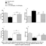 |
Figure 2: Effects of the combination and prednisone on oxidative stress parameters. The values are expressed as mean ± SEM, n = 5. *p< 0.05, **p< 0.01, ***p< 0.001 significant difference when compared to negative control group. |
Discussion
The aim of this study was to assess the subacute anti-inflammatory and anti-arthritis effects of the combination. The anti-inflammatory effect was observed by a significant reduction (p<0.05) in granuloma formation, exudate volume and leucocyte infiltration in the air pouch. The administration of plant extracts would affect the physiology of many organs such as the liver and kidneys. It is important to carry out a biochemical assessment in a sub-acute toxicity study 26. Biochemical assessments included blood levels of creatinine, urea, transaminases, cholesterols and triglycerides. The only changes observed were in ALT and cholesterol. The combination induced a significant decrease (p < 0.05) in blood levels of ALT and cholesterol. Similar results were obtained with extracts of Celosia trigyna and Eleophorbia drupifera leaves in rats 26,27. ALT and cholesterols are considered to be a more specific indicator of the hepatoprotective effect and lipid balance respectively, which would reflect the existence of hepatoprotective properties of the combination. The main endogenous markers of renal function are creatinine and urea. Their increases or decreases may reflect, respectively, renal failure or muscle atrophy 28. Our results showed no significant change with the administration of 2000 mg/kg of the combination in male and female rats. Other authors have found similar results 29. Serum AST, ALT, cholesterol and triglyceride levels and urea and creatinine levels were analyzed to identify possible hepatic and renal damage due to sub-acute treatment, a critical point for the development of new analgesic or anti-inflammatory drugs 30. The results suggest that the combination is not nephro- or hepatotoxic under our test conditions.
In inflammatory responses, an acute phase with vasodilatation and high permeability,
subacute phase with cell migration and a chronic phase, where fibrosis of the tissues is noted31. Subacute and chronic inflammation models such as air pouch were used to assess the transudative and proliferative components of inflammation 17. The results showed that granuloma formation was inhibited by the combinations. This inhibition followed this order: diclofenac > C1 > C3 > C2. Diclofenac showed significant inhibition (p < 0.05) compared to C2. The volume of fluid in the pouch was also significantly reduced by the combinations. C1 showed similar inhibition to diclofenac.
This reduction in granuloma formation and transudate would demonstrate the power of the extracts to inhibit the synthesis of macro-molecules and prevent the formation of granulomatous tissue 17,32. The getting leukocytes to the site of inflammation is an important parameter of the inflammatory response. Leukocyte migration results from an elaborate series of events, including cell adhesion and motility 3,33. The combinations inhibited the accumulation of leukocytes in the inflammatory fluid of the pouch. The results showed that the combinations especially combination C1 has effects on this carrageenan granuloma air pouch model and could therefore be a potential source of drugs against sub-chronic inflammation.
Polyarthritis is a chronic inflammatory illness that injures the joints and causes deformity, disability and premature death in most patients 34.
The induction of arthritis in rats, previously described 18 manifests itself as in humans. This model is frequently used to examine the effect of drugs against polyarthritis. After inoculation into rats, CFA induces polyarthritis in two phases. The acute phase, a maximum after 3 to 5 days, is attributed to the primary lesions, while the chronic phase occurs after 11 to 12 days, attributed to the secondary lesions and recognized by inflammation of the non-injected paw 21. In primary lesions, CFA-injected rats showed a significantly (P<0.05) reduced paw volume with the administration of combination C1. Arthritis scores were significantly (P < 0.01) elevated in the CFA control compared to combination C1 group. This suggests that the combination has anti-inflammatory properties and the ability to attenuate immune system responses. In the 19th century, Garrod discovered that hyperuricemia was the cause of gout and it is clear that serum uric acid may play a key role in inflammatory responses 35,36. The combinations significantly reduced (P < 0.001) the increase in serum uric acid levels.
Imbalance between oxidants and antioxidants induces hepatic damage causing lipid alterations that result in increased MDA levels. Antioxidant enzymes such as superoxide dismutase, catalase and glutathione peroxidase convert reactive oxygen species into harmless compounds that inhibit lipid peroxidation 37. The induction of arthritis even alters the antioxidant defense system, nearly depleting the vital lines of defense (GSH, SOD and CAT) against reactive oxygen species 38. Increased serum ALT and AST levels confirm liver damage. These were observed in the CFA control group. These results are in line with the results of previous studies 39–41. The combination C1 induced a significant (P < 0.01) increase in catalase activity, SOD and reduced glutathione accompanied by a significant (P < 0.01) decrease in MDA levels compared to CFA control. Similar results were also reported by other investigators 42–44. These results show that the combination C1 can reduce the deleterious effects of oxygen radical accumulation. Combination C1 therapy could be an alternative to combat oxidative stress. Previous studies have shown high contents of total polyphenols, flavonoids and tannins in the two hydroethanolic extracts of the two plants that form the combinations 14. Effect of the combinations on the two models of inflammation used in this study could be explained by the presence of total polyphenols, more precisely tannins, and the capacity of these two hydroethanolic extracts that form the combinations to inhibit pro-inflammatory enzymes. The antioxidant power of the combination is attributed to the flavonoids. 12,13.
Conclusion
Finally, the non-toxicity of the combination after daily administration of a dose of 2000 mg/kg for 28 days. The combination showed potential action against sub-acute inflammation and markedly reduced the symptoms of CFA-induced arthritis. The combination showed a significant decrease in inflammatory granuloma formation, exudate volume and leukocyte infiltration. The combination also significantly reduced the concentration of uric acid. Furthermore, the combination significantly prevented oxidative stress in arthritic rats. We can therefore conclude that the subacute anti-inflammatory and anti-arthritic activities of the combination are found to be very encouraging. Some compounds, including polyphenols and flavonoids were measured in the extracts forming the combination, which are known to have anti-inflammatory effects 13.Certainly, a clinical study is essential and highly recommended to validate the combination at 250 mg/kg of T. macroptera + 250 mg/kg of X. americana as an alternative treatment for inflammation.
Acknowledgment
The authors would like to thank the teams of the drug development laboratory, of Training, Research and Expertise Centre in Medicine Sciences, Joseph Ki-ZERBO University, Burkina Faso and the Point G University Hospital Center as well as the Department of Traditional Medicine of Bamako, Mali, for their support to this study.
Conflict of Interest
The authors declare that there are no conflicts of interest.
Funding Sources
There are no funding sources
References
- Hannoodee S, Nasuruddin DN. Acute Inflammatory Response. In: StatPearls. StatPearls Publishing; 2023. Accessed May 4, 2023. http://www.ncbi.nlm.nih.gov/books/NBK556083/
- Chen L, Deng H, Cui H, et al. Inflammatory responses and inflammation-associated diseases in organs. Oncotarget. 2017;9(6):7204-7218. doi:10.18632/oncotarget.23208
- Muller WA. Getting leukocytes to the site of inflammation. Veterinary pathology. 2013;50(1):7-22.
- Muller WA. Mechanisms of Transendothelial Migration of Leukocytes. Circulation Research. 2009;105(3):223-230. doi:10.1161/CIRCRESAHA.109.200717
- Furman D, Campisi J, Verdin E, et al. Chronic inflammation in the etiology of disease across the life span. Nat Med. 2019;25(12):1822-1832. doi:10.1038/s41591-019-0675-0
- Nunes C dos R, Barreto Arantes M, Menezes de Faria Pereira S, et al. Plants as Sources of Anti-Inflammatory Agents. Molecules. 2020;25(16):3726. doi:10.3390/molecules25163726
- Oguntibeju OO. Medicinal plants with anti-inflammatory activities from selected countries and regions of Africa. J Inflamm Res. 2018;11:307-317. doi:10.2147/JIR.S167789
- Agyare C, Obiri DD, Boakye YD, Osafo N. Anti-inflammatory and analgesic activities of African medicinal plants. Medicinal plant research in Africa. Published online 2013:725-752.
- Pham AT, Malterud KE, Paulsen BS, Diallo D, Wangensteen H. DPPH Radical Scavenging and Xanthine Oxidase Inhibitory Activity of Terminalia macroptera Leaves. Natural Product Communications. 2011;6(8):1934578X1100600819. doi:10.1177/1934578X1100600819
- Le NHT, Malterud KE, Diallo D, Paulsen BS, Nergård CS, Wangensteen H. Bioactive polyphenols in Ximenia americana and the traditional use among Malian healers. Journal of ethnopharmacology. 2012;139(3):858-862.
- Nadembega P, Boussim JI, Nikiema JB, Poli F, Antognoni F. Medicinal plants in Baskoure, Kourittenga Province, Burkina Faso: An ethnobotanical study. Journal of Ethnopharmacology. 2011;133(2):378-395. doi:10.1016/j.jep.2010.10.010
- BALLO M, TRAORE K, GUINDO AD, et al. In vitro inhibition of cyclooxygenases, anti-denaturation and antioxidant activities of Malian medicinal plants. Published online 2023.
- BALLO M, Youl E, Karim T, et al. study of antiradical activity, phospholipase A and 15-lipoxygenase inhibitory activity of eight malian medicinal plants used by traditional healers to treat inflammatory diseases. Indian Journal of Pharmacy and Pharmacology. 2022;9:174-179. doi:10.18231/j.ijpp.2022.031
- Ballo M, Youl EN, Haidara M, et al. Etude des constituants chimiques et activités antiradicalaires des extraits de huit plantes médicinales récoltées au mali. Pharmacopée et médecine traditionnelle africaine. 2021;20(2):72-79.
- Couto M, Cates C. Laboratory Guidelines for Animal Care. In: Pelegri FJ, ed. Vertebrate Embryogenesis: Embryological, Cellular, and Genetic Methods. Methods in Molecular Biology. Springer; 2019:407-430. doi:10.1007/978-1-4939-9009-2_25
- OECD. Test No. 407: Repeated Dose 28-Day Oral Toxicity Study in Rodents. Organisation for Economic Co-operation and Development; 2008. Accessed May 4, 2023. https://www.oecd-ilibrary.org/environment/test-no-407-repeated-dose-28-day-oral-toxicity-study-in-rodents_9789264070684-en
- Da FL, Keugni AB, Belemtougri GR, Fotio TLA, Dimo T. Acute and subacute anti-inflammatory activities of dichloromethane extract of Cassia alata (Linn.) leaves in wistar rats. African Journal of Traditional, Complementary and Alternative Medicines. 2018;15(1):174-182. doi:10.4314/ajtcam.v15i1
- Pearson CM, Wood FD. Studies of polyarthritis and other lesions induced in rats by injection of mycobacterial adjuvant. I. General clinical and pathologic characteristics and some modifying factors. Arthritis & Rheumatism. 1959;2(5):440-459. doi:10.1002/1529-0131(195910)2:5<440::AID-ART1780020510>3.0.CO;2-N
- Dutta S, Das S. A study of the anti-inflammatory effect of the leaves of Psidium guajava Linn. on experimental animal models. Pharmacognosy Res. 2010;2(5):313-317. doi:10.4103/0974-8490.72331
- Weng W, Wang F, He X, Zhou K, Wu X, Wu X. Protective effect of Corynoline on the CFA induced Rheumatoid arthritis via attenuation of oxidative and inflammatory mediators. Mol Cell Biochem. 2021;476(2):831-839. doi:10.1007/s11010-020-03948-8
- Zhang X, Dong Y, Dong H, Zhang W, Li F. Investigation of the effect of phlomisoside F on complete Freund’s adjuvant-induced arthritis. Experimental and therapeutic medicine. 2017;13(2):710-716.
- Sinha AK. Colorimetric assay of catalase. Analytical biochemistry. 1972;47(2):389-394.
- Beauchamp C, Fridovich I. Superoxide dismutase: improved assays and an assay applicable to acrylamide gels. Analytical biochemistry. 1971;44(1):276-287.
- Ohkawa H, Ohishi N, Yagi K. Assay for lipid peroxides in animal tissues by thiobarbituric acid reaction. Analytical biochemistry. 1979;95(2):351-358.
- Ellman GL. Tissue sulfhydryl groups. Archives of biochemistry and biophysics. 1959;82(1):70-77.
- Aimée S, Youssoufou O, Léonard D, Ilboudo S, Paténéma S, Bayala B. ACUTE AND SUBACUTE TOXICITIES OF THE AQUEOUS EXTRACT OF THE LEAVES OF CELOSIA TRIGYNA (L.). World Journal of Pharmaceutical Research. 2020;9:95-109. doi:10.20959/wjpr202015-19287
- Akpanabiatu MI, Igiri AO, Eyong EU, Eteng MU. Biochemical and Histological Effects of Eleophorbia drupifera Leaf Extract in Wistar Albino Rats. Pharmaceutical Biology. 2003;41(2):96-99. doi:10.1076/phbi.41.2.96.14242
- Gowda S, Desai PB, Kulkarni SS, Hull VV, Math AAK, Vernekar SN. Markers of renal function tests. N Am J Med Sci. 2010;2(4):170-173. Accessed May 5, 2023. https://www.ncbi.nlm.nih.gov/pmc/articles/PMC3354405/
- Kale OE, Awodele O, Akindele AJ. Subacute and subchronic oral toxicity assessments of Acridocarpus smeathmannii (DC.) Guill. & Perr. root in Wistar rats. Toxicol Rep. 2019;6:161-175. doi:10.1016/j.toxrep.2019.01.005
- Sauzem PD, Sant’Anna G da S, Machado P, et al. Effect of 5-trifluoromethyl-4,5-dihydro-1H-pyrazoles on chronic inflammatory pain model in rats. Eur J Pharmacol. 2009;616(1-3):91-100. doi:10.1016/j.ejphar.2009.06.008
- Rotelli AE, Guardia T, Juárez AO, de la Rocha NE, Pelzer LE. Comparative study of flavonoids in experimental models of inflammation. Pharmacol Res. 2003;48(6):601-606. doi:10.1016/s1043-6618(03)00225-1
- Chandrasekaran CV, Sundarajan K, Edwin JR, Gururaja GM, Mundkinajeddu D, Agarwal A. Immune-stimulatory and anti-inflammatory activities of Curcuma longa extract and its polysaccharide fraction. Pharmacognosy Research. 2013;5(2):71.
- Zuchtriegel G, Uhl B, Puhr-Westerheide D, et al. Platelets guide leukocytes to their sites of extravasation. PLoS biology. 2016;14(5):e1002459.
- Zhu L, Zhang Z, Xia N, et al. Anti-arthritic activity of ferulic acid in complete Freund’s adjuvant (CFA)-induced arthritis in rats: JAK2 inhibition. Inflammopharmacology. 2020;28:463-473.
- Choe JY, Kim SK. Association between serum uric acid and inflammation in rheumatoid arthritis: perspective on lowering serum uric acid of leflunomide. Clinica Chimica Acta. 2015;438:29-34.
- Jung SW, Kim SM, Kim YG, Lee SH, Moon JY. Uric acid and inflammation in kidney disease. American Journal of Physiology-Renal Physiology. Published online 2020.
- Jain A, Soni M, Deb L, et al. Antioxidant and hepatoprotective activity of ethanolic and aqueous extracts of Momordica dioica Roxb. leaves. Journal of ethnopharmacology. 2008;115(1):61-66.
- Tawfik MK. Combination of coenzyme Q10 with methotrexate suppresses Freund’s complete adjuvant-induced synovial inflammation with reduced hepatotoxicity in rats: Effect on oxidative stress and inflammation. International Immunopharmacology. 2015;24(1):80-87.
- Gardi C, Bauerova K, Stringa B, et al. Quercetin reduced inflammation and increased antioxidant defense in rat adjuvant arthritis. Archives of biochemistry and biophysics. 2015;583:150-157.
- Bonnet CS, Williams AS, Gilbert SJ, Harvey AK, Evans BA, Mason DJ. AMPA/kainate glutamate receptors contribute to inflammation, degeneration and pain related behaviour in inflammatory stages of arthritis. Annals of the rheumatic diseases. 2015;74(1):242-251.
- Akramas L, Leonavičienė L, Vasiliauskas A, et al. Anti-inflammatory and anti-oxidative effects of herbal preparation EM 1201 in adjuvant arthritic rats. Medicina. 2015;51(6):368-377.
- Ali EA, Barakat BM, Hassan R. Antioxidant and angiostatic effect of Spirulina platensis suspension in complete Freund’s adjuvant-induced arthritis in rats. PLoS One. 2015;10(4):e0121523.
- Fotio AL, Dimo T, Nguelefack TB, et al. Acute and chronic anti-inflammatory properties of the stem bark aqueous and methanol extracts of Sclerocarya birrea (Anacardiaceae). Inflammopharmacology. 2009;17:229-237.
- Saleem A, Saleem M, Akhtar MF, Shahzad M, Jahan S. Polystichum braunii extracts inhibit Complete Freund’s adjuvant-induced arthritis via upregulation of I-κB, IL-4, and IL-10, downregulation of COX-2, PGE2, IL-1β, IL-6, NF-κB, and TNF-α, and subsiding oxidative stress. Inflammopharmacology. 2020;28:1633-1648.

