
Manuscript accepted on :24-10-2022
Published online on: 13-01-2023
Plagiarism Check: Yes
Reviewed by: Dr. Ahmed Salah , Dr. Cherry Bansal
Second Review by: Dr. Narasimha Murthy , Dr. Sudhanshu Bharti
Final Approval by: Dr. Ian James Martin
A Sabah Afroze1 , R Tamilselvi2* and M Parisa Beham3
Department of ECE, Sethu Institute of Technology, Virudhunagar, Tamil nadu, India.
Corresponding Author E-mail: tamilselvi@sethu.ac.in
DOI : https://dx.doi.org/10.13005/bpj/2604
Abstract
Osteoarthritis (OA) is a disease categorized under arthritis and has impact on bone and cartilage dilapidation. Still now, OA is visualized only with X-ray and MRI scanning process. When a person exposed to X-rays will develop cancer later and it also causes tissue effects like cataracts, skin reddening and hair loss.To overcome this issue,a thermal camera(FLIR420bx) is used to capture the Knee OA from the subjects.The thermal imaging system is a non-contact,non-invasive passive imaging system that is the heat emmitted, or given off, by the human body.The medical evaluation of thermal images is validated and ground truth is received from skilled physicians which is time consuming and prone to error. Considering this fact, recently more image based diagnosis system is evolving for the early detection of OA. The success of this diagnosis system mostly relies on large number of OA/Non OA images to be trained. The novel dataset, titled as ‘Osthersit’, is a small seed thrown to afford OA thermal images of knee bones. It also consist OA affected images in thermograph modality. To have wider opening in many OA related ambiguity problems ,Osthersit dataset is made available to the researchers who work in the medical databases and can be retrieve from this web address: http://www.sethu.ac.in/osthersit/.
Keywords
FLIR toolbox; KL grading system; Thermal imaging; Osteoarthritis
Download this article as:| Copy the following to cite this article: Afroze A. S, Tamilselvi R, Beham M. P. OSTHERSIT- Dataset for Osteoarthritis Analysis using Thermal Images. Biomed Pharmacol J 2023;16(1). |
| Copy the following to cite this URL: Afroze A. S, Tamilselvi R, Beham M. P. OSTHERSIT- Dataset for Osteoarthritis Analysis using Thermal Images. Biomed Pharmacol J 2023;16(1). Available from: https://bit.ly/3HfZNEN |
Introduction
Osteoarthritis is the complicated disease which is showing it vigorous impact on millions of people in worldwide. It mostly occurs when the protective cartilage is cushions at the end of the bones while wears down over time. However, osteoarthritis [1] can damage any joint in the body, and the disorder most regularly affects joints in your hands, knees, hips and spine. OA is also called as mechanical condition which is characterized by the gradual wearing down of cartilage in bone joints. Then aging is the most common risk factor for osteoarthritis. Arthritis is not created by the normal wear and tear of bones. The primary osteoarthritis is caused by the breakdown of cartilage, which is a rubbery material that soothes the friction in your bone joints. The primary OA [2] can occur in any joint but generally affects your knees, hips, thumbs and fingers. Osteoarthritis is more common in older people. There is no curative solution for osteoarthritis, but the condition does not certainly get any worse over time. There are many numbers of treatments which helps to relieve the symptoms. The main treatments of OA are healthy lifestyle which includes maintaining a healthy weight and workout regularly. OA is analysed using radiography imaging which is considered as the gold standard method. But minor detachment is not visualized in the early stage. In figure 1, we can find the difference between normal knee image and osteoarthritis affected knee image.
 |
Figure 1: Normal knee vs Knee Osteoarthritis. |
For the grading of OA within the knee, the International Knee Documentation Committee (IKDC) system [3] is taken into account to have the foremost appropriate combination of interobserver precisions and correlation to knee surgical procedure findings. It was established by a gaggle of knee surgeons from Europe and America, who met within the year 1987 to develop a regular kindto live the results of knee ligament reconstructions. The Table 1, describes about the kellgren-Lawrence grading scale [4].
Table 1: Kellgren –Lawrence Grading Scale[4]
|
KLGrade |
Description |
| 0 | Normal without any detachments
|
| 1 | Minor variations in a doubtful manner
|
| 2 | Osteophytes, narrow space in the joint
|
| 3 | Multiple osteophytes
|
| 4 | Large osteophytes
|
A healthy immune system is found to be protective. It creates internal inflammation to get rid of infection and prevent the diseases. But in the inflammatory types of OA, the immune system doesn’t work accurately and it attacks the joints with uncontrolled inflammation and possibly causing the joint erosion. The aim of the treatment is to decrease the joint pain and stiffness. It improves the mobility and functions. Increase patients’ quality of life. There are many OA research scientists are learning about the cartilage, and lubricating surfaces in the joint. The cartilage regrowth process is undertaken using stem cells which are treated with molecules to support their growth. There are many people with osteoarthritis also experiencing depression, poor sleep, anxiety and fatigue and a general failure in quality of life. The figure 2, reports about the statistics of arthritis from the year 2000-2040.
 |
Figure 2: Statistics of OA. |
The OA is the second most common rheumatologic problem with a joint disease prevalence of [5] 22% to 39% in India. OA is more prevalent in women than men, but the generality increases enormously with age. Mostly in adults, it affects between 0.5%-1% of the population in India. Women’s are three to four times more regularly affected than men. In India, approximately 1% of the population suffers from rheumatoid arthritis. Therefore, as per calculations almost 10 lakh people suffer from this rheumatoid arthritis. Most ofthe people assume that arthritis is a disease which affects only in older people and the younger population will be safe from it. The obesity is the main important risk factor for osteoarthritis in women, also in men. Women’s who undergo menopause usually gain weight, and they have increased stress on the joints which helps to rise in osteoarthritis. Osteoarthritis affects 240 million people globally. In worldwide, the OA affects approximately 9.6% of men and 18.0% of women who is aged over 60 years[6] .In Infrared thermography, the thermal video imaging [7], is a process where thermal camera will captures the image and also creates an image of an object by using infrared radiation which is emitted from the object in a process, which is the examples of IR imaging[8] science. It is mainly used for detecting musculoskeletal, nervous and vascular system irregularities, inflammation screening, stroke and chronic diseases. In the full body imaging, it includes 22-28 views from head to toe. The thermography [9] is a type of infrared radiation technology which detects and records the temperature changes on the surface of skin. It can help screen for knee osteoarthritis. The thermal camera takes a picture of the areas of different temperature in the body. The thermal camera shows these patterns in a form of heatmap[14].
Benefits of Medical Thermography
It is simple to handle the camera.
It is 100% Safe while comparing to other devices.
Radiation free.
It is a painless process.
It is inexpensive while comparing to other diagnostic imaging procedures.
It is effective for women, men and children.
Thermography is an ideal tool for preventive medicine.
It is the perfect tool for early health screening.
Thermography is a test that uses an infrared camera to detect the heat patterns. Modern thermography discovers heat and blood flow in body tissues and may tip you off to areas of inflammation. The more inflammation occurs in the area where the heat produces rapidly.
Description of Thermal camera
The camera used in this proposed paper is FLIRT420BX, a thermal imaging camera, which comprise of 76800 Pixels (320 x 240). It provides a flexible, with the help of multiple hot spot measurements, high temperature range ,Wi-Fi connectivity , dew point measurements. This FLIR camera used to diagnose the osteoarthritis in patients. The term FLIR, which stands for “Forward Looking Infrared”, it mention to the technology that is used to create an infrared image of a scene without having to “scan” the scene with a moving sensor. The medical imaging with infrared cameras can ableto detect the presence of potential diseases, poor circulation, inflammations in the body, osteoarthritis and ability to see small differences in temperature within the human body.
Literature Survey
This section will discuss about the state of the art methods for the knee osteoarthritis classification. Haq and E. Murphy [10] proposed that OA is a prolonged disorder in the bone which results in the thinning of bones and reduction in space. This paper shows necessity of the bone research in related to various views of bone images and their severity indications. As the medical research needs more images for analysing and validation, this paper paves the solution for it. Ali Jalalv and and Mehrdad Anbarian [11] proposed aviconmotion analysis based on kinematic parameters with a 4 T-Series cameras. Only fifteen normal cases are visualized and thirty cases with pathological conditions are participated in this study. The results are validated according to Kellgren and Lawrence radiologic scale. OA focuses manly on any one joint as it has impact on other joints. Pawelwidera [12] recommenddedmultiplealgorithmsandlearningmethodconfigurationswhichhasmulti-classifierapproaches, feature choices and cost-sensitive learning to spot the simplest machine learning techniques. They inspected the behaviour of simplest machine learning models, with regard to the prediction errors [15], to substantiate their clinical applications. This may result in additional economical clinical trials. The ofiloskarrasavid [13] is projected based on the databases readily available (with only limited images) which focus only on treatment methods. Various treatment methods and other follow up only discussed. There is o technical description. Based on these gap in the database survey, a new database is proposed.
OSTHERSIT Database
In medical field and in challenging scenario addressing OA, there is a room for the research solution to OA related problems. This interested us to create a novel unique database called as OSTHERSIT database. We created this OSTHERSIT database, which is standard method for knee osteoarthritis assessments .OSTHERSIT is formed from thermal knee images which is composed from a radiologists from Trichy and Chennai. The FLIR thermal camera is used for capturing the knee regions in the human body. OSTHERSIT is raised from thermal scanned images of osteoarthritis. These images are collected from the standard scan centres in Tamil nadu after ethical clearance. This dataset consists of 100 OA thermal images which are collected from 30 cases. Among the 30 subjects, OSTHERSIT contains 9 male subjects and 21 female subject cases. Every subject consists of knee thermal image whereas few subjects consist of right side and left side of knee thermal images. In figure 3, it shows the Sample thermal images of Knee from Osteothermalsit dataset.
 |
Figure 3: Sample thermal images of Knee from Osthersit dataset |
All the images are also labeled uniquely to identify easily the gender and modality of the subjects. An example of thermal images of 3 subjects from the Osthersit dataset is shown in Fig.1 respectively. Fig.4 shows the sample of knee bone images of both left and right leg captures in X-ray modality. Osthersit dataset is developed by upcoming steps: 1) Design concepts 2) Labelling the thermal images 3) Annotations of the thermal images. Each steps are explained briefly.
Design concepts
Osthersit
Thermography is typically used for examining joints that are deep in the body and difficult to assess with normal x-ray in the diagnosis of arthritis. In this Osthersit dataset, thermal images are used as source and the images are derived from Indian community. Totally 75 thermal Knee images have been collected. Patient information are removed from the images and saved as image format. The size of all the images is 543KB with Dimensions of 320 x 240. The dataset provides the researchers a wide opening about the existences of possible views of the images.
Osthersit_Knee Pair
OA often affects small and large joints on both sides of the body (symmetrical), such as both knees, both hands and both wrists.
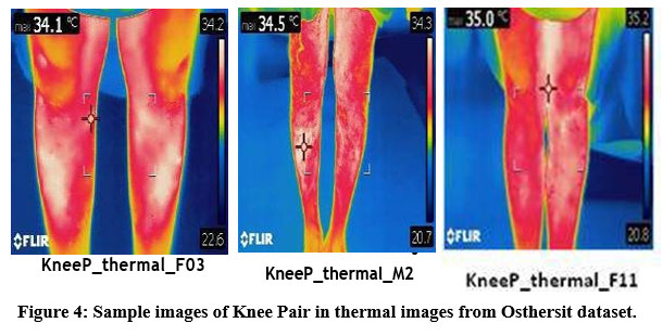 |
Figure 4: Sample images of Knee Pair in thermal images from Osthersit dataset. |
The size of all the images is 625 KB with Dimensions of 320 x 240. In the Osthersit_Knee Pair images, the label for an X-ray scan image of subject is given as: KneeP_thermal_F16.jpg; here, Knee refers to Knee pair, thermal refers to modality of image, F refers female, the gender of the subject and 12 is the subject ID.
Labeling the Thermal Image
Label leling is done as a user understanding simple way. This creates a simple use and ease understanding for the researchers. Researchers can able to find the subject ID, thermal scan ,region and gender of the subject. For example, the labelling of Osthersits of a subject is given as: Knee_thermal_F23.jpg and Knee_thermal_M10.jpg. Here, in the first case Knee refers to bone part, thermal refers to the modality of image, and the later F23 refer to female subject ID.
Annotations of the thermal images
All images are annotated by the radiologists where the images have been collected. Annotation is a necessity parameter is visualizing Osthersit provides a complete annotations along a careful analysis of Osthersits image. We manually annotated the following attributes for every knee thermal image.
Exclusive ID of the subject
Masculinity
Bone part
Age
Modality of the image
Size of the image with Dimension
In figure 5 , it shows the annotated image from Osthersit dataset.
 |
Figure 5: Sample of annotation of Thermal OA images. |
Conclusion
In this paper, a novel dataset for health dataset OSTHERSIT, a collection of osteoarthritis examined images for medical study. It helps to provide a standard dataset for the orthopaedic medical study and other aligned research in detecting the arthritis at an early stage. This dataset can also be explored to identify the bone mineral density and calcium contents to classify the osteoporosis risk conditions. Salient features of this OSTHERSIT dataset are: a) 50 thermal Knee images as Osthersit_knee_non_OA b) 25 thermal knee scan images as Osthersit_knee_OA c) 25 thermal pair images named as Osthersit_Knee Pair. By establishing this dataset accessible to the Osteoarthritis research group ,weecxpect this Osthersit will serve as a public domain research resource in healthcare. This Osthersit dataset along with radiograph images with proper annotations will also be accessible for research publically. This dataset can be look on through the following website: http://www.sethu.ac.in/osthersit/.
Ethical approval
The database “OSTHERSIT Database” used in this paper is a standard database derived from the Pixel scans ,Trichy and ethically approved by the committee. The database followed the standard operating rules and otherformalities and can be freely downloaded and used for Medical Research.
Conflict of Interest
There is no conflict of interest.
Funding Sources
There is no funding sources.
References
- Ashok Kumar ,SapnaGoel,Ritu Gupta,”Osteoarthritis Research in India: A Scientometric Assessment of Publications Output during 2007-16”,Vol.7, issue 3,2017.
CrossRef - Juan c mora , Rene Przkora , “Knee osteoarthritis: pathophysiology and current treatment modalities” ,Vol.11, 2018.
CrossRef - Ali Anshami , Knee osteoarthritis related pain: a narrative review of diagnosis and treatment , Vol.8,2014.
CrossRef - Mark D. Kohn BA, Adam A. Sassoon MD, Navin D. Fernando MD , “Classifications in Brief Kellgren-Lawrence Classification of Osteoarthritis” , DOI 10.1007/s11999-016-4732-4 ,2016.
CrossRef - G. Quicke , P.G. Conaghan k, N. Corp , G. Peat , “Osteoarthritis year in review 2021: epidemiology & therapy”, doi.org/10.1016/j.joca.2021.10.003,2021.
CrossRef - Tomasz Sosnowski, Grzegorz Bieszczad and Henryk Madura , “Image Processing in Thermal Cameras”, doi.org/10.1007/978-3-319-64674-9_3,2018.
CrossRef - Nikola Borojevi ,DarkoKolari ,Simeon Grazio , “Thermography hand temperature distribution in rheumatoid arthritis and osteoarthritis” , Vol 113, No 4, 2011.
- Haq ,E. Murphy , “Osteoarthritis”,vol.79,issue993.
- Ali Jalalvand, Mehrdad Anbarian, Shahram Ahanjan, Behrouz Hajiloo, “The Effect of Knee Osteoarthritis on Excursions of Lower Limb Joints During Gait” , Vol.6(4) ,pp 233-241,2017.
CrossRef - Pawelwidera ,Paco MJ Welsing , “Multi-classifier prediction of knee osteoarthritis progression from incomplete imbalanced longitudinal data”, vol.10, 2020 .
CrossRef - heofilosKarasavvidis , Michael T. Hirschmann, “Home-based management of knee osteoarthritis during COVID-19 pandemic: literature review and evidence-based recommendations” , doi.org/10.1186/s40634-020-00271-5,2020.
CrossRef - Mihaela Antonina Calin , Gilda Mologhianu, “A review of the effectiveness of thermal infrared imaging
in the diagnosis and monitoring of knee diseases”, Infrared Physics and Technology,vol.69,pg 19-25,2015.
CrossRef - Hyun Jung Yang, Haeln Park, “Infrared thermal imaging in patients with medical collateral ligament injury of the knee – A Retrospective study”, Journal of Pharmacopuncture,vol.17(4),pg-50-54,2014.
CrossRef - Viney lochab,Jaspreet singh, “Thermal imaging in total knee replacement and its relation with inflammation makers”,Mathematical Biosciences and Engineering,vol.18,issue 6,2021.
CrossRef - B. Suma, U. Snekhalatha, “Automated Thermal Image Segmentation of Knee Rheumatoid Arthritis”, International Conference on Communication and Signal Processing, April 6-8, 2016

