
Manuscript accepted on :23-06-2022
Published online on: 06-09-2022
Plagiarism Check: Yes
Reviewed by: Dr. Liudmila Spirina
Second Review by: Dr. Radha Pujari
Final Approval by: Dr. Ian James Martin
S. Bhuvaneswari* , M. R. Suchitra, M. Beevi Farhana Noor , R. Madhumitha, and P. Gajalakshmi
Department of Chemistry and Biosciences, SASTRA Deemed University, Srinivasa Ramanujan center, Kumbakonam, Tamil Nadu, India.
Corresponding Author E-mail:bhunz2010@gmail.com
DOI : https://dx.doi.org/10.13005/bpj/2486
Abstract
Nowadays breast cancer in rheumatoid arthritis patients is the most controversial one. Rheumatoid arthritis is an inflammatory autoimmune disorder. RA may induce the order disease like cancer especially breast cancer in most women. Breast cancer starts when the breast cell begins to grow out of control. Inflammatory, anticancer drugs may show severe side effects. To overcome these herbal plants are used to make drastic changes in medical treatments. The main objective of this paper is assessment of Anti-inflammatory, anti-arthritic, anti-microbial and anti-cancer activity against the Michigan cancer foundation (MCF-7) human breast cancer cell line using the extracts Andrographis echioides. Anti-inflammatory and anti-arthritic activity show maximum inhibition of albumin denaturation, anti-proteinase, hypo-tonicity induced hemolysis, anti-lipoxygenase activity with standard drugs such as aspirin, diclofenac sodium and indomethacin at the concentration of 100µg/ml under in vitro condition. Thus the potential inhibition of protein (egg albumin) denaturation (85.80%) and for the human red blood cell (HRBC) membrane stabilization model 85.50% was observed at the concentration of 100µg/ml. The antioxidant and cytotoxicity activity of Andrographis echioides showed better inhibition in a dose-dependent manner against MCF-7 human breast cancer cell lines. The antimicrobial activity of Andrographis echioides showed maximum inhibition of E.coli (11mm), Enterococcus aerogenes and Staphylococcus aureus (10mm), and Candida albicans (11mm) at 100µg/ml concentration. The innovation of the present study revealed that the stem of Andrographis echioides have potential source for natural anti-arthritis, cytotoxicity, anti-microbial, antioxidant activity, anti-inflammatory and anticancer activities.
Keywords
Aspirin; Breast cancer Michigan Cancer Foundation (MCF-7); Hemolysis; Inflammation; Rheumatoid arthritis
Download this article as:| Copy the following to cite this article: Bhuvaneswari S, kumar P. S, Noor M. B. F, Madhumitha R. Screening of Stem Extracts of Andrographis echioides for Anti-Inflammatory, Anti-Arthritic, Anti-Microbial, and Anti-Cancer Activity against MCF-7 Human Breast Cancer Cell Lines. Biomed Pharmacol J 2022;15(3). |
| Copy the following to cite this URL: Bhuvaneswari S, kumar P. S, Noor M. B. F, Madhumitha R. Screening of Stem Extracts of Andrographis echioides for Anti-Inflammatory, Anti-Arthritic, Anti-Microbial, and Anti-Cancer Activity against MCF-7 Human Breast Cancer Cell Lines. Biomed Pharmacol J 2022;15(3). Available from: https://bit.ly/3KP4Agt |
Introduction
Rheumatoid arthritis is a persistent, inflammatory autoimmune illness which affects the joints. It also affects the tissue which surrounds the joints1. Many mediators like TNF-α, IL-2 and enzymes are involved in rheumatoid arthritis2, 3, 4. Currently, a combination of many diseases may develop during the RA disease. And one of the fair problems is cancer, especially breast cancer in most females5. Irregular host immune response is examined as a source for the development of a tumor. Due to the immune suppression is the increased chance of cancer6, 7. Current studies exhibited that the promising threat of breast cancer in RA, however, the occurrence of breast cancer in the population stays doubtful. Cancer is a proliferating disease combining physical, environmental, metabolic, chemical, and genetic factors8. In the US country, women Breast cancer is the major cause of death and the cell targeting drug development without affecting normal cells is a challenging task in the field of drug discovery9. Cytotoxic agents are expensive and induce severe side effects 10, 11. Several medicinal plant species and their phytochemicals inhibit the progression and development of cancer.
Andrographis echioides come under the family of Acanthaceae (GopuramThangi). It is an ayurvedic herb plant used in the treatment of many ailments such as anti-inflammatory, anti-arthritic, antimicrobial, anti-ulcer, antioxidant activity, hair problems, etc., Their phytochemical constituents like flavonoids, tannins, phenol, glycosides, terpenoids, saponins, steroids, etc., Andrographis echioides is a medicinally valuable species widely distributed in the tropical region in South Asian countries. This paper reveals that anti-inflammatory, anti-arthritic, anti-microbial and anticancer activity against breast cancer cell line (MCF-7) from Andrographis echioides stem extract.
Materials and methods
Plant collection
Andrographis echioides stem was procured and authenticated by botanist, Research associate, CARISM and SRC SASTRA Deemed to be University, Tirumalaisamuthiram, Thanjavur (Voucher no. SRC SASTRA 0005)
Preparation of ethanolic extract
The stem of Andrographis echioides was rinsed with running water, slice into small pieces and shade dried at 35-40̊C for a week, after that make it into a uniform powder and subjected for soxhlet extraction process by using ethanol as solvent12.
Methodology for the anti-inflammatory activity of ethanolic extract of Andrographis echioides
Inhibition of albumin denaturation
5ml of reaction mixture contains 0.2ml of egg albumin, different concentration of extract (20-100µg/ml) and 2.8ml of phosphate buffered saline. Double distilled water served as a control.
Then incubated at 37̊C for 15mins and heated at 70̊C for 5mins. Cooled it and the absorbance was observed at 660nm. Diclofenac sodium was taken as standard13, 14. The protein denaturation inhibition in percentage was calculated as below
Percentage of inhibition = (Control – Test/Control) × 100
Anti-proteinase action
2ml of reaction mixture contains 1ml trypsin, 1ml TrisHcl buffer (pH 7.4) and 1ml of different concentration of extract (20-100µg/ml). Then incubated at 37̊C for 5mins then add 1ml of casein and again incubated for 20mins. 1ml of perchloric acid was added to stop the reaction. The dark suspension was centrifuged and the supernatant absorbance was measured at 210nm15. The proteinase inhibitory activity in percentage was calculated.
Percentage of inhibition = (Control – Test / Control) × 100
Hypotonicity induced hemolysis
Different concentrations of extracts (20-100µg/ml) were taken. Then, 1ml of phosphate buffer, 2ml of hyposaline and 0.5ml of HRBC suspension of mixed solution served as control. Standard as a diclofenac sodium. Absorbance read at 560nm16. The hemolysis percentage was evaluated by taking hemolysis produced in the control as 100%
Percentage of protection = (Control – Test / Control) × 100
Anti-lipoxygenase activity
Different concentrations of extracts (20-100µg/ml) were dissolved in 0.25ml of 2M borate buffer pH 9 and then 0.25ml of lipoxygenase enzyme solution was added and incubated at 25̊C for 5mins. Then 1ml of linoleic acid solution was added and mixed well then absorbance was read at 234nm. Standard used as Indomethacin17. The percent inhibition was calculated from the following equation,
Percentage of inhibition = (Control – Test / Control) × 100
For IC50 value, dose response curve was plotted, in which 50% of maximum scavenging capacity was calculated. All tests were performed in triplicate and the average values are taken.
Evaluation of anti-arthritic activity
Inhibition of protein denaturation model
A 5ml reaction mixture containing 0.2ml of egg albumin, 2.8ml of phosphate buffer and 2ml of distilled water and various concentrations of extract were taken. Diclofenac sodium as the standard drug and the procedure was performed as the same18, 19. The samples were incubated for 15mins at 37̊C and heated at 70̊C for 5 mins. After cooling, the absorbance is measured using a UV-Visible spectrophotometer at 660nm. The protein denaturation inhibition in percentage was calculated using the following formula,
Percentage of inhibition = (Control – Test / Control) × 100
Human red blood cell (HRBC) membrane stabilization model
Assay of membrane stabilizing activity
Different concentrations of extracts, reference sample and control were taken, then mixed with 1ml of phosphate buffer, 2ml of hyposaline and 0.5ml of HRBC suspension. All the mixtures were incubated for 30mins at 37̊C. The liquid was decanted and the hemoglobin content was estimated by a spectrophotometer at 560nm. The hemolysis percentage was evaluated by taking the hemolysis produced in control as 100%20, 21. The percentage of HRBC membrane stabilization or protection was observed as follows,
Percentage of inhibition = (Control – Test / Control) × 100
Antioxidant activity of Andrographis echioides by DPPH assay
Different concentrations of ethanolic extracts of Andrographis echioides were taken. 500µl of DPPH enzymes was added to all the test tubes. The mixture was allowed to stand for 5mins and observed at 517nm. The absorbance zero was set with ethanol. Ethanol and DPPH solution mixture was taken as blank. This experiment was performed in a triplicate manner22. DPPH activity of the tested samples, reveals as a percentage of inhibition which were calculated as follows,
Percentage of inhibition DPPH activity = (Control – Test / Control) × 100
Test and blank sample marked as A and B. The graph was plotted as percent inhibition vs concentration of the sample. 50% of inhibition activity was measured for each solution.
In vitro anticancer studies of ethanolic extract of stem of Andrographis echioides
MTT assay
In vitro cytotoxicity was carried out using Andrographis echioides. The sample used for this activity is MCF-7 cells by 3-(4,5-dimethylthiazol-2-yl)-2,5-diphenyltetrazolium bromide (MTT) assay. By trypsinization, a MCF-7 cell line of 15ml was harvested. After, the harvested cells were plated in a 96-well tissue of DMEM (10% FBS and 1% antibiotic solution) culture plate with a density of 1×10⁵cells/well (200µl) and incubated at 37̊C for 24-48hrs. Then, the wells rinsed with PBS solution and handled with different concentrations of the herbal extract in non-serum DMEM medium. It was performed in a triplicate manner. By the continuation of the incubation period, MTT (20µl of 5mg/ml) was taken in each plate and the cells were incubated for next 2-4hrs for the formation of purple precipitates were viewed under an inverted microscope. Finally, the cells were washed with a PBS solution (200µl). The formazan crystals were dissolved, DMSO (100µl) was taken and the plate was shaken for 5mins. Each well absorbance was observed at 570nm using a microplate reader (Thermo Fisher Scientific, USA), and the percentage of viability of cell and IC50 value was calculated by Graph Pad Prism 6.0 software (USA) 23.
Collection of test organisms
The antimicrobial activity of Andrographis echioides, was carried out using three strains [Escherichia coli (MTCC 25922), Enterococcus aerogenes, Staphylococcus aureus (MTCC 27853)] were prepared as test organisms. The fungal test sample Candida albicans (MTCC 282) in clinical trials was taken. The microorganisms were collected from Microbial Type Culture and Collection (MTCC) at Chandigarh, India.
Antibacterial activity of ethanolic extract of Andrographis echioides (Disc diffusion method)
Antibacterial activity was performed using ethanolic extract of Andrographis echioides by disc diffusion method. Petri dishes (60mm) were taken and Muller Hinton Agar was inoculated with organisms. 10µl of ethanolic extracts of 20-100µg/ml was poured in a sterile disc. Then, the discs were placed on the agar plates and kept at room temperature for 30mins due to compound diffusion. Respective solvent used as control. Finally, sterile petri dishes were incubated for 24hrs at 37̊C and the inhibitory zone was measured in millimeters and the experiment was repeated twice24.
Determination of the antifungal activity of ethanolic extract of Andrographis echioides by disc diffusion method
The ethanolic extracts of Andrographis echioides were determined using the disc diffusion method. The petri dishes (60mm) were poured with Sabouraud’s dextrose agar (SDA) and inoculated with test organisms. 10µl of ethanolic extracts of 20-100µg/ml was poured in a sterile disc. Then, the discs were places on the agar plates and kept at room temperature for 30mins due to compound diffusion. Respective solvent used as control. Finally, sterile petri dishes were incubated for 72hrs. The inhibitory zone was measured in millimeters and the experiment was repeated twice25.
Result and discussion
The phytochemical analysis of ethanolic extract of Andrographis echioides indicated the occurrence of anthocyanins, saponins, flavonoids, tannin, steroids, cardiac glycosides, leuco anthocyanin, coumarin, glycosides, terpenoids, emodin, phenol, alkaloids, proteins and absence of anthraquinone, carbohydrates and phlorotannins. The tannin has highest quantity followed by alkaloids (0.163mg/g), saponins (0.007mg/g), flavonoids (0.015mg/g), phenol (0.046mg/g), tannin (0.055mg/g) and terpenoids (0.004mg/g) respectively.
In-vitro anti-inflammatory activity
Inhibition of albumin denaturation
Normally, Denaturation of protein describes the losses of secondary and tertiary structure of protein and also for biological protein because of external intensity or compounds as strong base and acid, a solvent etc., In a previous study, some of the plant extracts that have the ability to block the denaturation mechanism in proteins were investigated under anti-inflammatory activity27. From this study, Andrographis is effective and has the ability for the inhibition of denaturation of albumin as shown in figure 1. 52% of potential inhibition of Andrographis was determined at 100µg/ml. The standard drug of Diclofenac sodium inhibits up to 82% with 100µg/ml by comparison with control as predicted in Table 1.
Table 1: Albumin denaturation inhibition of Andrographis echioides extracts with standard drug Diclofenac sodium.
| Concentration(µg/ml) | % of albumin denaturation | |
| Andrographis echioides | Diclofenac
Sodium |
|
| 20 (µg/ml) | 39.13±0.34 | 53.16±0.22 |
| 40 (µg/ml) | 40.18±0.45 | 61.25±1.03 |
| 60 (µg/ml) | 48.24±0.67 | 73.10±1.56 |
| 80 (µg/ml) | 50.16±1.09 | 77.69±1.16 |
| 100 (µg/ml) | 52.21±0.65 | 82.00±0.87 |
 |
Figure 1: Inhibition of albumin denaturation. |
Anti-proteinase activity
Basically, neutrophils are the origin of serine protease and are present in lysosomes. In a previous study, the authors reported that the proteinase enzyme of leucocytes has the ability to damage the tissues of inflammatory actions and an important level of protection was developed for enzyme inhibitors. Different concentrations of proteinase inhibition activity were predicted in table 2. Andrographis showed potential inhibition of 52% at 100µg/ml and diclofenac sodium inhibits maximum of 69% at 100µg/ml as shown in figure 2.
Table 2: Anti proteinase activity of Andrographis echioides extracts vs Diclofenac sodium.
| Concentration(µg/ml) | % of Anti proteinase activity | |
| Andrographis echioides | Diclofenac sodium | |
| 20 (µg/ml) | 30.17±0.26 | 42.11±1.22 |
| 40 (µg/ml) | 37.45±0.28 | 51.09±0.67 |
| 60 (µg/ml) | 40.34±0.37 | 58.21±0.52 |
| 80 (µg/ml) | 43.90±1.09 | 66.18±1.09 |
| 100 (µg/ml) | 52.23±0.39 | 69.00±0.65 |
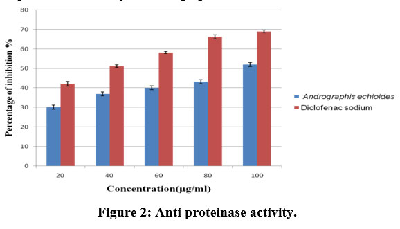 |
Figure 2: Anti proteinase activity. |
Hypotonicity-induced haemolysis
From this study, the result observed as different concentrations (20 to 100µg/ml) of Andrographis prevents the RBC membrane lysis by the solution of hypotonic as shown in table 3. At the concentration of 100µg/ml 51% of potential inhibition of Andrographis and 77% of inhibition of Diclofenac sodium was observed as shown in figure 3.
Table 3: Hypotonicity activity of Andrographis echioides extracts with standard drug diclofenac sodium.
| Concentration(µg/ml) | % of Hypotonicity induced haemolysis | |
| Andrographis echioides | Diclofenac sodium | |
| 20 (µg/ml) | 26.13±0.15 | 60.12±0.12 |
| 40 (µg/ml) | 33.09±0.18 | 62.91±1.07 |
| 60 (µg/ml) | 42.17±1.09 | 64.22±1.15 |
| 80 (µg/ml) | 46.21±0.20 | 75.43±0.16 |
| 100 (µg/ml) | 51.00±0.15 | 77.08±0.12 |
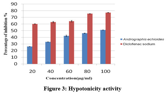 |
Figure 3: Hypotonicity activity. |
Anti-lipoxygenase activity
In screening methods, a medicinal plant contributes a great mechanism in the inhibition of lipoxygenase enzyme activity. In this experiment, Andrographis showed inhibition in a dose-dependent manner as predicted in table 4. As per the result, a well inhibition was observed at 100µg/ml of 52% of Andrographis and 72% of Diclofenac as observed in figure 4. It showed better anti-lipoxygenase activity.
Table 4: Anti-lipoxygenase activity of ethanolic extract of Andrographis echioides with standard drug diclofenac sodium
| Concentration(µg/ml) | % Anti lipoxygenase activity | |
| Andrographis echioides | Diclofenac sodium | |
| 20 (µg/ml) | 36.16±0.71 | 53.00±0.21 |
| 40 (µg/ml) | 38.19±0.90 | 59.12±0.82 |
| 60 (µg/ml) | 44.00±0.48 | 64.17±1.90 |
| 80 (µg/ml) | 48.21±1.09 | 68.23±1.65 |
| 100 (µg/ml) | 52.16±0.32 | 72.15±0.32 |
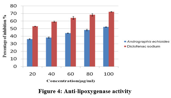 |
Figure 4: Anti-lipoxygenase activity. |
Evaluation of anti-arthritic activity
Protein denaturation inhibition
In this experiment both Andrographis (65% to 85%) and Diclofenac drug (84% to 97%) showed potential inhibition in a dose-dependent manner (20, 40, 60, 80 and 100µg/ml) as shown in table 5. The denaturation of protein inhibition was well observed as shown in figure 5. A previous study suggested that the inhibition of protein denaturation was analyzed for the study of Ashwagandha, Mikaniascandens and Coffee 26,27,28.
Table 5: Anti-arthritic activity of Andrographis echioides with standard drug diclofenac sodium by protein denaturation method.
| Concentrations (µg/ml) | Protein denaturation (%) | |
| Andrographis echioides | Diclofenac sodium | |
| 20 (µg/ml) | 65.79±0.36 | 84.65±0.55 |
| 40 (µg/ml) | 69.31±1.66 | 90.35±1.79 |
| 60 (µg/ml) | 73.13±1.78 | 91.49±1.64 |
| 80 (µg/ml) | 77.35±1.54 | 95.53±1.59 |
| 100 (µg/ml) | 85.80±0.38 | 97.43±0.28 |
Each values was obtained by calculating the average of three experiments and data are presented as mean ± SEM
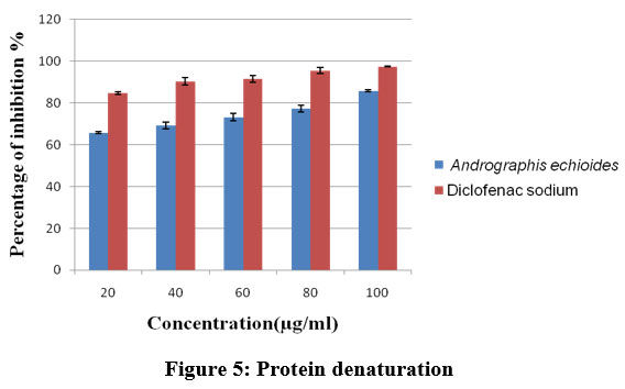 |
Figure 5: Protein denaturation. |
Membrane stabilization mechanism of Human red blood cell (HRBC)
The standard drug Diclofenac sodium of various concentrations (20, 40, 60, 80 and 100µg/ml) exhibited stabilization towards the HRBC membrane. The ethanolic extract of stem of Andrographis echioides at different concentrations (20,40,60,80 and 100µg/ml) acts as good stabilization of HRBC as shown in table 6. Andrographis and diclofenac have more effectiveness and are illustrated in figure 6.
In a previous study, the HRBC method was analyzed under in vitro condition. Thus the medicinal plant extracts has the ability to stabilize the membrane for the slowness of inflammatory reactions by protecting the liberation of constituents of lysosomes of activated neutrophils as bactericidal and protease enzymes, which leads to inflammation and damage ofextracellular liberations20. The membrane stabilization mechanism of HRBC is majorly involved in the study of Skimmiaanquetilia, Gendarussa vulgaris, Thunnus alalunga20, 29, and 30.
Table 6: Anti-arthritic activity of Andrographis echioides with standard drug diclofenacsodium using HRBC membrane stabilization method.
| Concentrations(µg/ml) | HRBC membrane stabilization assay (%) | |
| Andrographis echioides | Diclofenac sodium | |
| 20 (µg/ml) | 66.71±0.89 | 69.66±0.38 |
| 40 (µg/ml) | 70.00±1.69 | 72.42±1.25 |
| 60 (µg/ml) | 74.30±1.81 | 77.19±1.23 |
| 80 (µg/ml) | 82.59±1.83 | 85.95±1.47 |
| 100 (µg/ml) | 85.50±0.49 | 86.12±0.29 |
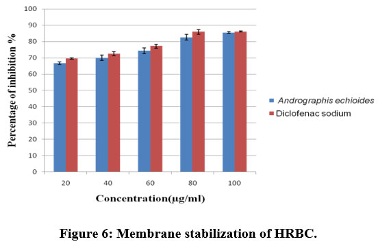 |
Figure 6: Membrane stabilization of HRBC. |
In vitro Antioxidant activity of Andrographis echioides by DPPH assay method
The ethanolic extracts of stem of Andrographis echioides contain the best antioxidant activity at high concentrations when compared with ascorbic acid. The plant showed 82.25% activity at 100µl/ml at the same time ascorbic acid gave 85.03 at the same concentration (Table 7 and figure 7).
The ethanolic extract showed the highest DPPH scavenging activity showed the best antioxidant activity when compared to other extract31, 32. Antioxidant, cytoprotective and anti-inflammatory properties have been proved by Andrographis echioides with flavonoids, tannins, terpenoids, gums, cardiac glycosides and phyosterols.
Table 7: Antioxidant activity of ethanolic extract of stem of Andrographis echioides by DPPH activity.
| Concentration(µg/ml) | Andrographis echioides | Ascorbic acid |
| 20µg/ml | 66.93±0.41 | 71.07±1.14 |
| 40µg/ml | 67.74±0.60 | 73.43±1.05 |
| 60µg/ml | 70.16±0.37 | 76.15±1.34 |
| 80µg/ml | 80.64±0.24 | 82.32±1.91 |
| 100µg/ml | 82.25±0.22 | 85.03±1.45 |
 |
Figure 7: Antioxidant activity. |
In vitro cytotoxicity effects of samples
The anti-cancer activity of Andrographis echioides was assessed by MTT assay. MCF-7 cell line was treated with different concentrations 20,40,60,80,100µg/ml of plant samples and incubation for 24hrs showed the percentage of cell viability (graph) of the cells in a dose dependent manner (figure 8) with IC50 value of 58.46µg/ml. This result is an agreement with a study carried out in previous study33. WHO reported the effectiveness of anticancer D. inoxia on human breast cancer cell line MCF-7. Thus the cytotoxicity study reveals the morphological change, characteristic, molecular and biochemical events present in an apoptotic cell. Another hand the cytotoxicity studies revealed that the Andrographis echioides exhibited dose-dependent toxicity on human breast cancer (MCF-7) cell lines. IC50 value for MCF-7 cells showed a 50% noticed cell death is observed34.
The anticancer activity was performed by using this plant genus (Andrographis paniculata) shown effective inhibition against the MCF-7 cell proliferation by using ethanolic extract35. Two different plants like Andrographis paniculata and Silybummarianum showed anticancer activity against five human cancer cell lines like human cervix (SiHa), human breast adenocarcinoma (MCF-7), ovary cancer cell line (ovcar-5), colon (HT-29) and liver (HepG2) by using a combination of this two plant36.
Table 8: Cell viability (%) test.
| Tested sample concentration (µg/ml) | Cell viability (%)
(in triplicates) |
Mean value (%) | ||
| Control | 100 | 100 | 100 | 100 |
| 100 μg/ml | 34.91 | 30.75 | 25.07 | 28.25 |
| 90 μg/ml | 45.20 | 52.20 | 55.26 | 48.89 |
| 80 μg/ml | 54.61 | 59.36 | 51.20 | 53.56 |
| 70 μg/ml | 62.48 | 63.58 | 62.05 | 60.71 |
| 60 μg/ml | 68.17 | 66.86 | 55.70 | 61.58 |
| 50 μg/ml | 71.02 | 75.83 | 76.14 | 63.67 |
| 40 μg/ml | 77.36 | 69.05 | 83.49 | 65.64 |
| 30 μg/ml | 87.43 | 88.96 | 94.87 | 94.43 |
| 20 μg/ml | 89.40 | 95.09 | 89.84 | 96.45 |
| 10 μg/ml | 112.59 | 100.87 | 91.90 | 99.45 |
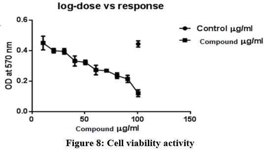 |
Figure 8: Cell viability activity. |
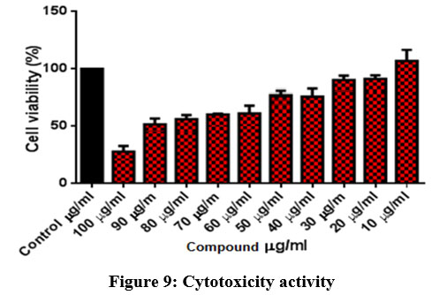 |
Figure 9: Cytotoxicity activity. |
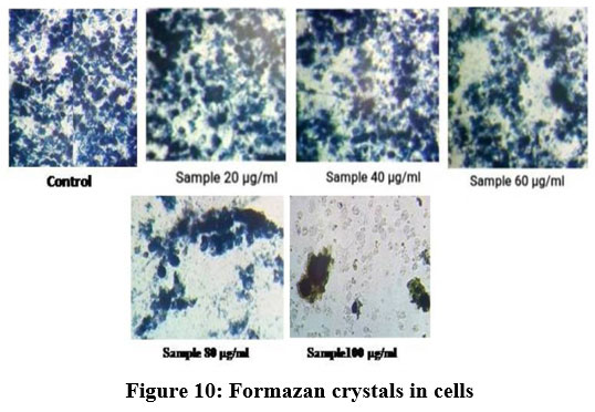 |
Figure 10: Formazan crystals in cells. |
Antimicrobial activity of Andrographis echioides
Antimicrobial activity of Andrographis against microbes was performed by disc diffusion method as predicted in Table 9, 10 and plate 1-5. The extracts has well inhibitory activity of bacteria like Escherichia coli(11mm), Enterococcus aerogenes and Staphylococcus aureus (10mm) and fungi like Candida albicans (9mm), Candida tropicalis (0mm) at concentration 100µg/ml. At concentration 80mg/ml, the extracts exhibited antibacterial activity all the three bacteria, but was more susceptible against Escherichia coli (10mm), Staphylococcus aureus (9mm), Enterococcus aerogenes (9mm) and antifungal activity against Candida albicans (8mm). However, the crude extract observed as good inhibitory actions against pathogens with various concentrations (60, 80 and 100µg/ml). If concentration increases from 20-100µg/ml, then the inhibitory action also gets increased in this study.
The antibacterial activity against gram-negative and gram-positive strains by using ethanolic extract of Andrographis echioides showed maximum activity against gram-negative strains like Salmonella typhi, Klebsiella pneumonia and Vibrio parahaemolyticus when compared to control drug ampicillin and also showed least activity against gram-positive bacteria like Bacillus subtilis and Staphylococcus aureus and moderate inhibitory activity against Aeromonas hydrophila. E.coli when compared to control drug ampicillin37.
Table 9: Antibacterial activity of ethanolic extract of Andrographis echioides.
| Plant extracts | Concentrations
(µg/ml) |
Escherichia
Coli(mm) |
Staphylococcus aureus(mm) | Enterococcus aerogenes(mm) |
| Extracts | 20µg/ml | 0 | 0 | 0 |
| 40µg/ml | 0 | 0 | 0 | |
| 60µg/ml | 8 | 0 | 0 | |
| 80µg/ml | 10 | 9 | 9 | |
| 100µg/ml | 11 | 10 | 10 | |
|
Ethanol |
10 µl/disc | 0 | 0 |
0 |
Table 10: Antifungal activity of ethanolic extract of Andrographis echioides.
| Plant extract | The concentration of ethanolic extract (µg/ml) | Organisms/zone of inhibition (mm) | |
| Candida albicans | Candida tropicalis | ||
| 20µg/ml | 0 | 0 | |
| 40µg/ml | 0 | 0 | |
| 60µg/ml | 0 | 0 | |
| 80µg/ml | 8 | 0 | |
| 100µg/ml | 9 | 0 | |
| Ethanol | 10 µl/disc | 0 | 0 |
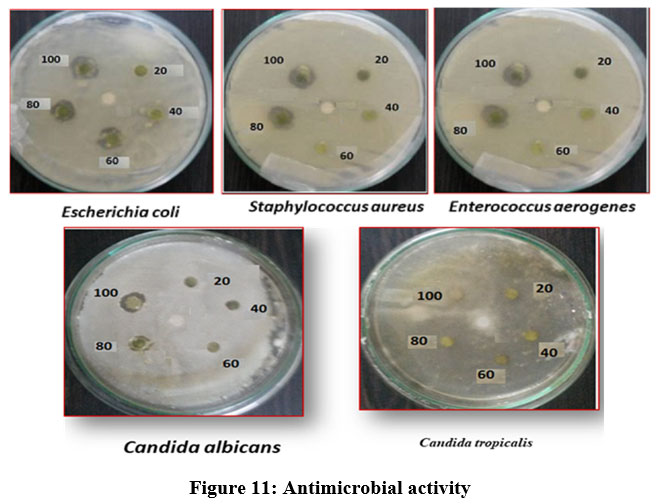 |
Figure 11: Antimicrobial activity. |
Conclusion
Thus the present work showed the ability of ethanolic extract of stem of Andrographis echioides shows the natural anti-arthritis, anti-inflammatory, anticancer activity under in vitro conditions and it may reduce the side effect of chemical drugs. More comprehensive studies related to this compound will enhance pharmaceutical exploration in the field of cancer activities.
Acknowledgment
There was no funding agency for the concerned work, and all the expenditures were carried out by the authors themselves, although we are thankful to the Head of the department for allowing us to work on the project in reference.
Conflict of interest
The authors declare no conflict of interest.
Funding Sources
There is no funding source.
Reference
- Billingham M.E. Models of arthritis and the search for anti-arthritic drugs. PharmacolTher., 1983; 21(3):389-428.
CrossRef - Mythilypriya R, Shanthi P and Sachdanandam P. Efficacy of Siddha formulation Kalpaamruthaa in ameliorating joint destruction in rheumatoid arthritis in rats. Chem boil interact., 2008; 176(3):243-251.
CrossRef - Berbert A. A, Kondo C. R. M, Almendra C. L, Matsuo T andDichi I. Supplementation of fish oil and olive oil in patients with rheumatoid arthritis. Nutrition., 2005; 21(2):131-6.
CrossRef - Nair V, Singh S and Gupta Y. K. Evaluation of the disease-modifying activity of Colchicum luteum Baker in experimental arthritis. J. Ethnopharmacol., 2011; 133(2):303-307.
CrossRef - Shin H. Y. Jeong H. J, Na H. J, Hong S. H, Lee S. K, Lee K.N, Song Y. S, Kim H. M and Shin T. Y. Daeganghwal-tanginhibits the stem cell factor-induced migration and inflammatory cytokines secretion in mast cells. J. Ethanopharmacol., 2003; 85(1): 157-161.
CrossRef - Kore K. J, Shete R. V and Desai N.V. Anti-Arthritic activity of Hydroalcoholic extract of LawsoniaInnermis against adjuvant arthritis.Int. J. Drug Dev. and Res., 2011; 3(4): 217-224.
- Holliday K. L, McWilliams D. F, Maciewicz R. A, Muir K. R, Zhang W and Doherty M. Lifetime body mass index, other anthropometric measures of obesity and risk of knee or hip osteoarthritis in the goal case-control study. Osteoarthritis Cartilage. 2011; 19(1):37-43.
CrossRef - Rahman S, Salehin F and andIqbal A. In vitro antioxidant and anticancer activity of young Zingiberofficinale against human breast carcinoma cell lines. BMC Complement Altern Med., 2011, 11:76.
CrossRef - Schwartzman G, Ratain M. J, Cragg G. M, Wong J. E, Saijo N, Parkinson D. R, Fujiwara Y, Pazdur R, Newman D.J, Dagher R and Di Leone L. Anticancer drug discovery and development throughout the world. J Clin Oncol., 2002; 20:47S-59S.
- Kim D. W, Hong G. H, Lee H. H, Choi S. H, Chun B. G, Won C. K, Hwang I. K and Won M. H. Effect of colloidal silver against the cytotoxicity of hydrogen peroxide and naphthazarin on primary cultured cortical astrocytes. Int J Neurosci., 2007; 117(3): 387-400.
CrossRef - Yeruva L, Elegbede J. A and Carper S. W. Methyl jasmonate decrease membrane fluidity and induces apoptosis via tumor necrosis factor receptor 1 in breast cancer cells. Anticancer Drug., 2008; 19(8): 766-776.
CrossRef - Deepti R, Sushila R, Permender R, Aakash D, Sheetal A and Dharmender R. HPTLC densitometric quantification of stigmasterol and lupeol from Ficusreligiosa. Arab. Chem., 2015; 8: 366-371.
CrossRef - Vane J. R. Inhibition of prostaglandins synthesis as a mechanism of action for Aspirin-like drugs. Nature New Biology., 1971; 231: 232-235.
CrossRef - Chen Y. F, Jobanputra P, Barton P, Bryan S, Fry-Smith A, Harris G and Taylor R. S. Cyclooxygenase-2selective non-steroidal anti-inflammatory drugs (etodolac, meloxicam, celecoxib, rofecoxib, etoricoxib, valdecoxib and lumiracoxib) for osteoarthritis and rheumatoid arthritis: a systematic review and economic evaluation. Health technolAssess., 2008; 12(11): 1-278.
CrossRef - Schaffer D, Florin T, Eagle C, Marschner I, Singh G, Grobler M, Fenn C, Schou M and Curnow K. M. Risk of serious NSAID-related gastrointestinal Events during long-term exposure: a systematic review. Med J Aust., 2006; 185: 501-506.
CrossRef - Scott D. L, Wolfe F and Huizinga T. W. Rheumatoid arthritis. Lancet.,376(9746). 2010; 1094-1108.
CrossRef - Donahue K. E, Gartlehner G, Jonas D. E, Lux J. L, Thieda P, Jonas B. L, Hansen R. A, Morgan L. C and Lohr K. N. Systematic review: comparative effectiveness and harms of disease-modifying medications for rheumatoid arthritis. Ann Intern Med., 2008; 148 (2): 124-34.
CrossRef - Chandra S. C, Bhadra S, Roy S, Saha S. K, Islam S and Bachar S. C. Analgesic and anti-inflammatory activities of ethanolic root extract of Swertiachirata. Jordan J. Biol. Sci., 2005; 5 (1): 31-36.
- Sangeetha M, Kousalya K, Lavanya R, Sowmya C, Chamundeeswari D andMaheswara U.C. In vitro anti-inflammatory and anti-arthritic activity of leaves of Cleodendroninerme. Res. J. Pharm. Biol. Chem. Sci., 2011; 2(1); 822-827.
- Azeem A. K, Dilip C, Prasanth S. S, Shahima V. J. S, Kumar S and Naseera C. Anti-inflammatory activity of the glandular extracts of Thunnusalalunga. Asian Pac. J. Trop. Med., 2010; 794-796.
CrossRef - Chippada S. C andMeena V. Antioxidant, an anti-inflammatory and anti-arthritic activity of Centellaasiatica J. Chem. Biol. phys., 2011; 1(2): 260-9.
- Braca A, Sortino C, Politi M, Morelli P and Mendez J. Antioxidant activity of flavonoids from Licania licaniaeflora. J Ethanopharmacol. 2002; 79(3): 379-381.
CrossRef - Kim H. J, Park S. Y, Lee H. M, Seo D. I and Kim Y. M. Antiproliferative effect of the methanol extract from the roots of Petsites japonicas on Hep3B hepatocellular carcinoma cells in vitro and in vivo. Exp. Ther. Med., 2015; 9(5): 1791-1796.
CrossRef - Karumaran S, Nethaji S and Rajakumar R. Antimicrobial and antioxidant activity of leaf extracts of Aeglemarmelos.Pelagia Research Library., 2016; 7(3): 205-208.
- Vivek M. N, SachidanandaSwamy H. C, Manasa M. Pallavi S, Kambar Y, Asha M. M, Chaithra M, PrashithKekuda T. R, Mallikarjun N, Onkarappa R. Antimicrobial and antioxidant activity of leaf and flower extract of Caesalpinia pulcherrima, Delonixregia and Peltaphorumferrugineum. J. Appl. Pharm. Sci., 2013; 3(8): 064-071.
- Sangita C, Chatterjee P, Dey P and Bhattacharya S. Evaluation of the anti-inflammatory effect of Ashwagandha: A preliminary study in vitro. Pharmacogn. J., 2012; 4(29): 47-9.
CrossRef - Chandra S, Chatterjee P, Dev P and Bhattacharya S. Preliminary in vitro assessment of the anti-inflammatory property of Mikaniascandens flower extract. J. Adv. Pharm. Educ. Res., 2012; 2(1): 25-31.
- Chandra S, Chatterjee P, Dey P and Bhattacharya S. Evaluation of in vitro anti-inflammatory activity of coffee against the denaturation of the protein. Asian Pacific Trop Biomed., 2012; 12: S178-S180.
CrossRef - Kumar V, Bhat Z. A, Kumar D, Khan N. A and Chashoo I. A. Evaluation of the anti-inflammatory potential of leaf extracts of Skimmiaanquetilia. Asian Pac J Trop Biomed., 2012; 627-30.
CrossRef - Saleem T. K. M, Azeem A. K, Dilip C, Sankar C, Prasanth N. V and Duraisami R. Anti-inflammatory activity of the leaf extracts of Gendarussa Vulgaris Asian Pac J Trop Biomed., 2011; 1(2): 147-149.
CrossRef - Murugan A, Dr., Elangovan K andRanjith Singh A. J. A. Evaluation of Andrographis echioides for their phytochemical and in vitro antioxidant properties. Indo Am. J. Pharm., 2017; 7(05): 8779-8786.
- Mathivanan D andSuseem S. R. Chemical and biological evaluation of Andrographis echioides leaf extracts collected from the Vellore district in Tamil Nadu, India. Pac. Sci. Rev., 2016; 18 (2): 138-144.
CrossRef - Gajendran B, Chinnasamya A, Durai P, Raman J and Manikandan R. Biosynthesis and characterization of silver nanoparticles from Daturainoxia and its apoptotic effect on human breast cancer cell line MCF-7. Mater. Lett., 2014; 122 (122): 98-102.
CrossRef - Liu Q, Jiang H. In vitro cytotoxicity of Tanacetumvulgare mediated silver nanoparticles against breast cancer (MCF-7) cell lines. Biomed Res. Int., 2017; 28(3): 1354-1358.
- Sholihah M. M, Indarto D and Pramana T. Y. The inhibitory effect of Andrographis paniculata extract on proliferation of breast cancer cell line. IOP Conference Series: Mater. Sci. Eng., 2019; 546: 1-8.
CrossRef - Singh S, Mehta A, Baweja S, Agarwal L and Mehta P. Anticancer activity of Andrographis paniculata and Silybummarianum on Five human cancer cell lines. Pharmacol. Toxicol. 2013; 8(1): 42-48.
CrossRef - Punnagai K, David D. C, Latharani M and Radhika R. Evaluation of Antibacterial activity of ethanolic extract of Androgaphis echioides – An in vitro Int. J. Phytopharm., 2016; 7(4): 214-217.
CrossRef

