
Manuscript accepted on :06-04-2022
Published online on: 09-05-2022
Plagiarism Check: Yes
Reviewed by: Dr. Salman Ahmed
Second Review by: Dr. Chetana Chandrashekar
Final Approval by: Dr. Ayush Dogra
Priyanka Nitin*  , Sreeshyla H S and Usha Hegde
Department of Oral Pathology and Microbiology, JSS Dental College and Hospital, A constituent college of JSS Academy of Higher Education and Research Mysuru
Karnataka, India.
.
Corresponding Author E-mail: dr.priyankanitin@jssuni.edu.in
DOI : https://dx.doi.org/10.13005/bpj/2424
Abstract
Fibrous histiocytoma, as the name suggests, is a lesion which is prominently composed of a combination of fibroblastic & histiocytic cells. Fibrous histiocytoma can usually be seen as either cutaneous types or involving deep tissues. The diagnosis of fibrous histiocytoma is only confirmed after surgical excision. It is imperative to differentiate this lesion from the other aggressive forms of fibrohistiocytic neoplasms. Atypical Fibrous Histiocytoma (AFH) comes in the spectrum of fibrous histiocytic lesion ranging from benign to malignant lesions. Atypical Fibrous Histiocytoma in oral cavity is distinctly uncommon. This uncommon occurrence of the lesion needs to be made known of, to prevent inappropriate treatment. Hence, here we present an uncommon case report of Atypical oral fibrous histiocytoma which presented clinically as epulis fissuratum/ Inflammatory fibrous hyperplasia
Keywords
Atypical Oral Fibrous Histiocytoma; case report; Epulis, Tumor; Fascicles, Immunohistochemistry
Download this article as:| Copy the following to cite this article: Nitin P, Sreeshyla H. S, Hegde U. Atypical Oral Fibrous Histiocytoma - An Uncommon Histiocytic Lesion Presenting as Inflammatory Fibrous Hyperplasia. Biomed Pharmacol J 2022;15(2). |
| Copy the following to cite this URL: Nitin P, Sreeshyla H. S, Hegde U. Atypical Oral Fibrous Histiocytoma - An Uncommon Histiocytic Lesion Presenting as Inflammatory Fibrous Hyperplasia. Biomed Pharmacol J 2022;15(2). Available from: https://bit.ly/3w4u4B3 |
Introduction
The phrase ‘fibrohistiocytic’ is descriptive in nature. It is usually used to describe lesions composed of cells resembling normal histiocytes and fibroblasts.1 The review of literature suggests that fibrous histiocytoma is a true neoplasm with a definite growth potential but limited capacity for aggressive behaviour. 2 Fibrohistiocytic lesions are common in the skin and rarely seen in the oral cavity. 2,3 Numerous lesions come under the spectrum of fibrohistiocytic tumors. They range from being benign, intermediate to malignant. Sometimes, a lesion may be categorized as Atypical, showing features of both the types. Diagnosis becomes difficult in such cases as there is always a chance that it can be misdiagnosed. Here we present an unusual case report of a fibrohistiocytic lesion in the oral cavity, where the provisional diagnosis was that of denture epulis, but histopathologically turned out to be Atypical Fibrous Histiocytoma(AFH). Such uncommon cases need to be reported because they may be otherwise be treated inappropriately.
Case Report
A 65-year-old patient visited our institute with a complaint of a mass in the lower jaw and an ill-fitting lower denture. The medical and dental histories were noncontributory. On examination, completely edentulous maxillary and mandibular arch were found. A non-tender, flabby pedunculated mass was noted on the alveolar ridge in 34-35 regions. No other unusual changes were observed in the oral cavity. Based on history and clinical details, a provisional diagnosis of Epulis fissuratum was given. The flabby mass was biopsied and sent for histopathological examination.
On gross examination, the tissue measured around 3×1 cm. It was firm in consistency & cream in color. Histopathologically, H & E stained sections revealed epithelium and connective tissue stroma. The epithelium was stratified squamous parakeratinized in nature showing hyperplasia, acanthosis, elongation and widening of rete ridges. The connective tissue was hypercellular in a hyalinized and collagenous stroma. Sheets of tumor cells were seen just below the hyalinized connective tissue separating it from the overlying epithelium. The tumor cells were spindle in shape showing storiform pattern.2 Short intersecting fascicles of eosinophilic fibroblastic cells were noted. Also, plump pleomorphic spindle and polyhedral cells with hyperchromatic nuclei, atypia and few mitotic figures were noted. Few cells in connective tissue showed brownish pigments which were demonstrated as melanin pigmented with Masson`s fontana stain. The clinical presentation and the clear-cut features of tumor cells showing both benign and atypical features led to the diagnosis of Atypical Fibrous Histiocytoma. (Fig 1) Atypical fibroxanthoma, Dermal leiomyosarcoma, Sarcomatoid carcinoma, Nodular melanoma, Dermatofibrosarcoma protuberans, and Angiosarcoma are some of the histological differential diagnoses considered for this case.
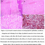 |
Figure 1: (A). H and E stained sections reveal hyperplastic epithelium, acanthosis, elongation and widening of rete ridges, hyalinized connective tissue stroma and sheets of tumor, cells (40x). (B). H and E stained sections reveal short intersecting fascicles of eosinophilic fibroblastic cells (100x). |
Discussion
Fibrous histiocytoma is a neoplastic lesion composed of a combination of cells resembling fibroblasts and histiocytes, that are arranged in sheets or short fascicles. The presence of inflammatory cells is variable. Presence of foam cells and siderophages are also noted. 2 Fibrohistiocytic lesions are commonly seen in the skin. Their occurrence is rare in the oral cavity. These lesions are classified as benign, intermediate and malignant based on clinical and histopathological features. There are some lesions which are benign but show atypical features wherein they are considered as a separate entity as they may show varying prognosis.
Atypical Fibrous Histiocytoma (AFH) comes under the umbrella of Fibrohistiocytic lesions. This term was coined by Kaddu. In very few cases of fibrous histiocytoma, border line histological features that include significantly more atypia and mitotic activity than the usual type may be seen. These lesions are termed as Atypical Fibrous Histiocytoma. The diagnosis should be made judiciously only to those cases in which there is a clear-cut background of classic fibrous histiocytoma displaying increased atypia and increased mitotic activity including atypical forms.2
Atypical Fibrous Histiocytoma can be seen at any anatomical site at any age.1,2 The etiology is obscure. The tumor is usually associated with a history of trauma or chronic infection, Chronic irritation.4,5 Spontaneous development have also been reported for those located within the oral cavity.
Regarding the pathogenesis, various theories have been put forth. Atypical Fibrous Histiocytoma was considered as an inflammatory fibrohistiocytic process in the past. The tendency for recurrence and rare metastases argues against a reactive lesion. Recently clonal chromosomal abnormalities have been observed which favors neoplastic origin. 2
Kram and his co-workers proposed another hypothesis, that Atypical Fibrous Histiocytoma is a heterogeneous process. They stated that the neoplastic element was made of histiocytic cells and the reactive element was made of fibroblastic cells. Otherwise, alternatively, they proposed that, the lesion represents a true neoplasm, wherein the neoplastic cells (histiocytic cells) are masked by predominent reactive cells (fibroblastic cells). 2
Histological findings demonstrate a well-defined unencapsulated lesion with epithelial hyperplasia, an interposed grenz zone.2,6 Hyalinized connective tissue stroma separated sheets of tumor, cells. Short intersecting fascicles of eosinophilic fibroblastic cells are seen. Plump pleomorphic spindle and polyhedral cells with mild atypia and few mitotic figures are noted. There is a spectrum of features, wherein the lesions show focal mild pleomorphism to those lesions revealing marked pleomorphism. Hemorrhagic areas, hemosiderin deposits, and necrosis can also be seen. 2,6 The above features were noted in our case with additional finding of melanin pigments but no necrosis.
Electron microscopic studies shows two distinct cell types. One type of cell resembles fibroblasts with organized lamellae of rough endoplasmic reticulum, few or no lipid droplets, and no phagolysosomes. The other rounded cell resembles histiocytes with numerous cell processes, mitochondria, and phagolysosomes.4
Atypical fibroxanthoma, Dermal leiomyosarcoma, Sarcomatoid carcinoma, Nodular melanoma, Dermatofibrosarcoma protuberans, and Angiosarcoma are some of the histological differential diagnoses to be considered for fibrohistiocytic lesions. Various immunohistochemical markers can be utilized to differentiate Atypical Fibrous Histiocytoma from other lesions. (Table 1, Table 2)1-4,6,7. The criteria for differentiating Atypical Fibrous Histiocytoma from other lesions is based more on architectural features than cytological features. In the present case, Immunohistochemistry was not done as the histological features were clearly evident under light microscope.
Table 1: Immunohistochemical markers for differential diagnosis of AFH 1-4,6,7
| Lesion | Marker | Reactivity | AFH |
| Atypical fibroxanthoma | MiB 1 | Strongly Positive | Less than 10% of cells |
| Dermal leiomyosarcoma | Desmin | Positive | Focally Positive |
| Sarcomatoid carcinoma | Cytokeratin | Positive | Negative |
| Nodular melanoma | S-100 | Positive | Negative |
| Dermatofibrosarcoma protuberans | CD 34 | Strongly Positive | Focally Positive |
| Angiosarcoma | CD31, CD34 | Positive | Focally Positive |
Table 2: Other immunohistochemical markers for the diagnosis of AFH 1,2,3,6
| Markers | Reactivity |
| Vimentin | Positive |
| EMA | Negative |
| HMB 45 | Negative |
| Alpha smooth muscle actin | Focally Positive |
| CD 68 | Variable |
| Factor XIII a | Variable |
| S-100 | Negative |
Atypical fibrous histiocytoma is always excised with clear margins to prevent recurrence. When incompletely excised, it tends to recur. Follow up of our case showed no recurrence. When compared to regular fibrous histiocytoma, Atypical Fibrous Histiocytoma shows increased recurrence.2,6,9 Rarely, it metastasizeses.6,8,10
The limitations of this case report is deficient review of literature resulting from insufficient number of published cases of Atypical Fibrous Histiocytoma.
Conclusion
To conclude, Atypical Fibrous Histiocytoma is a distinct lesion but it is not well known. It should be considered in the differential diagnosis of fibrous growth of gingiva and oral mucosa. It requires thorough histopathological examination to prevent inapt treatment as it can be mistaken for a malignant tumor. In the present case, the patient came for treatment of ill-fitting denture due to flabby tissue clinically mimicking epulis fissuratum, which was diagnosed as Atypical oral fibrous histiocytoma. Upon recall, the patient showed no recurrence.
Conflict of Interest
There is no conflict of interest.
Funding Sources
There is no funding Sources.
References
- Skoulakis C.E, Papadakis C.E, Datseris G.E, Drivas E.I, Kyrmizakis D.E, Bizakis J.G. Subcutaneous benign fibrous histiocytoma of the cheek. Case report and review of the literature. ACTA otorhinolaryngologica italica 2007;27: 90-93.
- Kram A, Stan´czyk J, Woyke S. Atypical Fibrous Histiocytoma and Atypical Fibroxanthoma: Presentation of Two Cases. Pol J Pathol 2003,54;4:267-271.
- Menditti D, Laino L, Mezzogiorno A, Sava S, Bianchi A, Caruso G, Di MaioL and Baldi A. Oral benign fibrous histiocytoma: two case reports. Cases Journal 2009,2:9343.
CrossRef - George A, Pynadath MK, Jayapalan CS, Noufal A, Manjunath GA and Nair RB Benign Fibrous Histiocytoma of Maxillary Gingiva. Dentistry 2014;4(5): 227.
- Sharma H, Alam S, Upadhyay S, Yadav H, Longani P, Kohli P. Benign Fibrous Histiocytoma of the Buccal Mucosa: A case Report.Journal of Dental Sciences & Oral Rehabilitation 2013;38-40.
- Abdelkrim SB, Belajouza C, Jomaa W, Beizig N, Said ZB, Mokni M, Nouira R and Sriha B. Atypical Cutaneous Fibrous Histiocytoma: An Unusual and Misleading Variant of Fibrous Histiocytoma. Case Reports in Pathology 2011:1-3.
CrossRef - Usha Hedge, Sujeeth K Shetty, Huchanahalli Sheshanna Sreeshyla, Vidya Gowdappa Doddawada. Dermatofibrosarcoma Protuberans – A recurrent lesion with unusual presentation in the parotid region. Journal of Clinical and Diagnostic research 2014;8(3):130-131
CrossRef - Edward H, Soulem, D E and Enriquez P. Atypical fibrous histiocytoma, malignant Fibrous histiocytoma, malignant Histiocytoma, and epithelioid sarcoma. A Compayative Study of 65 Tumors. Cancer 1972;30:128-143
CrossRef - Shankar V. Atypical fibrous histiocytoma. Pathology Outlines.com website. http://www.pathologyoutlines.com/topic/softtissueatypfh.html. Accessed January 8th, 2020.
- Guillou L, Gebhard S, Salmeron M, Coindre JM. Metastasizing Fibrous Histiocytoma of the Skin: A Clinicopathologic and Immunohistochemical Analysis of Three Cases. Mod Pathol 2000;13(6):654–660.
CrossRef

