
Manuscript accepted on :13-08-2021
Published online on: 25-08-2021
Plagiarism Check: Yes
Reviewed by: Dr Ramaiah Maddi

Second Review by: Dr. Aktsar Roskiana Ahmad
Final Approval by: Dr. Fai poon
Emad K. Abbas1, Hussein H. Echrish2, Sabaa A. Mohammed3
1Department of human anatomy, College of Medicine, University of Basra, Iraq
2Department of Histopathological Teaching Hospital University of Basra, Iraq
3Department of Pharmacognosy and Medical Plants College of Pharmacy, University of Basra, Iraq
Corresponding Author E-mail: emad66.k_iraq@yahoo.com
DOI : https://dx.doi.org/10.13005/bpj/2261
Abstract
Background:Turmeric is typically used as a spicy food preservative and colorant. It has been proved that curcumin has a wide range of biological effects including anti-inflammatory, anti-viral, anti-fungal, and curcumin activity that can improve antibiotic activity on the wounds. Objectives: To evaluate the effects of Curcumin with and without antibiotics on skin wound treatment. Materials and Methods: The protocol was approved by the animal house in medical college / Basra university. This study used nine male rabbits aged about 6 months and an average weight of (1.083 g). Each group consists of 3 rabbits: control group (normal saline) A, topical curcumin in group B, topical curcumin, and tetracycline ointment in group C. Regular treatments were given to rabbits in therapeutic groups. Result: The lowest Mean ± SDof swelling of suturing area was noted in both groups that treated by curcumin alone (9.07 ± 0.97 vs 15±1 mm, p value = 0.002) and that treated with curcumine and antibiotic (9.1±0.9vs 15±1 mm, p value = 0.002) versus the control group ( that treated by normal saline) and the lowest Mean ± SD of elevation of suture line was noted in both group that treated by curcumin alone (2.63 ± 0.06 vs 4.07±0.21 mm, p value >0.001) and that treated with curcumin and antibiotic (2.7 ± 0.2 vs 4.07±0.21 mm, p value =0.001) versus control group. There is no significant statistical difference between the Mean ± SD neither of swelling of suture area nor of elevation of suture lines of groups that treated by curcumin alone and group that treated with curcumin and antibiotic [(9.07 ± 0.97 vs 9.1 ±0.9, p value=0.97),(2.63±0.06 vs 2.7 ± 0.2, p value=0.61) respectively]. The histopathological evaluation is consistent with morphological changes as at day 3 of wound healing in both groups that treated by curcumine with and without antibiotic, there is formation a thin layer of keratin and absence of features that indicate delay wound healing such as hemorrhage, inflammatory cell infiltrate of (Neutrophils, macrophages and lymphocytes) and debris, which are detected in control group. Furthermore, at day 7 of control group, there is decrease of inflammation, presence of gap between the two edgesof the wound but no keratin formation. No clear histopathological difference in wound healing between tested groups that treated by curcumin with and without antibiotic. Conclusion: There issignificant clinical and histological evidences that the curcumin not only prevent delay of wound healing but it is also enhanced wound healing. No significant difference in using curcumin alone or combine it with local antibiotic.
Keywords
Antibiotic; Curcuma long; Curcumin; Wound healing
Download this article as:| Copy the following to cite this article: Abbas E. K, Echrish H. H, Mohammed S. A. The effect of Curcuminvs. Curcumin combined Antibiotic Ointmentson the Healing of skin wound. Biomed Pharmacol J 2021;14(3) |
| Copy the following to cite this URL: Abbas E. K, Echrish H. H, Mohammed S. A. The effect of Curcuminvs. Curcumin combined Antibiotic Ointmentson the Healing of skin wound. Biomed Pharmacol J 2021;14(3). Available from: https://bit.ly/3jl8F05 |
Introduction
Wounds are the main cause of physical illness,healing is a method of survival and an attempt to restore normal anatomical structure and function1 .Therefore,the property of living organisms is wound healing, in addition,wound cure is a process chain needed to extract invaded pathogens from the wounded tissue of the body and to completely or partially restructure the injured tissue,in general, wound healing takes place in three complex and overlapping interrelated stages, namely inflammation, granulation, and remodeling2. Restoring damaged tissue is important for all surgical manipulations3. In general, wound healing is divided into three interconnected complex and overlapping phases: inflammation, granulation, and remodeling,collagen formation occurs at a rapid rate in the early stages of wound healing, accompanied by collagen breakdown in the remodeling process, where collagen cross-linking occurs and tensile strength increases,several factors influence the wound healing process, varying as age to nutritional status and neuropathy after wounding4. The cure of the wound is blocked by immune-suppressants, cytotoxins, and non-steroidal anti-inflammatory drugs,antibiotic, analgesic, and herbal medications, however, improve wound healing5.
Various natural ingredients, plant ingredients that are composed of active principles such as triterpenes, alkaloids, flavonoids, and biomolecules facilitate the wound healing process 4.
Turmeric is an ancient spice of Curcuma longa rhizomes, which is in the genus of ginger (Zingiberaceae), for decades, turmeric has been used for medicinal purposes in India, Turmeric is one of the most effective healers of nature 6,7.Curcumin gives turmeric the yellow color and is now known for most medical properties, 2-5 %of turmeric is estimated to be curcumin8.
The characteristics of curcumin are antibiotics and antioxidants9.And speed up the re-epithelization of cells such as myofibroblast, fibroblast, and macrophages required for the cure of wounds 10.Curcumin reduces the level of pain and inflammation and promotes homeostasis by selectively inhibiting lipoxy and cyclooxygenase (COX) in the arachidonic acid cascadecombination 11.Turmeric is considered an easily available antiseptic for cuts, burns, and bruises in Ayurvedic medicine,turmeric is good antibiotic dermatology used in India, the application of turmeric powder eliminates bacterial, fungal infections12.Combination antibiotic therapy is progressively used to lower the risk of antibiotic resistance in bacteria and to boost antibacterial range throughout therapy13.A few experiments have shown that antibacterial efficacy can be enhanced by combining antibiotics with crude plant extracts 14,15.
Skin wound healing is a fascinating process and not just for mammals, it is an evolutionary advantage, due to its essential functions as a physical, chemical, and bacterial barrier, skin wound healing is a significant step for wound survival, while certain interactions during the healing phase are essential, redundancy is high and other cells or mediators may assume roles and signals without significant complications,in preserving the physiological homeostasis of the human body, the integrity of healthy skin is an important factor 16.There are many scoring or grading methods for evaluation of wound healing. One of these was used in a study that evaluate ethanolic extract of curcumin (curcuma Longa) on wound healing in black Bengal goats5, it was precise, clear and simple represented way of wound healing. For these reasons was chosen in this study. This scoring is divided to three grades:
Grade I: presence of hemorrhage and debris
Grade II: No debris, still there is hemorrhage with inflammatory cell infiltrate inside the tissue such as neutrophils, lymphocytes and macrophages.
Grade III: decrease of inflammation, gap between the edges of the wound and then formation of keratin layer.
Aim of this study: To evaluate the effects of Curcumin with and withoutantibiotics on skin wound treatment.
Material and Methods
Animal modules and diagnostic samples
The protocol was approved by theanimal housein medical college / BasraUniversity. This study used 9 male rabbits aged about 6 months and an average weight of (1.083 g). They were kept in an air-conditioned house in stainless steel cages. This study was design to compare and evaluate the effects of curcumin with and without antibiotic on healing of surgical wound in rabbits in three groups Each group consists of 3 rabbits:control that treated with normal saline group A, topical curcumin in group B, topical curcumin, and tetracycline ointment in group C. Regular treatments were given to rabbits in therapeutic groups. The swelling of the suture area was evaluated at day 3 as the maximum of swelling achieved at the day 3, and the elevation of suture line was evaluated at day 7 which is the day at which skin suture was removed. Measurements of swelling areas were recorded in millimeter (mm) by using slide calipers. T-test was used to evaluate the statistical differences. Skin samples of all rabbits were taken at day 3, 7and14of the injury. Skin biopsy was taken including the wound site and then fixed in formalin for histological examination. After embedding in paraffin, slices were taken and stained by Hematoxylin and Eosin and Mallory Tricom for histological examination a modern Leica light microscope (1000X) magnification provided with a camera (0.5X), as well as a (10X) computerized magnification which provided a high-resolution image.
Turmeric paste preparation
Turmeric roots were collected from the nearby Iraqi market and then grinded by an electrical mill to a fine powder. The paste was prepared by water to (50g) of turmeric, water was gradually applied to the paste until it was semi-solid and then used on the injury directly. Tetracycline Ointment: was bought from a pharmacy in Basra and used on the wound.
Surgery procedure
Anesthetized Rabbits use an anesthetic (Lidocaine 2 %) solution to place the rabbit in a prone position and sterilize the lower back skin with a 10 % povidone-iodine solution. The cut was around 1 cm in the skin and sutured with 2-0 non-absorbed silk.
Result and Discussion
The lowest Mean ± SD of swelling of suturing area ( at day 3) was noted in both groups that treated by curcumin alone (9.06 ± 0.97 vs 15±1 mm, p value = 0.002, table 1) and that treated with curcumine and antibiotic (9.1 ±0.9 vs 15±1 mm, p value =0.002, table 1) versus the control group that treated by normal saline and the lowest Mean ± SD of elevation of suture line (at day 7) was noted in both group that treated by curcumin alone (2.63 ± 0.06 vs 4.07±0.21 mm, p value >0.001, table 1) and that treated with curcumin and antibiotic (2.7 ± 0.2 vs 4.07±0.21 mm, p value =0.001, table 1), and this could indicate less inflammation in group B and C as compared with group A. This result could support the reports that describe the wound healing is a complex process depend on many cell types and many mediators [2] and the role of curcumine could be through different mechanisms as will described below.Another interesting result was, no significant statistical difference between the Mean ± SD neither of swelling of suture area nor of elevation of suture lines of groups that treated by curcumin alone and group that treated with curcumin and antibiotic [(9.06 ± 0.97 vs 9.1 ±0.9, p value=0.97), (2.63 ±0.06 vs 2.7 ± 0.2, p value=0.61) respectively, table 1].
Table 1: represent Mean ± SD of the morphological difference (swelling of the suturing area and elevation of suture line) in three groups (A control group, B treated with curcumin and C treated with Curcumin and tetracycline Ointment).
| Group | Mean± SD of swelling of suturing area at Day 3 (mm) | Mean±SD of elevation of suture line at Day 7 (mm) |
| Group A | 15± 1 | 4.06±0.21 |
| Group B | 9.06±0.97 | 2.63±0.06 |
| Group C | 9.1±0.9 | 2.70±0.2 |
Another essential result of this study, as shown in the (Table 2 and Figure 1) at day 3 of group A (control group), the grade of healing was between grade I and II because there is hemorrhage, inflammatory cell infiltrate mainly neutrophils, macrophages, lymphocytes and still there is debris while at day 3 of group B and C, the grade of healing was grade III (there is no hemorrhage and no debris and no inflammatory cell infiltrate and there is mature keratin layers formation (Figure 2) that indicate complete healing was achieved within 3 days in group B and C.
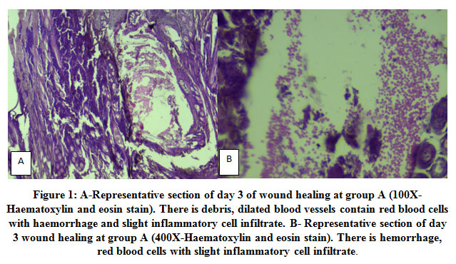 |
Figure 1: A-Representative section of day 3 of wound healing at group A (100X- Haematoxylin and eosin stain). There is debris, dilated blood vessels contain red blood cells with haemorrhage and slight inflammatory cell infiltrate. B- Representative section of day 3 wound healing at group A (400X-Haematoxylin and eosin stain). There is hemorrhage, red blood cells with slight inflammatory cell infiltrate. |
 |
Figure 2: A-Representative section of day 3 of wound healing at group B (400X-Haematoxylin and eosin stain). There is full layer of keratin formation. B- Representative section of day 3 of wound healing at group B (40X-Haematoxylin and eosin stain). There is full layer of keratin formation. |
Further more, at day 7 of group A (control group), the grade of healing of was between grade II and III because there is decrease of inflammation, presence of gap between the two edges of the wound but no keratin layer formation (Table 2 and Figure 3). This indicate complete healing still was not achieved within 7 days after injury. While at day 7 of both group B and C, the grade of healing was grade III because mature keratin layer was formed (Figure 4).
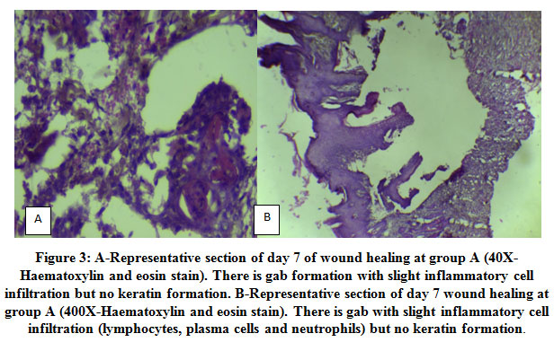 |
Figure 3: A-Representative section of day 7 of wound healing at group A (40X-Haematoxylin and eosin stain). There is gab formation with slight inflammatory cell infiltration but no keratin formation. B-Representative section of day 7 wound healing at group A (400X-Haematoxylin and eosin stain). There is gab with slight inflammatory cell infiltration (lymphocytes, plasma cells and neutrophils) but no keratin formation. |
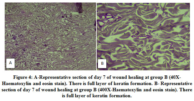 |
Figure 4: A-Representative section of day 7 of wound healing at group B (40X-Haematoxylin and eosin stain). There is full layer of keratin formation. B- Representative section of day 7 of wound healing at group B (400X-Haematoxylin and eosin stain). There is full layer of keratin formation. |
Moreover, at day 14 of group A (control group), the grade of healing was grade III because there is mature keratin formation (Table 2 and Figure 5). While at both group (B and C), the process of healing was already reached the last stage of grade III at day 7 as there is formation of mature keratin (Table 2 and Figure 6).
Table 2: Represent histological grade of wound healing in three groups that mentioned above.
| Group | Day | Grade of healing |
| Group A | 3 | Grade I to II |
| 7 | Grade II to III | |
| 14 | Grade III | |
| Group B | 3 | Grade III |
| 7 | Grade III | |
| 14 | Grade III | |
| Group C | 3 | Grade III |
| 7 | Grade III | |
| 14 | Grade III |
 |
Figure 5: Representative section of day 14 of wound healing at group A (100X-Mallory triple stain). There is full layer of keratin formation. |
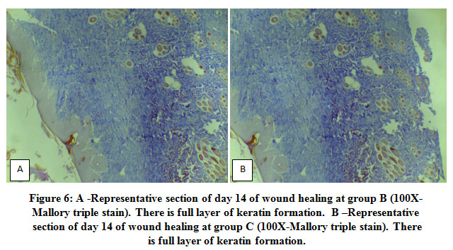 |
Figure 6: A -Representative section of day 14 of wound healing at group B (100X- Mallory triple stain). There is full layer of keratin formation. B –Representative section of day 14 of wound healing at group C (100X-Mallory triple stain). There is full layer of keratin formation. |
This indicate that the histopathological evaluation is consistent with morphological changes because there is hemorrhage, inflammatory cell infiltrate mainly neutrophils, macrophages, lymphocytes and still there is debris at day 3 of control group. Furthermore, at day 7 of control group there is decrease of inflammation, presence of gap between the two edges of the wound but no keratin formation. While at day 3 of both groups that treated by curcumin with and without antibiotic the wound healing was achieved and reach to thin layer of keratin at day 3 and then to mature keratin at day 7. There is no clear histopathological difference in wound healing between tested groups that treated by curcumin with and without antibiotic. This indicate that curcumin alone (independent on adjuvant antibiotic) play a core role in the enhancement of wound healing. As the wound healing is a complex process [2]. The role of curcumin in enhancement of wound healing is more complex 5.
Many individual factors could affect healing such as age, neutritional factors, presence of neuropathy and others and as mention above 4, but in the current study all these factors are standardized because all nine rabbits that submitted in this study have the same individual features.Therefore, the effects of curcumin on wound healing could be due other mechanisms like anti-inflammatory, anti-bacteriological effect of curcumin9,11,12and antifungal13. The current study reveals an interesting finding that, the role of curcumine not only due to prevention of factors that delay wound healing such as anti-inflammatory, antifungal and antibacterial effect, it also enhance wound healing through different mechanism such as enhancement of granulation tissue formation, contraction of the wound, enhancement tissue re-modeling , releasing of chemotaxic agents, releasing of growth factors and re-epithelization of cells such as myofibroblast, fibroblast and macrophages required for the cure of the wound10. Therefore, might be all or more than one mechanism could be performed by curcumin. Although, some microbilogical study, postulated the synergestic effect of some antibiotic with extract of some plants (curcumin was not used)15. One of an interesting finding of this study is that, there is no additive role of combination of tetracycline ointment with curcumin as compare with curcumine alone on histopathological and morphological healing of the wounds. The result of this study was comparable to the result of other study as Sukandaret al. (2016) postulated that combination of penicillin or ampicillin with curcumin could have synergestic effect against stapyllococusaureus, but this synergestic effect was not detected with tetracycline14.
It is well known that combination of antibiotic therapy is progressively used to lower the risk of antibiotic resistance in bacteria and boost antibacterial range throughout therapy14,15. Therefore, the result of current study could indicate that curcumine could has more than one broad spectrum anti-bacterial agents or could contain new anti-bacterial agent that didn’t used before. Then the bacteria cannot resist the antibacterial effect. Therefore, the fundamental finding of this study is the curcumin alone (independent of adjuvant antibiotic) play a core role in the enhancement of wound healing.
References
- Wang L, Qin W, Zhou Y, Chen B, et al. Transforming growth factor β plays an important role in enhancing wound healing by topical application of Povidone-iodine. Scientific Reports. 2017; 7(1):991.
CrossRef - Gonzalez ACDO, Andrade ZDA, Costa TF, Medrado ARAP. Wound healing – A literature review. An Bras Dermatol. 2016; 91(5): 614-620.
CrossRef - Majumder M, Kamaath JV. Herbal concept of Wound healing. Journal of Pharmaceutical Research. 2005; 4: 06-09
- Panchatcharam M, Miriyala S, Gayathri VS, Suguna L. Curcumin improves wound healing by modulating collagen and decreasing reactive oxygen species. Moll Cell Biochem. 2006; 290(1): 87-96
CrossRef - Miah et al. Clinical evaluation of ethanolic extract of curcumin (CurcumaLonga)on Wound healing in Black Bengal goats. J. Adv. Vet. Anim. Res. 2017; 4(2): 181-186
CrossRef - Dosoky NS, Setzer WN. Chemical composition and biological activities of essential oils of Curcuma species. Nutrients. 2018; 10(9): 7-10
CrossRef - Chopra RN, Nayar SL, Chopra IC. Glossary of Indian medical plants. New Delhi Publication and Information Directorate. 1999: 111-113
- 8. Prasad S, Aggarwal BB. Turmeric, the golden spice. Herbal Medicine: Biomolecular and Clinical Aspects. 2nd edition. 2011:263-288
CrossRef - Venkatasubbu GD, Anusuya T. Investigation of curcumin nanocomposite for wound dressing. International Journal of Biological Macromolecules. 2017; 98: 366-378
CrossRef - Goel A, Kunnumakkara AB, Aggarwal BB. Curcumin as “Curecumin” from the kitchen to the clinic. Biochemical Pharmacology. 2008; 75:787-809.
CrossRef - Majeed M. Curcuminoids antioxidant nutrients. Piscataway, NJ: Nutriscience Pubs, Inc. 1995; 13: 50-54
- Bhowmik D, Chiranjib KP, Kumar S, Chandria M, Jayakar B. Tumeric: A Herbal and Traditional Medicine. Scholar Research library.2011;2(4): 373-383
- Sukandar EY, Kurniati NF, Puspatriani K, Adityas HP. Antibacterial activity of Curcumin in combination with tetracycline against Staphylococcus aureus by disruption of the cell wall. Res. J. Med. Plants. 2018; 12(1): 1-8
CrossRef - Sukandar EY, Kurniati NF, Anggadiredja K, Kamil A. In vitro antibacterial activity of Kaempferiapandurateroxb. And Curcuma xanthorrhizaroxb. Extracts in combination with certain antibiotic against MSSA and MRSA. Int. J. Pharm. Sci. 2016; 8: 108-111
- Nascimento GF, Locatelli J, Freitas PC, SlivaGL. Antibacterial activity of plant extracts and phytochemicals on antibiotic- resistant bacteria. Brazilian Journal of Microbiology. 2000; 31: 247-256
CrossRef - Sorg H, Tilkom DJ, Hager S, Hauser J, Mirastschijski U. Skin wound healing: An update on the current knowledge and concepts. Eur. Surg. Res. 2017; 58:81-94
CrossRef

