Manuscript accepted on :03-02-2021
Published online on: 17-02-2021
Plagiarism Check: Yes
Reviewed by: Dr. Sasho Stoleski
Second Review by: Dr. Ahmed Salah
Final Approval by: Dr. Alessandro Leite Cavalcanti
1Department of Oral Pathology, Senior Lecturer, Tagore Dental College and Hospital, Chennai. Tamil Nadu, India.
2Department of Oral Pathology, Professor and Head, Manipal College of Dental Sciences, Mangalore, Karnataka, India.
Corresponding Author E-mail : dr.prasanna1oralpath@gmail.com
DOI : https://dx.doi.org/10.13005/bpj/2152
Abstract
Proper histopathological grading and typing of a tumour plays a significant role in evaluating and assessing the clinical management and prognosis of the tumour. Microscopically sometimes it is difficult in categorizing a tumour into benign or malignant as histopathology does not depict all the features which are of diagnostic and prognostic value. The present study aims :To evaluate the proliferative index of the oral epithelial cells taken from a buccal smear. Materials and method : A total of 90 subjects were included with 30 subjects in each category of normal, smokers and tobacco chewers. The smears were collected from buccal mucosa and applied on the glass slides followed by fixation with alcohol for 30 min and staining of the slides with AgNOR staining as proposed by Bukhari et al (2007). Results: The AgNOR number where more in smokers when compared to normal subjects and was statistically significant. Similarly in chewers it was also comparatively higher when compared to normal and statistically significant. But the AgNOR counts between smoker and tobacco chewer, showed a mean difference of 0.6 and was not statistically significant.
Keywords
AgNOR; Buccal Smears; Exfoliative Cytology; Proliferative Index; Silver Dots
Download this article as:| Copy the following to cite this article: Prasanna S, Srikant N. Silver Dots – A Screening and Prognostic Aid in Oral Health. Biomed Pharmacol J 2020;14(1). |
| Copy the following to cite this URL: Prasanna S, Srikant N. Silver Dots – A Screening and Prognostic Aid in Oral Health. Biomed Pharmacol J 2020;14(1). Available from: https://bit.ly/3jXtkpj |
Introduction
Detection of oral cancerous lesion at an early stage will help in improving the survival and morbidity rate of patients suffering from cancer. So a technique which is less invasive and as an alternate to scalpel biopsy, exfoliative cytology at this juncture can be considered as apt alternate which is less invasive and painful, well accepted by the patients.1
Exfoliative cytology is the field involving study of exfoliated cells having a prominent role in diagnosis and screening of a disease or lesion. When a cell in a deeper epithelial layer loses its adhesion property in diseased state, these cells are exfoliated separately or along with cells of superficial layer.1
Any regulatory process of proliferation and synthesis of protein is controlled by a mastermind behind, in case of cells it’s the nucleus2. The nucleus has a specialized region called the nucleolar organizer regions (NORs), a chromosomal loop of DNA regulating ribosomal synthesis. Few proteins in the nucleolar region react better with silver stains, so termed as AgNOR protein3 which are acidic, non-histone in nature. AgNOR-P are best visualized on routine histopathological and cytological samples using silver solution4 as they reflect the cellular proliferation activity and degree of protein synthesis.3
Quantitative and qualitative changes of NORs can imply the degree of cell nucleolar activity in hyperplastic and neoplastic conditions. Actively proliferating cells have impaired nucleolar association and, therefore, exhibit a higher AgNOR count, regardless of the ploidy state of the cell. Recent histopathologic studies of NORs have resulted in successful diagnosis, categorization and prognostication of various benign and malignant lesions. Counting is the most widely used method for evaluating AgNORs because of technique simplicity and reproducibility.4Any type of screening test which works on biomarkers are amenable to automation, there by resulting cost savings and potential for applications in the developing world.5
Aim of the Study
The study aimed at performing AgNOR staining procedure from buccal smears collected from normal, smokers and tobacco chewer subjects to assess the proliferative index of the oral epithelium in the corresponding groups.
Materials and Methods
A total of 30 subjects were taken in each category of normal, smokers and tobacco chewers with an age range of 20 – 70years, comprising a total of 90 subjects. The subjects were collected from those attending the outpatient department for a routine dental check-up and treatment. The smokers had the habit of smoking > 5 cigarettes per day, the tobacco chewers had the habit of using smokeless form of tobacco for more than 5 years with a frequency of consuming it > 4 times a day.
Patients who were included in the study subjects didn’t had any oral lesions. The smears were collected by scraping with wooden spatula along the buccal mucosa and smear was applied on the glass slides and fixing the slide in alcohol for 30 min followed AgNOR staining as proposed by Bukhari et al (2007).
Preparation of Working solutions:-
The preparation of AgNOR staining solution and staining procedure for the present study was done according to methodology prescribed by Bukhari et al (2007)3
Solution A
The solution was prepared by dissolving 500 mg gelatin powder in 25ml deionized water at 37 c and then 250 μl formic acid is added. Continuous shaking of the glass ware for about 10 min at 37 c was sufficient to dissolve the gelatin and a clear solution is obtained.
Solution B
It consist of silver nitrate and deionized water. 50% w/v concentrated solution of silver nitrate in deionized water.
The final working solution was prepared by mixing one part of solution A with two parts of solution B and filtered using a filter paper into glass bottle and used immediately. Solution was prepared when required to avoid wastage and considering cost factor.
The slides are covered with the silver solution and kept in dark place for 20 – 30 minutes and dehydrated with alcohol (50%,70%,80%,96%,100%) for 5 min each and clarified with xylene for 5 min.5 The slides were dried in dark place and coverslip mounted with DPX.
The AgNOR counting in the present study was done, where the buccal cells having good staining clarity, no overlapping of cells were considered and criteria for AgNOR count, 3 or more black dots in the nucleus were considered to have more cellular and proliferative activity. The counting is done at 40x magnification.
The datas are subjected to ANOVA test which will help in determining whether there is any significant variation between each groups and Posthoc Tukey Test to detect where the exact difference is present.
Results
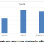
Table 1 shows comparison of score between the three groups shows that smoker group has the highest value of 10.83 and normal has the least value of 7. This difference is statistically Significant with a test value of 93.404* and p value of <0.001.
Table 1: Comparison between normal, smoker and tobacco chewer.
| Groups | N | Mean | Std. Deviation | Welch Statistics (*)/F (ANOVA) | P value | |
| Score | Normal | 30 | 7 | 0.695 | 93.404* | <0.001 |
| Smoker | 30 | 10.83 | 1.802 | |||
| Tobacco chewer | 30 | 10.23 | 1.654 | |||
| Total | 90 | 9.36 | 2.23 |
 |
Graph 1: Comparing mean values of normal subjects, smoker and tobacco chewers. |
Table 2 shows Posthoc Tukey tests comparing normal and smoker groups shows a mean difference of -3.833* and is statistically significant with a p value of <0.001. Comparing normal and tobacco chewer groups shows a mean difference of -3.233* and is statistically significant with a p value of <0.001. Comparing smoker and tobacco chewer groups shows a mean difference of 0.6 and is not statistically significant with a p value of 0.258.
Table 2: Posthoc Tukey Test comparing normal ,smoker and tobacco chewer.
| Dependent Variable | (I) group | (J) group | Mean Difference (I-J) | Std. Error | P value |
| Score | Normal | Smoker | -3.833* | 0.379 | <0.001 |
| Tobacco chewer | -3.233* | 0.379 | <0.001 | ||
| Smoker | Tobacco chewer | 0.6 | 0.379 | 0.258 |
Discussion
Cancer especially oral squamous cell carcinoma is quite common in India due to adverse use of tobacco in smoking and smokeless form.6 Oral squamous cell carcinoma has a poor prognosis inspite of advances in therapy, so diagnosing it at an early stage and treating them is the main tool in improving patient survival rate. Generally scalpel biopsy is used for taking specimen which is invasive and traumatic to the patient both psychological and physical, so they are employed only in severe suspected lesions and not in all conditions.7
To control the rapidly developing situation, technique’s which are less expensive, non-invasive, and well accepted by the patient needs to be developed and those which can be repeated frequently. Exfoliative cytology is an easy procedure that can be carried out at outdoor patient department as a chair side procedure to diagnose malignancy at early stage and in regular periodic check-up. In 1920, aspiration and exfoliative cytology was introduced by Johannes Muller (1801 – 1858), a pathologist in Berlin to show cancer cells in microscope on scrapings from the cut surface of surgically excised tumors.8
The silver staining procedure which is used for identification of NORs has been frequently utilized in formalin fixed, paraffin embedded specimens but in this study we have used it in exfoliative cytological smears. Jahanshah Salehinejada et al(2007)8 has showed in their study that in cytologic smears the analysis of AgNORs is more accurate as whole nucleus can be assessed as in tissue sections and has used AgNOR technique successfully in oral smears9.
In this study we have considered 3 or more black dots in the nucleus to have more cellular and proliferative activity (Fig.1).In the present study the AgNOR count was more in smokers when compared to normal subjects which was statistically significant and the results were in accordance with Jahanshah Salehinejada et al (2007)8, Patricia Campos Fontes (2008)5, Sampaio et al (1999)9. A thorough literature search showed very few studies were conducted with tobacco chewers like those of Nikhil I Malgaonkar (2016)10 and as that of the present study. The results showed that the AgNOR counts in chewers were also comparatively higher when compared to normal which was statistically significant and was in accordance with Sachin Jindal et al (2013)11 , Mohan B.C.,Angadi P.V (2013)12, Park NH (2000)13..
While comparing the smokers and tobacco chewers in the present study, it showed a mean difference of 0.6 and which not statistically significant .To the best of our knowledge and extensive search in literature, no studies were available comparing the smokers and tobacco chewers.
Conclusion
Inspite of advances in diagnostic techniques, simple, non-invasive, less expensive and valid diagnostic procedures are needed, exfoliative cytology is one such procedure. With AgNOR staining in these exfoliative smears can help in diagnosing early lesions. This technique will be useful in mass screening and as a chair side procedure. So subjects with adverse habits can be monitored regularly with this procedure and chance of cancer occurrence can be reduced.
Acknowledgment
None
Conflict of Interest
there is no conflict of interest
Funding Source
None
References
- Kaur M., Saxena S., Samantha YP., Chawla G., Yadav G. (2013).Usefulness of Oral Exfoliative Cytology in Dental Practice. Journal of Oral Health Community Dentistry, 7(3), 161 – 165.
CrossRef - Sandhya Panjeta Gulia., Emani Sitaramam., Karri Prasada Reddy. (2011).The Role of Silver Staining Nucleolar Organiser Regions (AgNORs) in Lesions of the Oral Cavity. Journal of Clinical and Diagnostic Research, 5(5),1011-1015.
- Mulazim Hussain Bukhari.(2007).Modified method of AgNOR staining for tissue and interpretation in histopathology. Int. J. Exp. Path,88, 47–53.
CrossRef - Patricia Campos Fontes et al, Comparison of Exfoliative Pap Stain and AgNOR Counts of the Tongue in Smokers and Nonsmokers, Head and Neck Pathol (2008) 2:157–162.
CrossRef - Makesh Raj L. S., Monica C. Solomon., Karen Boaz. (2018). Invasive Tumour Front, Agnor’s Characters in Prognostication of OSCC: A suggested standard cut off among Indian patients. International Journal of Current Advanced Research, 7(8), 14634-14638.
- Ignacio Gonzalez Segura et al, Exfoliative cytology as a tool for monitoring pre – malignant and malignant lesions based on combined stains and morphometry techniques, J Oral Pathol Med (2015), 44 : 178 – 184.
CrossRef - A. Singh et al, Role of exfoliative cytology in oral lesions: with special reference to rule out malignancy, Journal of college of Medical Sciences-Nepal, 2010, Vol.6, No-2, 29-37.
CrossRef - Evaluation of AgNOR Staining in Exfoliative Cytology of Normal Oral (Buccal) Mucosa: Effect of Smoking, Jahanshah Salehinejada et al, Journal of Mashhad Dental School, Mashhad University of Medical Sciences, 2007; 31(Special Issue): 22-24.
- Sampaio Hde C, Loyola AM, Gomez RS, Mesquita RA. AgNOR count in exfoliative cytology of normal buccal mucosa. Effect of smoking. Acta Cytol. 1999;43:117–20.
CrossRef - Malgaonkar NI, Dagrus K, Vanaki SS, Puranik RS, Sharanesha MB, Tarakji B. Quantitative analysis of agnor counts of buccal mucosal cells of chewers and non chewers of gutkha: A comparative cytologic study. J Can Res Ther 2016;12:228-31
CrossRef - Sachin Jindal et al, Alteration in buccal mucosal cells due to the effect of tobacco and alcohol by assessing the silver-stained nucleolar organiser regions and micronuclei, J Cytol. 2013 Jul-Sep; 30(3): 174–178.
CrossRef - Mohan B.C. · Angadi P.V. ,Exfoliative Cytological Assessment of Apparently Normal Buccal Mucosa among Quid Chewers Using Argyrophilic Nucleolar Organizer Region Counts and Papanicolaou Staining, Acta Cytologica 2013;57:164–170 .
CrossRef - Park NH, Kang MK. Genetic instability and oral cancer. Electron J Biotechnol. 2000;3:66–71.
CrossRef


