
Manuscript accepted on :30-Dec-2020
Published online on: 12-01-2021
Plagiarism Check: Yes
Reviewed by: Dr. Flavio Palmieri
Second Review by: Dr. Asim Faraz
Final Approval by: Dr. Francesca Gorini
Sarika A Panwar1 , Mousami V Munot2, Suraj Gawande3 and Pallavi S Deshpande4
1Department of Electronics and Telecommunication Engineering, AISSMS-IOIT,Pune-411001
2Department of Electronics and Telecommunication Engineering, PICT, Pune
3Department of Application Engineer, Design Tech Systems Pvt. Ltd, Pune
4Department of Electronics and Telecommunication Engineering, SAE, Kondhwa, Pune
Corresponding Author Email: vrushalimendre@gmail.com
DOI : https://dx.doi.org/10.13005/bpj/2124
Abstract
Introduction: The World Brain Tumor Day is seen on eighth June, in a year. Despite exhaustive research in the medical field, the prevalence of this deadly disease is increasing globally with over new 28,000 braintumor cases being reported annually, in India alone. Recent advancements in the field of machine learning facilitate minimally invasive, efficient and reliable procedures for the diagnosis of Brain tumor. Objective: This research intends to design and devlop a reliable framework for accurate diagnosis of brain tumor mainly meningioma type, gliomatype and pituitary cerebrum tumor utilizing Magnetic Resonance Imaging (MRI), one of the most mainstream non-obtrusive procedure Methods: In the proposed system, pre-trained AlexNet is used to classify meningioma, glioma and pituitary brain tumor. The concept of transfer learning is applied using AlexNet for extracting the features from brain MRI images.The AlexNet contains eight layers in which the first five are convolution layer and the remaining three are fully connected layers. The last layer is a softmax layer which gets the output from fully connected layers. The ReLU non-linearity is applied to the output of every convolution and fully connected layer. The idea of transfer learning is applied utilizing AlexNet for computing thefeatures from brain MRI pictures. The AlexNet contains eight layers in which the initial five are convolution layers and the staying three layers are fully connected layers. a softmax is the last layer , which is feeded by the fully connected layers. The ReLUactivation function is applied to the output of convolution layer and fully connectedlayer Result: The proposed system framework recorded the best order precision of 100 % to classify the brain tumor when validated using a practical dataset. Conclusion: The proposed work presents accurate and automatic brain tumor classification using transfer learning. The features extracted using AlexNet has proven to be efficient in obtaining good discriminative power in diagnosis of brain tumor
Keywords
Brain Tumor Diagnosis; CNN; Deep Network
Download this article as:| Copy the following to cite this article: Panwar A. S, Munot V. M, Gawande S, Deshpande S. P. A Reliableand an Efficient Approach for Diagnosis of Brain Tumor Using Transfer Learning. Biomed Pharmacol J 2021;14(1). |
| Copy the following to cite this URL: Panwar A. S, Munot V. M, Gawande S, Deshpande S. P. A Reliableand an Efficient Approach for Diagnosis of Brain Tumor Using Transfer Learning. Biomed Pharmacol J 2021;14(1). Available from: https://bit.ly/3q9LExu |
Introduction
An abnormal development of tissue in the brain or central spine is termed as brain tumor. A tumor in the brain can destroy proper brain function. Tumor can be malignant or benign. Benign tumors are not cancerous. They won’t invade surrounding tissue or spread elsewhere. Whereas Malignant brain tumors hold cancer cells and many times do not encompass clear boundaries. As per the statistics of American Society of Clinical Oncology (ASCO) in this year i.e. 2020, an anticipatednumber of adults is 23,890 which includes about 13,590 males and 10,300 females in the United States will be detected with primary cancerous tumors of thespinal cord and brain .Around about 3,540 kids under the age of 15 will also be diagnosed with a brain or CNS tumor this year[1]. Accurate spotting of a brain tumor is aintricate process. Magnetic Resonance Imaging (MRI scan) is the non invasive first step in diagnosis. Once an MRI Scancertifiespresence of a tumor in the brain, then to determine the type of brain tumor is next step.this is carried out from a sample of tissue received through biopsy which is an invasive procedure. Though MRI is a non-invasive imaging technique, it showes some limitations. The consistency and reliability in the diagnosis depends on quality of image and the skill/proficiency of the radiologist in interpreting the images. These limitations has given motivation to conduct an extensive research in developing a framework of Computer Aided Diagnosis [CAD] systems, so as to acquire diagnostically accurate results using the image processing and pattern recognition methods. Enormous amounts of medical imaging data as MRI images are generated during image acquisition helps in complete examination of human organs achievable [2]. However this research area requires more accuracy as it is a health diagnosis related problem.
During the past few years deep learning has acquired a huge attention by showing promising results in the various domains such as human speech recognition, character recognition from handwritten script, image classification, image detection and segmentation. A subset of machine learning, deep learning models are designed, where the data are fed into the deep learning algorithm and it automatically learns what features are most useful to determine the output. Deep learning is typically used for classifying images, identifying objects in images, and enhancing images and signals. Deep learning models take time to train. But pretrained networks can shorten training through transfer learning. Applications of deep learning are prevalent in medical diagnosis [3].
Christian et al. [4] proposed a deep Convolutional Neural Network architecture for classification and detection in the ImageNet Large-Scale Visual Recognition Challenge 2014(ILSVRC14). GoogleNet, a 22 layers deep network is used and its quality is assessed in classification and detection.
Ali et al. [5] reviewed brain tumor segmentation methods with focus on deep learning. Present state is assessed and future elaborations to deploy brain tumor segmentation based on MRI into daily clinical routine are addressed.
Renhao Liu et al. [6] used pre-trained CNN to compute the features to get deep feature representations for brain tumor MRI. The researchers reported better classification accuracy of 95%than conventional feature extraction methods.
Talo et al. [7] proposed a deep transfer learning techniquefor classifying between normal and abnormal brain MR images using Convolutional Neural Network (CNN) based ResNet34. The proposed research work quotes 100% accuracy in classification.The results were based on n total 613 MR images.
Siyuan Lu et al. [8] Presented a “pathological brain detection”method withAlexNet and transfer learning showing 100.00% accuracy, Authors [7] claims reduction of the AlexNet retaining time.
Naz et al. [9] used Matlab 2015a for feature extraction and Weka 3.8.3 version for classification. Automatic segmentation and detection of brain tumor is done using Convolution Neural Network. The authors have worked on tumor detection with 93.7% accuracy.
Javeria Amin et al. [10] proposed ainnovative method to classify the brain tumor MR images and non tumor images. The pre-trained CNN model is used wherein feature learning is executed using Alex net and Google net. Assessment of the projected model is carried out on medical image computing and computer-assisted intervention (MICCAI) challenge datasets.
Basheer et al. [11] proposed CNN based classifier to identify the benign and malignant brain tumor. The dataset consisted of 40 images and a classifier performance validated the suitability of the proposed classifier with 93 % and 100% accuracy for healthy and diseased data set respectively.
Deepak et al. [2] presents an accurate system for brain tumor classification. The projected system used Convolutional Neural Network (CNN) with transfer learning to extract features from brain MRI images. The proposed system reports the classification accuracy of 92.3%.The CNN model accuracy is also compared with SVM and KNN classifiers on deep CNN features. SVM classifier reported the accuracy of 97.8% and KNN classifier reported the accuracy of 98%. Authors in [12] further report the limitations of deep transfer learning as there was considerable misclassification of samples from the class meningioma which they claim can be corrected by further tuning of the transfer learned model.
Swati et al. [12] proposed a method for brain tumor classification using transfer learning with fine-tuning. The authors experimented on AlexNet, VGG16, and VGG19. VGG19. They claims having achieved better performance on VGG19 (94.82%) than AlexNet (89.95%) and VGG16 (94.65%)
Malathi et al. [13] proposed segmentation of brain tumor using CNN architecture. parameters also causes the reduction of over fitting. The proposed method detects enhancing tumor and specifying tumor to the actual tumor region only.
Kalaiselvi et al. [14] presented six different CNN models with varying number of layers, epochs model, dropout model, stopping criteria and batch normalization. Authors claimed to get the satisfactory results on a model having five layers with Stopping Criteria, Batch Normalization and Dropout Model.
Limitations of existing methods are summarized below: None of the existing methods proposed earlier could achieve more than 98% accuracy which is very important in medical diagnostic systems. The Performance of actual state of art methods are insufficientfor the deployment of the system. Most of the earlier methods manually segment the tumor region before classification. Deepak et al. [2] observed misclassification of instances of the class meningioma. Authors also report over fitting with smaller training data.
Objective of the proposed system is to explore the concept of CNN based transfer learning to classify brain tumor into three classes as meningioma, glioma and pituitary tumor with pretrained networks named GoogleNet and AlexNet using fine tuning strategy.
The rest of the paper is organized as follows: Section 2 describes the Deep Learning Concept with main focus on transfer learning and pre-trained networks. Section 3 focuses on databases used in the proposed system with detailed methodology used. Results of the experimentation and performance evaluation are discussed in section 4.Section 5 contains the conclusion.
Deep Transfer Learning
Deep Learning is viewed as subclass of Machine Learning where the multiple layered architecture is used to extract the higher level features like image features such as identification of digits , letters and faces from the input [15]. In transfer learning concept we utilize existing knowledge acquired from one task to solve the problem on a target task. Transfer learning is beneficial when there is inadequate amount of training data [16].
Deep learning can solve complex problems with quite good results but at the cost of training time and more data samples than traditional machine learning algorithms. To solve this problem of abundant data requirement and high training time in case of complex problems, pretrained networks are used which forms the basis of transfer learning in the context of deep learning [16].
Pretrained network is a saved network created by someone else to solve similar problems. Pre-trained networks are trained on a large dataset. Using the transfer learning approach, pretrained models can be used for a given classification task by doing its customization. The learned feature map on a large data set can be used for other tasks without starting from scratch and it can also be beneficial in case of data scarcity [17]. Various pretrained networks for image classification are AlexNet [18,20], Visual Geometry Group (VGG-16) [19], VGG-19 [19],GoogleNet [4] etc. The pretrained network features may or may not work well for different tasks. For new tasks to work well, in a pretrained network the final layers (classification or regression) can be replaced or selective retraining of the previous layers can be done. Configurable architecture of Deep Networks has hyper parameters. Initial layers in deep networks extract generic features and higher layers focus on task specific features [16].
Materials and Methods
In the proposed system, pre-trained AlexNet is used to classify meningioma, glioma and pituitary brain tumor.The AlexNetcomprise eight layers in which the initial five layers are convolution layers and the last three layers are fully connected type . The last layer is a softmax layer which gets the output from fully connected layers. The fully-connected layers are attached to the all neurons in the previous layer. The ReLUactivation function is applied to the output of every convolution and fully connected layer.
 |
Figure 1: Re-use of pre-trained AlexNet |
The Fig.1 is about reusing of a pre-trained model i.e. Alexnet. AlexNet is a CNN designed to perform classification of image data. The Alexnet is basically trained on millions of images and it is able to identify an image into thousand different object categories. The original AlexNet can categorize an image into various items like animals,Coffee mugs and keyboards etc.The network takes an image at the input layer with predefined size and predicts the class of the image in the form of a label. The network learns the high level features from the provided image. Fig. 1 shows the flow of how to prepare Alexnet for transfer learning.
Initially the AlexNet is loaded in its original form.Originally the last three layers viz fully connected layer, a softmax layer and a classification layer ofAlexNet are designed for 1000 object categories. These layers must be replaced and fine tuned for classification of brain tumor into classes i.e meningioma, glioma and pituitary brain tumor. Replacement of the last three layers of Alexnet is followed by training of the network on MRI brain tumor images with three tumor classes. Finally the evaluation of CNN model performance using AlexNet on unseen brain tumor images is done.
Fig. 2 shows overall modification of Alexnet for brain tumor classification problem. Last three layers of the Alexnet are modified to make it compatible for brain tumor classification.
 |
Figure 2: Overall Modification of Alexnet for Brain Tumor Classification. |
Results
MATLAB R2020a has been used to implement the proposed classification model. The execution environment has a specification of 8GB RAM and Intel i5-72000 @2.70 GHz CPU.
Dataset used is publicly available dataset of on Figshare [21].The dataset consists of 300 MRI images of patients diagnosed with brain tumors of three different types: meningioma, glioma and pituitary brain tumor. The pituitary gland is connected directly to part of the brain called the hypothalamus. The 100 MRI images of pituitary tumors have been collected. Glioma brain tumor occurs in different age groups, with 75 to 84 yearolds. The 100 MRI images of glioma tumors have been collected. Meningioma tumors grow in meninges surrounding the spinal cord and brain. The 100 MRI images of meningioma tumor have been collected. All images are the T1-weighted contrast-enhanced MRI images.Fig.3 shows the MRI images of pituitary, Glioma and Meningioma.
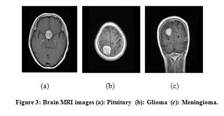 |
Figure 3: Brain MRI images (a): Pituitary (b): Glioma (c): Meningioma. |
The original image size of each image is 625*698*3.Input brain images to AlexNet are resized to fixed size of. 227*227*3. It ensures the AlexNet output is of a size 1*1*N vector, where N is channel dimension having value 3. Resizing of an image may result in information loss which may ultimately affect the accuracy.
Image preprocessing flow has been explained in fig. 4. Image resizing is done by normalizing the image intensity values in the range of [0 1].
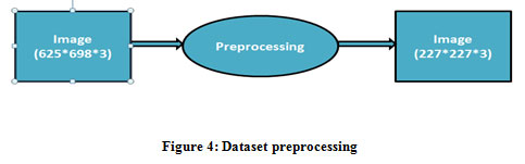 |
Figure 4: Dataset preprocessing |
To develop AlexNet network model for brain tumor classification, initially training and testing dataset is prepared followed by setting of training options. The MRI images of brain tumor have been divided into training and testing dataset. The percentage of split used is 85%-15% i.e. 255 images are given to AlexNet for training. Remaining 45 images are provided to the trained CNN for performance evaluation of the model for unknown data.
Once the dataset is ready, the training and tuning of hyper-parameters is done for modified AlexNet. Setting of training options is very important in which we define hyper-parameters of the network. Hyper-parameters are used in fine tuning of the network. Solver used is SGDM (Stochastic Gradient Descent with Momentum) with momentum of 0.9000. Initial Learn Rate is 0.001 with ‘piecewise’ LearnRateSchedule. MiniBatchSize is 64. ExecutionEnvironment is ‘cpu’. Epochs are 20 with 3 iterations per Epochs. Total iterations are 60.
Training process and effect of hyper-parameter tuning is observed from the training progress report. Training progress report shows two performance evaluation parameters, they are training accuracy and training loss. These parameters give the knowledge of how well network convergence has been achieved on training image dataset.
The original AlexNet is a series network with 25 layers and depth of network is 8. This includes, input layer, 7 rectified linear units (ReLU) layer, 2 normalization layers, 3 pooling layers, 2 dropout layers, 1 softmax layer and 8 learnable weights layers which contains 5 convolution layers and 3 fully connected layers.To use AlexNet for transfer learning, the final three layers i.e. fully connected,softmax and classification output layers are modified for brain tumor diagnosis.
Table 1 presents the input data normalization over entire training period. It shows that as numbers of epochs are increased, the training accuracy also reached to 100% and the loss is minimized to 0.0006. Base learning rate is constant during entire training period.
Table 1: Initializing Input Data Normalization
| Epochs | Iteration
number |
Time Elapsed
(hr:mn:ss) |
Mini-Batch Accuracy | Mini-Batch
Loss |
Base Learning Rate |
| 1 | 1 | 00:00:08 | 12.50% | 2.9395 | 0.0010 |
| 17 | 50 | 00:04:24 | 96.88% | 0.0310 | 0.0010 |
| 20 | 60 | 00:05:12 | 100.00% | 0.0006 | 0.0010 |
For classification of brain tumor in three predefined classes, the data set is partitioned as training subset, validation subset and testing subset. Validation subset is used to test the modified AlexNet model in training phase so that to get the insight on how the AlexNet model adapts to an independent data set. Initially the whole dataset is divided in two parts: Part I-80% samples and Part II-20% samples. Part I i.e. 240 samples are further splitted into two subsets i.e. training subset and validation subset. In first round 70% and 30% of 240 samples are used for training and validation respectively and 20% of 300 samples are reserved for testing. Percentage accuracy and predictions for each class are shown in Table 4. Many iterations of validation are carried out withdiverse partitions are as shown in Table 2.
Table 2: Performance for training and cross validation subset
| Part-I (80% samples) | Part-II(20% samples) | %Accuracy | Predictions | |||
| Training Subset | Validataion Subset | Test Set | Meningioma(20) | Glioma(20) | Pituitary(20) | |
| 70 | 30 | 20 | 96.67 | 20 | 18 | 20 |
| 60 | 40 | 20 | 91.67 | 20 | 17 | 18 |
| 80 | 20 | 20 | 96.67 | 20 | 18 | 20 |
| 90 | 10 | 20 | 100 | 20 | 20 | 20 |
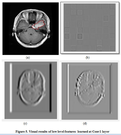 |
Figure 5: Visual results of low level features learned at Conv1 layer |
To classify the brain tumor in three classes, pretrained AlexNet model extracts features. The network learns these features during training process. There are five convolution layers in the proposed model. Initial layers have small receptive field so those layers learn low-level features such as edges and lines. The later layers have larger receptive fields so those layers learn high-level features specific to brain tumor MRI image.
Activation function is present in the end or in between CNN layers. It decides whether the neuron would fire or not. It is a non linear transformation applied over the input image. The transformed output of a Convolutional layer is then sent to the next layer of network as input. Activations of different layers of the network can be visualized. channelsare the 2-D arrays in each layer of a convolutional neural network. The network learns useful and significant features from the image. In proposed model Conv1 layer has 96 channels. Fig.5 (a) is an input image of the network and fig.5 (b) is the output activation of Conv1 layer where in each square block in the given grid is the output of channel of a convolution layer. A pixel with darker shade represents strongest negative activation and that with bright shade represents strongest positive activation.Fig.5 (c) displays low level features of a activation channel whereas fig.5 (d) indicate low level feature at strongest activation channel. A Conv5 layer is the deepest layer in proposed model which learns the complicated features known as high level features. A Conv5 layer has two groups of 128 activation channels. Few of the activation channels are shown in fig. 6 (a) and fig.6 (b), are specific activation channels whereas fig. 6 (c) is strongest one.
Conventional machine learning algorithms require manual feature extraction whereas the proposed model overcomes this problem by automated feature extraction and classification of brain tumor images in three classes with 100% accuracy. Therefore, proposed model outperforms all state-of-the-art methods of classification of brain MR images.
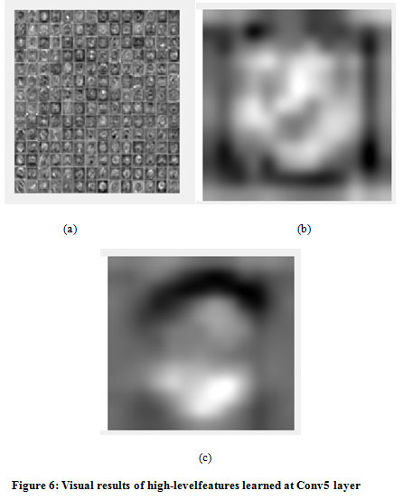 |
Figure 6: Visual results of high-level features learned at Conv5 layer |
There are several parameters used to evaluate the performance of the CNN. Classification accuracy (training and testing) is one of the very important parameters which can even identify the over fitting in a model. If training accuracy is much higher than the testing accuracy, then it can be known that the model is over fitted. Fig. 7 shows the graph of test accuracy approaching to 100% over 60 iterations for 90% training, 10% validation and 20% test set.
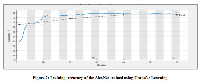 |
Figure 7: Training Accuracy of the AlexNet trained using Transfer Learning |
A loss function is also an optimization parameter used to evaluate network model. It measures the squared error between a network’s output and desired output [22]. Fig.7 presents the behavior of loss function over 60 iterations.Fig.8 shows some sample instances which indicate that all classifications are correct. This proves that the features extracted using AlexNet has got the good discriminative power for classification of brain tumor.
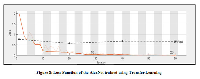 |
Figure 8: Loss Function of the AlexNet trained using Transfer Learning |
Table 3 shows the comparison of the proposed work with earlier published work. A direct comparison may not necessarily justify their effectiveness and efficiency but a general idea of the performance might be obtained.It can be observed from table 3 that Talo et al. [7] and Lu Siyuan et al. [8] has got the comparable accuracy with the proposed work i.e. 100% for CE-MRI brain tumor images. But Talo et al. [7] classified the brain MRI Normal image from Abnormal Image without considering the types of brain tumor.Similarly and Lu Siyuan et al. [8] also classified the images in only two classes as Normal and Pathological image. The proposed work achieved 100% accuracy with three class output and outperformed all state-of-the –art deep learning methods for Contrast Enhanced MRI brain tumor images.
Table 3: Comparison of proposed method with other related work
| Work | No. of Images | Method used | Accuracy |
| Renhao Liu [6] | 22 cases with 4 images of each case | CNN-F Architecture | 95.45% |
| Talo, Muhammed [7] | 613 MRI images | ResNet 34 Architecture | 100% but classification in two classes Normal and Abnormal MRI Image |
| Lu, Siyuan et al. [8] | 215 MRI images | AlexNet | 100 % Accuracy but classified the images in two classes as Normal and Pathological image |
| Amin et al.[10] | BRATS Dataset | Fusion of AlexNet and GoogleNet | 99.89% but classified the images into two classes as benign and malignant tumor |
| Basheera [11] | 40 images | CNN-S Architecture | 93% Accuracy but classified the images into two classes as benign and malignant tumor |
| Deepak et al. [2] | Figshare dataset | GoogleNet Architecture | 98% Accuracy
Classified the tumor as Meningioma, Pituitary or Glioma Tumor |
| Zar Nawab Khan et al. [12] | Figshare dataset | VGG-19 Architecture | 94.82%Accuracy
Classified the tumor as Meningioma, Pituitary or Glioma Tumor |
| Kalaiselv [14] | BRATS 2013 dataset | CNN Model with six different structures | 96-99% Accuracy |
| Proposed Method | Publicly Dataset | AlexNet | 100% Accuracy
Classified the tumor as Meningioma, Pituitary and Glioma Tumor |
Conclusion and Future Scope
The proposed work presents accurate and automatic brain tumor classification using transfer learning. The concept of transfer learning is applied using Alex Net for extracting the features from brain MRI images. The system recorded the best classification accuracy. The proposed method requires minimum preprocessing of MR images. It must be emphasized that the proposed approach used the Publicly available data set. Most of the reported literature is focused on classifying the MRI images as Benign or Malignant (two class problem). The reported approaches for classifying tumor have limited accuracies. This research presents a facilitating tool to the medical practitioners that not only classifies the image but further aids the process of diagnosis by reliably identifying the type of tumor.
References
- https://www.cancer.net/cancer-types/brain-tumor/statistics
- Deepak, S., and P. M. Ameer. “Brain tumor classification using deep CNN features via transfer learning.” Computers in biology and medicine 111 (2019): 103345.
CrossRef - Sahiner, Berkman, et al. “Deep learning in medical imaging and radiation therapy.” Medical physics1 (2019): e1-e36.
CrossRef - Szegedy, Christian, et al. “Going deeper with convolutions.” Proceedings of the IEEE conference on computer vision and pattern recognition. 2015.
CrossRef - Işın, Ali, CemDirekoğlu, and MelikeŞah. “Review of MRI-based brain tumor image segmentation using deep learning methods.” Procedia Computer Science 102 (2016): 317-324.
CrossRef - Liu, Renhao, et al. “Exploring deep features from brain tumor magnetic resonance images via transfer learning.” 2016 International Joint Conference on Neural Networks (IJCNN). IEEE, 2016.
CrossRef - Talo, Muhammed, et al. “Application of deep transfer learning for automated brain abnormality classification using MR images.” Cognitive Systems Research 54 (2019): 176-188.
CrossRef - Lu, Siyuan, Zhihai Lu, and Yu-Dong Zhang. “Pathological brain detection based on AlexNet and transfer learning.” Journal of computational science 30 (2019): 41-47.
- Naz, Sadaf, and Nitesh Kumar. “An Efficient Brain Tumor Detection system using Automatic segmentation with Convolutional Neural Network.” (2019).
- Amin, J., Sharif, M., Yasmin, M. et al. A New Approach for Brain Tumor Segmentation and Classification Based on Score Level Fusion Using Transfer Learning. J Med Syst 43, 326 (2019).
CrossRef - Basheera, Shaik, and M. Satya Sai Ram. “Classification of brain tumors using deep features extracted using CNN.” Journal of Physics: Conference Series. Vol. 1172. No. 1. IOP Publishing, 2019.
CrossRef - Swati, Zar Nawab Khan, et al. “Brain tumor classification for MR images using transfer learning and fine-tuning.” Computerized Medical Imaging and Graphics 75 (2019): 34-46.
CrossRef - Malathi, M., and P. Sinthia. “Brain Tumour Segmentation Using Convolutional Neural Network with TensorFlow.” Asian Pacific journal of cancer prevention: APJCP7 (2019): 2095.
CrossRef - Kalaiselvi, T., et al. “Deriving tumor detection models using convolutional neural networks from MRI of human brain scans.” International Journal of Information Technology (2020): 1-6.
- Deng, L.; Yu, D. (2014). “Deep Learning: Methods and Applications”(PDF). Foundations and Trends in Signal Processing. 7 (3–4): 1–199.
CrossRef - Sarkar, D. “A Comprehensive Hands-on Guide to Transfer Learning with Real-World Applications in Deep Learning.” (2018).
- Alom, Md Zahangir, et al. “The history began from alexnet: A comprehensive survey on deep learning approaches.” arXiv preprint arXiv:1803.01164 (2018)
- Krizhevsky, A., and I. Sutskever. “& Hinton, G.(2012). ImageNet classification with deep convolutional neural networks.” Advances in neural information processing systems.
- Dave Gershgorn (18 June 2018). “The inside story of how AI got good enough to dominate Silicon Valley”. Quartz. Retrieved 5 October 2018.
- Simonyan, Karen, and Andrew Zisserman. “Very deep convolutional networks for large-scale image recognition.” arXiv preprint arXiv:1409.1556 (2014).
- https://figshare.com/articles/brain_tumor_dataset/1512427#:~:text=This%20brain%20tumor%20dataset%20contains,be%20found%20in%20readme%20file
- Brownlee, J. “Loss and Loss Functions for Training Deep Learning Neural Networks.” Machine Learning Mastery 23 (2019).

