
Manuscript accepted on :7-Dec-2020
Published online on: --
Plagiarism Check: Yes
Reviewed by: Nermina Rizvanovic
Second Review by: Ahmed Salah
Final Approval by: Dr. Gul Ozcan
I Dewa Ayu Inten Dwi-Primayanti1* , Susy Purnawati1 and Wayan Sukanata2
1Department of Physiology, Faculty of Medicine. Udayana University. Denpasar, Indonesia.
2Department of Animals Husbandry. Udayana University. Denpasar, Indonesia.
Corresponding Author E-mail:dwiprimayanti@unud.ac.id
DOI : https://dx.doi.org/10.13005/bpj/2064
Abstract
Cigarette smoke contains more than 4000 constituents including tar, nicotine, carbonic monoxide, heavy metals, etc.Cigarette smoke is a source of exogenous free radicals. The accumulation of free radicals causes an imbalance of the endogenous antioxidant body system triggering oxidative stress as a risk factor for various degenerative diseases. The purpose of this study was to determine the effect of giving stingless bee honey to of malondialdehyde (MDA) and superoxide dismutase (SOD)levels in serum and hepatic tissue as a marker of oxidative stress in white rats exposed to cigarette smoke. This study was an experimental study with a posttest only control group design. This research was conducted in vivo at the Integrated Biomedical Laboratory, Faculty of Medicine, Udayana University. The research sample was 30 adult male Wistar rats aged 12-14 weeks, weight 200-400 gr. In this study the sample divided into 2 groups, the control group (exposed to cigarette smoke for 4 weeks of research but not given honey) and the treatment group (exposed to cigarette smoke and given stingless bee honey (Kele-honey) for 4 weeks as well). Statistical analysis using SPSS statistics program, the test used was the average comparative analysis by T-independent test(Mann WhitneyTest for nonparametric) to compare MDA and SOD means of rat serum and hepatic tissue exposed to cigarettes smoke. The results showed that the mean of MDA serum among the control group was 0.2247 ± 0.1192, and among the treatment group was 0.2394 ± 01058, ( p>0.05). The mean of MDA hepatic tissue among the control group was 0.5951 ± 0.1029, and among the treatment group was 0.6721 ± 0.6721, (p>0.05). The results showed that the mean of SOD serum among the control group was 1.0724 ± 0.3446, and among the treatment group was 0.6166 ± 0.2841, (p<0.05). The mean of SOD hepatic among the control group was 1.0174 ± 0.3249, and among the treatment group was 0.6721 ± 0.0847, (p<0.05). The result indicated that mean of MDA both in serum and hepatic, of the treatment group higher than the control group, the differences are not significant. Whereas, the mean of SOD both in serum and hepatic, of the intervention group lower than the control group, the differences are significant.
Keywords
Cigarettes Smoke; MDA; SOD; Stingless Bee Honey (Kele-honey)
Download this article as:| Copy the following to cite this article: Dwi-Primayanti I. D. A. I, Purnawati S, Sukanata W. Effect of Kele Honey (Trigona Sp) in Malondyaldehide and Superoxide Dismutase Serum and Hepatic Tissue of White Rats (Rattus Norvegicus) Exposed to Cigarettes Smoke.Biomed Pharmacol J 2020;13(4). |
| Copy the following to cite this URL: Dwi-Primayanti I. D. A. I, Purnawati S, Sukanata W. Effect of Kele Honey (Trigona Sp) in Malondyaldehide and Superoxide Dismutase Serum and Hepatic Tissue of White Rats (Rattus Norvegicus) Exposed to Cigarettes Smoke.Biomed Pharmacol J 2020;13(4). Available from: https://bit.ly/37drKuO |
Introduction
Cigarette smoke has been implicated in cardiopulmonary disease and other chronic diseases. Cigarette smoke contains more than 400 hazards such as tar, nicotine, CO2, and others, that can generate free radical (1). Free radicals are molecules with one or more unpaired electrons in their outermost orbit, which are very unstable and reactive. The free radical with anunpairedelectron, therefore,taking an electron from another molecule, making the molecule donates electron converts into a free radical (chain reaction).Free radicals are contributed to aging and certain degenerative diseases. Free radical accumulation can cause a free radical–antioxidants imbalance causing a state of oxidative stress, which results inoxidative damage.A previous study exhibited that cigarette smoke reduces SOD serum in healthy mice and increase MDA levels (2). Oxidative damage caused damage to lipids in cell walls, which is called lipid peroxidation. Lipid peroxidation can cause cell damage, and not only cell walls, but also adversely affect protein, RNA, and DNA(3).A previousstudy has indicated that cigarette smokegenerates oxidative stress as seen from ahigh level of malondialdehyde; a marker of lipid peroxidation, in rat, compared to controls(4). Cigarette smoke produces ROS (Reactive Oxygen Species) cause oxidative stress through the mechanism of destruction of membrane lipids from cells. The formed of lipid radicals will react with oxygen to form lipid peroxyl radicals and lipid peroxides and MDA which are soluble in water and can be detected in the blood(5).
Antioxidants prevent damage cells by giving up electron without becoming free radical.Humans have enzymaticand nonenzymatic antioxidant systems, and synergistically protect the cells from free radical damage. Enzymatic antioxidants are endogenous antioxidants involved glutathione peroxidase (GSH-Px), glutathione reductase (GSH-R), catalase (CAT) and SOD. The enzymes work by protecting the tissue from oxidative damage caused by free radicals such as superoxide anions (O2-.), hydroxyl radicals(.OH), and hydrogen peroxide (H2O2).However, it should be understood that endogenous antioxidants cannot work individually, especially under conditions of oxidative stress, which must still be supported by antioxidants from dietary intake. Consumption of foods rich in antioxidants is required to induce the endogenous antioxidant body systems to maintain cellular antioxidant status against oxidative damage. The non-enzymatic antioxidant systemcan be found abundantly in vegetables or fruits, include vitamin C, E, carotenoids, natural flavonoids, and others(5).
Honey has been used throughout the worldfor centuries not only as a healthy food resource but also as traditional medicine. Honey is a natural product from beescontains glucose, fructose, and several disaccharides, trisaccharide, vitamins, minerals,proteins, and organic acids(6). Literature shows that honey has anti-inflammatory, antimicrobial properties, accelerate wound healing, antitumor, antidiabetic and antioxidant effects. Other pharmacological investigations address the other effects of honey such as gastric protection, improve blood profile, improve risk factors for metabolic and cardiovascular disease, and as a hepatoprotective agent(7). The therapeutic effect of honey variesbased on the type of honey used (8)(9)(10).Manyproperties of honey have been approved, including antioxidants. The honey’s ability as an antioxidant, due to phenolic compounds such as flavonoids and phenols acids (11). Polyphenols contain many subunits such as procyanins, anthocyanins, phenolic acids, flavonoids, tannins, and lignin(5).These properties are important in breaking free radicals chain preventing stress oxidative as a risk factor for chronic degenerative diseases(12).As an antioxidant flavonoid canscavenging peroxyl radicals, prevent lipid peroxidationand catalytic breakdown of hydrogen peroxide.Besides, working as an antioxidant, flavonoids also help modulate the body’s endogenous antioxidant system against free radicals.
Bee produces various products such as bee bread, pollen, royal jelly, propolis, beeswax, and natural honey. In some areas in Bali, breeders have begun to develop natural honey from a stingless bee (trigona Sp). Thus, stingless bee honey called Kelehoney by local residents. Kelehoney is a pure bee product of the stingless bee, bees that are smaller than Apis bee.Kelehoney is different from a honey bee that produced by the Apisbees genus(11,13,14). Stingless bee honey for this study was taken from farms in the Gianyar-Bali. Stingless bee honey has a more acidic taste, and prices tend to be more expensive because it is traditionally bred by the community (15).This study used natural honey commonly consumed by the residents, even though not consumed regularly.The use of stingless bee honey products as an effort in developing the local beekeepersfrom an economic perspective. In health, it is expected that by utilizing the natural antioxidant properties of stingless bee it is can prevent various diseases associated with oxidative stress, in this study especially caused by cigarette smoke.
Physiologically, the liver as major organs besides the kidneys that play a role in clearing materialof metabolites, and xenobiotics, as well as various toxins and other harmful substances(16). Smoking may affect various adverse effects on organs that have no direct contact with the smoke such as the liver (17).In this study, an investigation was conducted on the effect of kele-honey on the levels of MDA and SOD of serum and hepatic tissue of rat exposed to cigarette smoke.
Research Metode
This research was used natural honey of stingless bee (locally name Kele-Honey) which is collected from beekeepers in Gianyar-Bali, without extraction and adding other ingredients. Honey has been analyzed in the Food Analysis Laboratory, Faculty of Agricultural Technology, Udayana University.The results analysis showed that the stingless bee honey (Kele-honey) had an antioxidant capacity of 287.55 mg / L GAEAC, with a total phenol of 152.76 mg / 100g GAE and flavonoid content of 92.90 mg / 100g. In this preliminary study, thedose of honey was 2.5 gr/kgof body weight.
This study was an experimental study with a posttest only control group design. This research was conducted in vivo at the Integrated Biomedical Laboratory, Faculty of Medicine, Udayana University. Research conducted from September 2019 to January 2020. The research sample was 30 adult male Wistar rats aged 12-14 weeks, weight 200-400 gr. In this study, the sample will be divided into 2 groups, each of which amounted to 15 rats. A first group is a control group in which the rats will be exposed to cigarette smoke for 4 weeks of research but not given honey. Whereas, the second group of mice also received the same exposure to cigarette smoke, and given honey intake for 4 weeks of research (same period as cigarette smoke exposure, but at different hours). Honey is given every day at a dose of 2.5 gr/kg BW. Both groups were given the same food intake. Both, the control group and the treatment group were given daily exposure to cigarette smoke, by blowing cigarette smoke (4cigarettes) into the cage once a day during the treatment. There were 4 mice in a cage. All mice in both the control and treatment groups will be tested for MDA and SOD in serum and hepatic tissue, using the ELISA examination method. The experimental protocol was applied by the Ethical Committee Facultyof MedicineUdayana University.
Analisis data
Statistical analysis using IBM SPSS statistics 25program, the test used was the average comparative analysis by T-independent test(Mann-WhitneyU-testwere performedfor a variable with skewed distribution) to compare MDA and SOD means of rat serum and hepatic exposed to cigarettes smoke.
Serum Preparation
Mice were anesthetized with keymin and at a dose of 40 mg per kg BW and zylazine at a dose of 5 mg per kg BW. The blood sampling through the sinus orbitalis is collected using a 3 ml vacuum tube (vacutainer). The blood wascentrifuged at a speed of 3500 rpm for 5 minutes.
Hepatic Tissue Preparation.
After taking blood, proceed with the procedure of liver necropsy.First, animals are sacrificed by dislocating the cervical vertebrae. Furthermore, fixed and disinfecting using 70% alcohol in the abdominal area. The procedure continued with surgery on the abdominal cavity, and hepatic organ is taken. The material was fixed with 10% formalin PBS and stored at -20 degrees.
Determination of Superoxide Dismutase (SOD), Malondialdehyde (MDA) in Rat’s Hepatic tissue, and serum.
The excised hepatic tissue weighing about 2 mg were homogenized with ice-cold saline and centrifuged at 3000 rpm for 20 min to obtain the supernatant. The expressions of SOD and MDA in serum and hepatic tissues were measured by Malondialdehyde Elisa Kit (Bioassay Technology Laboratory) and Superoxide dismutase (Cu-Zn) Elisa Kit (Bioassay Technology Laboratory). All of the procedures were performed by the same operator according to the manufacturer’s protocol. The absorbent was read at a wavelength of 450 nm.
Result
This study usedRandomized Post Test Only Control Group Design, which wasusing 30 healthy Wistar rats divided into two groups. Group 1 was the control group (exposed to cigarettes smoke only and no Honey) and intervention group (exposed to cigarettes group withhoney). The analysis of honey intervention was tested based on the MDA and SOD mean of serum and hepatic between the control group and the intervention group.
The results showed that the mean of MDA serumamong the control group was 0.2247 ± 0.1192, and among the intervention group (Kele honey) was 0.2394 ± 01058. Analysis with Mann-Whitney test showed that the value of Z = 0,623, p=0.533 ( p>0.05). The mean of MDA hepatic among the control group was 0.5951 ± 0.1029, and among the intervention group ( Kele honey) was 0.6721 ± 0.6721. Analysis with T-independent testshowed that the value of t = 2.238, p=0,33( p>0.05). The result indicated that mean of MDA both in serum and hepatic, of group intervention higher than the control group, however, the difference between the group regarding the stingless honey bee ( Kele-honey) intake was not significant. The mean level of MDA serum and hepatic can be seen in figure 1. a below.
The results showed that the mean of SOD serum among the control group was 1.0724 ± 0.3446, and among the intervention group (Kele honey) was 0.6166 ± 0.2841. Analysis with the T-independent test showed that the value of T = 3.952, p=0.000( p<0.05). The mean of SOD hepatic among the control group was 1.0174 ± 0.3249, and among the intervention group ( Kele honey) was 0.6721 ± 0.0847. Analysis with Mann-Whitney test showed that the value of Z = 3.952, p=0,003 ( p<0.05). The result indicated that mean of SOD both in serum and hepatic, of group intervention lower than control group, the differences are significant. The mean level of SOD serum and hepatic can be seen in figure 1. b below.
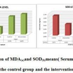 |
Figure 1: Comparison of MDA(a)and SOD(b)means( Serum and Hepatic tissue) Betweenthe control group and the intervention group. |
Discussion
One burning cigarette produces about 5,000 mg of gas (92%) and solid particles (8%). Cigarette smoke content free radicals. Free radicals are a form of reactive oxygen species (ROS) that highly reactive(18).Free radicals can be formed endogenously (derived from normal responses to intracellular or extracellular biochemical processes);and exogenously (derived from pollution, food and other materials such as cigarettes). Under stress conditions, the formation of reactive oxygen compounds is higher than its destruction, triggering the defense of the body’s antioxidant system.There is a gas phase of cigarette smokethat is rich in free radicals, and non-radical oxidants, such as superoxide radicals (.O2–), hydroxyl radicals (.OH), peroxyl (.ROO), and hydrogen peroxide (H2O2). The main target of free radicals is protein, unsaturated fatty acids and lipoproteins, and DNA. The most vulnerable to free radical attacks are unsaturated fatty acids. ROS can attack polyunsaturated fatty acids (PUFA) in cell membranes, causing lipid peroxidation(19).Lipid peroxidation is the initiation of a chain reaction by hydrogen or oxygen radicals, resulting in oxidized unsaturated fatty acids (PUFA).MDAis an oxidation product of unsaturated fatty acids by free radicals(18)(12).
The results showed that no significant differences between the control and treatment group in MDA both serum and hepatic, indicating that lipid peroxidation was not significantly affected by the intervention. Indeed, the stingless bee honey (Kele-honey) (given every day at a dose of 2.5 gr/kg BW) treatment in this studywas not necessarily reduced MDA levels as oxidative stress biomarkers. The results of this study are similar to the study of Shi et al which showed no significant difference in the serum MDA levelof mice exposed to alcohol after given honey(7).Increased free radicals will induce lipid peroxidation. MDA is a biomarker of the lipid peroxidation process that occurs due to an attack of free radicals on lipids, mainly polyunsaturated fatty acids (PUFAs)(20).This resulting study was contradictive to previous in vitro study that showed MDA levels were significantly reduced by the honey intervention(12).
Furthermore, there were significant differences in SOD levels in both serum and hepatic however SOD levels in the treatment group were lower than those control groups. This indicates thatthe treatment ofstingless bee honey (Kele-honey) in this study did not have a significant impacton supporting the endogenous antioxidant system increasement. The Kele-honeyused in this study had antioxidant total phenol (152.76 mg / 100g GAE) and a flavonoid (92.90 mg / 100g. Similar to Tualang honey, Manuka honey and other with antioxidant phenolics and flavonoid activities (6).A previous study exhibited that, there was a significant increase in lipid peroxidation along with a decrease in SOD in mice exposed to cigarettes smokes. In the event of ROS generation is higher than the scavenging activities will induce oxidative stress, which results in damage to the structure and function of cells(4).
Normally, cells have a defense mechanism against damage caused by free radicals, including antioxidant enzymes (thioredoxins, SOD, CAT, and GSH-Px) and antioxidants nonenzymes (ascorbic acid, vitamin E, and glutathione). However, an increase in ROS production beyond the antioxidant defense system causes oxidative stress or cellular damage.SOD is an enzyme that catalyzes superoxide radicals (O2•) to hydrogen peroxide (H2O2) and water through the reduction and oxidation reactions of manganese (Mn), zinc (Zn), and copper (Cu). An increase or decrease in SOD levels can indicate a change in oxidative status in the body.The mechanism of antioxidant flavonoids that is, directly scavenger free radicals by donating hydrogen atoms, forming chelate bonds with metals such as Fe2+ and Cu+ to prevent oxygen metabolism and radical formation. Flavonoids inhibit enzymes that produce free radicals such as xanthine oxidase, protein kinase C,lipoxygenase, cyclooxygenase, microsomal monooxygenase, mitochondrial succinoxidase, and NADPH oxidase(10). SOD, as a free radical scavenging enzyme along with GSH-Px, and GST, etc. are the first line of defense against oxidative damage (7).Based on this mechanism, low SOD levels in the treatment groupmight be related to the antioxidant activity of honey.The decrease in SOD that occurs, indicates that the antioxidant capacity is low in scavenging ROSor preventing the free radical’s generation. It can be assumed that it is necessary to increase the dose of honey administration as well as the treatment as consideration, of course,scientific re-research needs to be done.
Several studies explain that honey has high antioxidant activity. Honey contains polyphenolic compounds, flavonoids, and flavonols which play an important role in repairing oxidative stress. Phenol is very effective as an antidote to peroxyl radicals(10).As antioxidants, phenolic compounds have the ability as a reducing agent(5).Based on these, theoretically,it is expected that honey can prevent an increase and even decrease MDA levels associated with its antioxidant activity. However, this study was shown in inverse results.The composition and physicochemical properties of honey varybased on the type of plant from which bees consume nectar(6).Not only the type of flora, but also climatic conditions, and geographical areas affect the physical and chemical properties of honey(11)(12). Moreover, the composition of honey is also strongly influenced by how the honey is processed, and storage techniques(21).Although the consumption of stingless be honey (kele honey) as antioxidant-rich food, in this study the serum concentration of SOD was even lower significantly. It is assumed that antioxidant supplementation might be affected by many factors such as a degree of absorption, intake of other nutrients, homeostatic regulation, etc.
This is well-known that antioxidants can against free radicals through several ways such aspreventing the formation of new free radicals, breaking the chain reaction, repairing damage caused by free radicals.Antioxidant compounds work by donating one electron to the oxidant molecule so that it becomes less reactive and more stable.Kele honey contains phenolic and flavonoid properties that are responsible for stabilizing ROS through the release of hydrogen from one of the hydroxyl groups(21).
Conclusion
Stingless bee honey (Kele-honey) with a dose of 2.5 mg/kgBWinhibited not a significant increase of MDA levels and inhibited the significant decrease of SOD level both in serum and hepatic tissue of Wistar exposed to cigarettes smoke.
Financial Support
The research financial was supports from the Lembaga Penelitian dan PengabdianKepada Masyarakat, Udayana University, Denpasar, Bali.
Authors’ Contribution
IDAIDP, SP, IWS conceived the idea. IDAIDP and IWS performed the experiments. IDAIDP and SPwere conduct data analyzed. All authors wrote the manuscript.
Conflicts of Interest
There are no conflicts of interest.
References
- Primayanti D, Aman, Bagiada A. Ipomoea Batatas Syrup Decrease Malondialdehyde and Increase Nitrous Oxide Plasma Levels Amongst. 2012;1(3):125–30.
- Hu JP, Zhao XP, Ma XZ, Wang Y, Zheng LJ. Effects of cigarette smoke on aerobic capacity and serum MDA content and SOD activity of animal. Int J Clin Exp Med. 2014;7(11):4461–5.
- Khan SU, Khan RA, Khan WU. Phytochemical Screening and In vitro Antioxidant Activities of Methanolic Extract of Nigella sativa Seeds. World Appl Sci J [Internet]. 2017;35(6):971–5. Available from: http://www.idosi.org/wasj/wasj35(6)17/19.pdf
- Mohamed M, Sulaiman SA, Jaafar H, Salam KN. Antioxidant protective effect of honey in cigarette smoke-induced testicular damage in rats. Int J Mol Sci. 2011;12(9):5508–21.
CrossRef - Can Z, Baltaş N, Keskin S, Yıldız O, Kolaylı S. Properties of Antioxidant and Anti-Inflammatory Activity and Phenolic Profiles of Şevketi Bostan (Cnicus benedictus L.) Cultivated in Aegean Region from Turkey. Turkish J Agric – Food Sci Technol. 2017;5(4):308.
CrossRef - Ahmed S, Othman NH. Review of the medicinal effects of tualang honey and a comparison with Manuka honey. Malaysian J Med Sci. 2013;20(3):6–13.
- Shi P, Chen B, Chen C, Xu J, Shen Z, Miao X, et al. Honey reduces blood alcohol concentration but not affects the level of serum MDA and GSH-Px activity in intoxicated male mice models. BMC Complement Altern Med. 2015;15(1):1–6.
CrossRef - Alvarez-Suarez JM, Tulipani S, Romandini S, Bertoli E, Battino M. Contribution of honey in nutrition and human health: A review. Med J Nutrition Metab. 2010;3(1):15–23.
CrossRef - Pereira C, Grácio D, Teixeira JP, Magro F. Oxidative Stress and DNA Damage: Implications in Inflammatory Bowel Disease. Inflamm Bowel Dis. 2015;21(10):2403–17.
CrossRef - Varsha VK, Eswaran U, Priya V, Bhargava HR. A Comparative Study of the Biochemical , Antioxidative and Anti-microbial Activity of Apis and Trigona Honey Collected from Different Geographical Areas of India. World Appl Sci J. 2015;33(1):160–7.
- Rao PV, Krishnan KT, Salleh N, Gan SH. Biological and therapeutic effects of honey produced by honey bees and stingless bees: A comparative review. Brazilian J Pharmacogn [Internet]. 2016;26(5):657–64. Available from: http://dx.doi.org/10.1016/j.bjp.2016.01.012
CrossRef - Hilary S, Habib H, Souka U, Ibrahim W, Platat C. Bioactivity of arid region honey: An in vitro study. BMC Complement Altern Med. 2017;17(1):1–10.
CrossRef - Erejuwa OO, Sulaiman SA, Ab Wahab MS. Honey: A novel antioxidant. Molecules. 2012;17(4):4400–23.
CrossRef - Erejuwa OO, Sulaiman SA, Ab Wahab MS. Effects of honey and its mechanisms of action on the development and progression of cancer. Molecules. 2014;19(2):2497–522.
CrossRef - Rosyidi D, Eka Radiati L, Minarti S, Mustakim M, Susilo A, Jaya F, et al. Perbandingan Sifat Antioksidan Propolis pada Dua Jenis Lebah (Apis mellifera dan Trigona sp.) di Mojokerto dan Batu, Jawa Timur, Indonesia. J Ilmu dan Teknol Has Ternak. 2018;13(2):108–17.
CrossRef - Silverthorn DU. Human Physiology. An Integrated Approach Fifth Edition. 8th ed. USA: Pearson Education.Inc; 2010. 215–446 p.
- Wannamethee SG, Shaper AG. Cigarette smoking and serum liver enzymes: The role of alcohol and inflammation. Ann Clin Biochem. 2010;47(4):321–6.
CrossRef - Indasari EN, Marhendra APW, Wardhana AW. Extract Bee Propolis (Trigona sp) for Preventive Increase Protease Activity and Defect of Trachea Histology in Rats (Rattus norvegicus) Exposed to Cigarette Smoke. IOP Conf Ser Earth Environ Sci. 2019;391(1).
CrossRef - Kamceva G, Arsova-Sarafinovska Z, Ruskovska T, Zdravkovska M, Kamceva-Panova L, Stikova E. Cigarette smoking and oxidative stress in patients with coronary artery disease. Maced J Med Sci. 2016;4(4):636–40.
CrossRef - Wiraguna AAGP, Dianasari R, Pangkahila W. The Topical Skin Application of Purple Corn Extract (Zea mays) Inhibited the Increase in MMP-1 Levels and Decreased Collagen in Wistar rats (Rattus norvegicus) Exposed to UV-B Rays. Biomed Pharmacol J. 2019;12(1):297–304.
CrossRef - Cianciosi D, Forbes-Hernández TY, Afrin S, Gasparrini M, Reboredo-Rodriguez P, Manna PP, et al. Phenolic compounds in honey and their associated health benefits: A review. Molecules. 2018;23(9):1–20.
CrossRef

