
Manuscript accepted on :15-Jun-2020
Published online on: --
Plagiarism Check: Yes
Reviewed by: Hanefi Özbek
Second Review by: Cherry Bansal
Swee-Hua Erin Lim1*, Maitha Ahmed Almakhmari1, Shaima Ibrahim Alameri1, Swee-Yee Chin2, Aisha Abushelaibi1, Chun-Wai Mai 2,3 and Kok-Song Lai1
1Health Sciences Division, Abu Dhabi Women’s College, Higher Colleges of Technology, Abu Dhabi, United Arab Emirates, 41012
2Department of Pharmaceutical Chemistry, School of Pharmacy, International Medical University, Kuala Lumpur, Malaysia, 57000
3Centre for Cancer and Stem Cells Research, Institute for Research, Development and Innovation, International Medical University, Kuala Lumpur, Malaysia, 57000
Corresponding Author E-mail: lerin@hct.ac.ae
DOI : https://dx.doi.org/10.13005/bpj/1984
Abstract
Clinacanthus nutans is widely used as an antimicrobial, anti-inflammatory, anti-pyretic and cytotoxic agent in Asian countries such as Thailand and Malaysia. Our previous study suggested C. nutans may exhibit anti-inflammatory effects. However, there are limited studies at current for the antimicrobial affect for this plant; hence this study was carried out to investigate the minimum inhibitory concentration (MIC) of C. nutans polar and non-polar leave and stem extracts against twelve gram- negative and positive bacteria. Briefly, the plant parts were separated, dried and powdered, then the extracts were prepared as polar and non-polar extracts from leaves and stem. The result showed inhibition effects in all of the bacteria tested. Our findings from this study indicated that non-polar leaves extracts are more potent as an antimicrobial agent compared to polar leaves, non-polar stem and polar stem. Future studies are warranted for further identification of bioactive compounds in C. nutans with anti-bacterial activities.
Keywords
Antibacterial; Antimicrobial activity; Anti-inflammator; Bacterial resistance; Clinacanthus nutans; Natural product.
Download this article as:| Copy the following to cite this article: Lim W. H. E, Almakhmari M. A, Alameri S. I, Chin S. Y, Abushelaibi A, Mai C. W, Lai K. S. Antibacterial Activity of Clinacanthus nutans Polar and Non-Polar Leaves and Stem Extracts. Biomed Pharmacol J 2020;13(3). |
| Copy the following to cite this URL: Lim W. H. E, Almakhmari M. A, Alameri S. I, Chin S. Y, Abushelaibi A, Mai C. W, Lai K. S. Antibacterial Activity of Clinacanthus nutans Polar and Non-Polar Leaves and Stem Extracts. Biomed Pharmacol J 2020;13(3). Available from: https://bit.ly/3iL0PJq |
Introduction
Despite more aggressive antimicrobial stewardship being promoted, the evolution of bacterial infections fueled by excessive antibiotic pressure is still a pressing problem in the clinical setting.1 Due to limitations in the development of new antibiotics, researchers are turning to natural products to aid in drug discovery.2-3
Clinacanthus nutans in the Acanthaceae family, also known as ‘belalai gajah’ is grown in tropical weathered countries in Asia such as in Malaysia, Thailand and China.4 C. nutans was used traditionally by Asians as an antifungal,5 cytotoxic,6 anti-inflammatory,7 antioxidant,8 and anti-pyretic9 agent. In addition, C. nutans had been used to treat certain diseases like skin rash, diabetes and insect bites.10 Traditionally, the dried leaves of C. nutans had been mixed with fresh juices or green tea, or the leaves are put directly on the infected skin with nettle rashes. Fazil et al. (2016)9 and Mai et al. (2009)11 described the antiproliferative effects of flavonoids from C. nutans fresh and dried leaves against lung cancer cells, Both, the fresh and the dried leaves possessed similar effect9, 11 In addition, Alam et al. (2016)12 proved that the stem of C. nutans showed a wide range of properties such as anti-viral and anti-inflammatory activities extracted from ethanol 80%, while the leaves show a broad cytological activity, cholinergic modulation and anti-papillomavirus infectivity from petroleum ether extract.12 C. nutans leaves showed an inhibition effect on fungi like P. oryzae5 and antibacterial activity on the gram-positive bacteria was greater than the gram-negative bacteria; this study was done using stem and leaves (crude extracts) on three bacteria, that are Bacillus cereus, Escherichia coli and Salmonella enterica.6
Our previous study identified that the polar leaves C. nutans extracts exhibited anti-inflammatory effects by inhibiting the lipopolysaccharides (LPS) induced Toll-like receptor-4 (TLR-4) activation, nitric oxide accumulation, inflammatory cytokines reduction and TLR-4 related inflammatory proteins expressions.13 Therefore, in our study, we will investigate the antibacterial effect in gram-positive and gram-negative bacteria using the polar and non-polar stem and leaves extracts.
Material and Methods
Bacteria
The collection of bacteria was purchased from Institute of Medical Research, Kuala Lumpur, Malaysia. The bacteria are: Bacillus cereus, Bacillus subtilis, Enterobacter, Escherichia coli, Enterobacter aerogenes, Enterococcus faecalis, Klebsiella pneumoniae, Proteus vulgaris, Pseudomonas aeruginosa, Staphylococcus aureus, Staphylococcus epidermidis, Staphylococcus saprophyticus. They were maintained on tryptic soy agar (TSA; Oxoid, UK), while performing the antimicrobial testing, Mueller Hinton broth (Oxoid, UK) was used.
Preparation of the Extracts
The plant was collected from previous study and extracted using the same procedure.13 Briefly, the plant was dried, powdered and separated into leaves and stem bark. The powdered stem and leaves were extracted with non-polar solvents (diethyl ether and hexane) or polar solvents (dichloromethane and methanol) by immersion it in the solvent at room temperature for three days. Four extracts were prepared, namely non-polar leave extracts (LN), polar leave extracts (LP), non-polar stem extracts (SN) and polar stem extracts (SP). The extracts were filtered and then we removed the solvents by rotary evaporator at 60°C. The concentrations of each extract used for testing were 0.25, 0.5, 1, 2, 4, 8, 16, 32 mg/mL.
Broth Microdilution Test
The broth microdilution method was used to help to determine the minimum inhibitory concentration (MIC). Testing was carried out based on the CLSI M07-A8 guidelines,14 but with some changes to aid visualization: resazurin (7-hydroxy-3H-phenoxazin-3-one 10-oxide) (Sigma Aldrich, USA) was added to a final concentration of 0.002%. In summary, dilutions were done in triplicate, each well containing 40µL of bacterial suspension (final concentration of 2-8 x 10⁵ cfu/mL) with 50µL of test compound that inoculated with 10µL of resazurin. Gentamicin (Bioworld, USA), at 32µg/mL for the highest concentration (Oxoid, UK) and DMSO at 5% at highest concentration were used as negative and positive controls respectively. Disks were obtained from Oxoid, UK. The plates were incubated for 18h at 37°C. The MIC value was taken at the lowest concentration at the resazurin color change from purple to pink or colorless indicating the reduction in the growth of the bacteria, resazurin (7-Hydroxy-3H-phenoxazin-3-one 10-oxide). Pink demonstrates bacteria that are alive, while the blue demonstrates the death of bacteria.
Statistical Analysis
All data were presented as mean ± standard deviation (S.D.) from at least three independent experiments. SPSS (version 18.0) for Windows was used to conduct the one way Analysis of Variance (ANOVA) post hoc Dunnett’ t-test, in which statistically significant was defined as p-value was not more than 0.05.
Results and discussion
All four extracts from C. nutans showed growth inhibition in all twelve bacteria species as extract concentration increased (Figure 1). LN had the strongest inhibitory effect in all bacteria compared to LP and SP, as well as SN. Figure 1 shows the effects of different antibiotic’s concentrations extracted from the C. nutans LN on twelve species of bacteria. The effective concentration ranged between 0.25 mg/mL which was the lowest concentration and 1 mg/mL. For example, P. mirabilis viability was dropped to 41% while P. mirabilis and K. pneumoniae dropped to slightly above 30%. As for Enterococcus faecalis, it dropped to only 20% viability. E. coli being the most effected showed viability at percentage of 5% at 16mg/mL, followed by P. vulgaris with 7.18% and B. subtilis with 8.16%. Lastly, the viability percentage of all the bacteria species increased slightly when the concentration of the antimicrobial doubled to 32 mg/mL. Table 1 represents the MIC50 and MIC90 values in mg/mL of LN extracted from C. nutans.
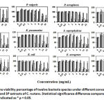 |
Figure 1: The viability percentage of twelve bacteria species under different concentrations of LN, LP, SN and SP extracts of C. nutans. Statistical significance difference compared to vehicle control is indicated as * p < 0.05 |
Table 1: MIC50 and MIC90 values in mg/mL for LN and LP extracts of C.nutans.
| Bacteria | Leaf Non Polar (LN) | Leaf Polar (LP) | ||
| MIC50 (mg/mL) | MIC90 (mg/mL) | MIC50 (mg/mL) | MIC90 (mg/mL) | |
| S. saprophyticus | 4.76 | 6.99 | 6.66 | 9.14 |
| S. epidermidis | 5.80 | 7.05 | 7.52 | 9.70 |
| S. aureus | 4.75 | 7.05 | 5.58 | 9.44 |
| P. aeruginosa | 4.34 | 6.94 | 6.66 | 10.04 |
| P. vulgaris | 5.17 | 7.24 | 7.95 | 11.10 |
| P. mirabilis | 5.81 | 7.83 | 8.82 | 12.49 |
| E. faecalis | 5.98 | 7.20 | 6.86 | 10.03 |
| E. coli | 4.39 | 6.79 | 6.04 | 9.48 |
| E. aerogenes | 5.94 | 7.87 | 8.93 | 12.30 |
| B. subtilis | 5.08 | 6.88 | 7.05 | 9.36 |
| B. cereus | 5.26 | 7.19 | 6.57 | 8.62 |
| K. pneumoniae | 5.65 | 7.72 | 8.29 | 11.29 |
For the LP extracts, a dose-response viability rate was observed in twelve species of bacteria. Figure 1 compares the viability percentage changes after treating the bacteria with different concentrations of LP extracts. Concentration levels of 0.25 to 4 mg/mL show an almost constant viability percentage of 81% for S. aureus. However, the percentage fell noticeably from 81% to 20.26% at 16 mg/mL. Bacterial species like S. epidermidis, B. subtilis, E. coli, P. aeruginosa showed a drop in viability percentage when antimicrobial concentration increased from 4 to 6 mg/mL. However, for the bacteria species like E. faecalis, P. mirabilis, K. pneumoniae and P. vulgaris their viability percentage decrease when the concentration of the antimicrobial increased from 8 to 16 mg/mL. B. subtilis shows to be the most effected by the treatment compared to the others, the viability showed an early drop in percentage, it was 141% at 2mg/mL and reached a value of 26% at 16 mg/mL. Table 1 shows the MIC50 and MIC90 values in mg/mL of LP extracted from C. nutans.
For the activity of SN extract, the reduction of growth was not significant for all bacteria tested compared to other extracts (Figure 1). Table 2 shows the MIC50 and MIC90 values in mg/mL of SN extracted from C. nutans. The highest reduction of viability was to 62.73% at 32mg/mL for P. vulgaris which is higher than the other extracts. The polar stem was more effective than non-polar, because the highest reduction in growth compared to non-polar stem was to 12% at 32mg/mL for E. coli. The growth of B. cereus was low in 0.25 to 1 mg/mL but then it peaks at 2 mg/mL to 209% and then sharply went down at 4 mg/mL to 83%. Table 2 represents the MIC50 and MIC90 values in mg/mL of SP extracted from C. nutans.
Table 2: Represents the MIC50 and MIC90 values in mg/mL of SP extracted from C. nutans.
| Bacteria | Stem Non Polar (SN) | Stem Polar (SP) | ||
| MIC50 (mg/mL) | MIC90 (mg/mL) | MIC50 (mg/mL) | MIC90 (mg/mL) | |
| S.saprophyticus | 22.58 | 33.50 | 6.71 | 9.17 |
| S. epidermidis | 15.50 | 20.61 | 7.60 | 9.97 |
| S. aureus | 13.23 | 19.30 | 6.10 | 10.23 |
| P. aeruginosa | 21.58 | 35.95 | 6.27 | 9.48 |
| P. vulgaris | 10.86 | 15.85 | 8.49 | 11.80 |
| P. mirabilis | 46.08 | 69.18 | 9.10 | 13.31 |
| E. faecalis | 22.23 | 34.99 | 6.53 | 9.72 |
| E. coli | 20.46 | 35.53 | 6.06 | 9.63 |
| E. aerogenes | 19.78 | 29.66 | 9.18 | 12.98 |
| B. subtilis | 14.05 | 19.84 | 7.17 | 9.72 |
| B. cereus | 22.50 | 32.94 | 7.25 | 9.57 |
| K. pneumoniae | 30.81 | 46.88 | 8.19 | 11.70 |
In the present study, leaves and stems extracts obtained from C. nutans were evaluated as an antimicrobial agent against twelve bacteria. The C. nutans is a herb known for its medical properties. This plant grows in a tropical climate, in countries like Malaysia and Thailand. The C. nutans therapeutic characteristics has spurred the interest and attention of many researchers. This medical herb has numerous chemical compounds that possess antibacterial activities such as, β-sitosterol, stigmasterol and flavonoids.15 The population of South East Asia traditionally used this plant to treat various medical conditions such as insect bites, skin rashes, herpes infection, inflammation, and cancer. Moreover, the C. nutans plant has been shown to possess antimicrobial activity. Thus, it is used to treat various skin conditions that are caused by bacterial colonization in Thai traditional medicine.16
The results of our experiment showed that leaves extract was more potent antimicrobial compared to stems at the validity of 5% in concentration of 16 mg/mL. In a past study, Justicia adhatoda from the Acanthaceae family species was tested for the antimicrobial effects of the methanolic and leaf extract of the plant. The results indicated that the antimicrobial effects of the leaf extract surpassed the methanol extract.17 Moreover, the same study was conducted on Crossandra infundibuliformis (Acanthaceae family), and the results illustrated that the ethanol extracted from the leaf of the plant has better antibacterial activity compared to others.18 Hence, this confirmed that the leaf extracted from C. nutans is stronger than the stems since all plants are from the same family.
The non-polar extracts overall indicated a more effective antimicrobial effect compared to polar extract at validity of 5% in concentration of 16 mg/mL. In a past study, the non-polar extracts showed more effective antimicrobial activity against gram-positive bacteria.19 In addition, another study showed that chloroform is the best solvent that has been extracted from non-polar solvents from Angelica archanglicafor biological activity.20
In our study, the antimicrobial activity of C. nutans extracts was more effective against the gram-negative bacteria than the gram-positive. In fact, the inhibitory results found against gram-negative bacteria were unexpected, due to the fact that usually they are more resistant compared to gram-positive bacteria. In general, the cell wall of the gram-positive bacteria mainly consists of peptidoglycan, and it represents 90% of the cell wall. On the other hand, the gram-negative bacteria have a more complex cell wall. Similarly, to the gram-positive bacteria, the gram-negative bacteria cell wall is composed of peptidoglycan. However, peptidoglycan represents 5-10% of the total cell wall. In addition, the gram-negative bacteria have an outer layer that is known as lipopolysaccharide (LPS).21 C. nutans extracts may possibly have more effect to LPS than the peptidoglycan. However, Andrographis paniculata (Acanthaceae family), a plant from the same family as C. nutans showed antibacterial effects towards gram-positive bacteria. This may be because the mesh-like peptidoglycan layer is more accessible to the extracts which permeates the cell wall. The outer membrane of the gram-negative cell wall may contribute to their resistance against C. nutans as the membrane is composed of lipopolysaccharide which restricts entry of the plant extract.
Conclusion
In this study, the results revealed that LN of C. nutans have a stronger antibacterial activity than LP, NS and NP. The gram-negative bacteria were more sensitive to the extracts compared to gram-positive bacteria. Due to limited studies on C. nutans extracts, further studies must be conducted to elucidate the pharmacologically active compounds from this plant. This may lead to the development of a new generation of antibiotics.
Acknowledgment
The authors are grateful to the Advanced Technology Entrepreneur Program (ADAPT) grant from Majlis Amanah Rakyat (MARA), International Medical University and the HCT Interdisciplinary Research Grant (113118) from the Higher Colleges of Technology for supporting this study.
Conflict of Interest
The authors declare they have no conflicts of interests.
Funding source
HCT Interdisciplinary Research Grant (113118) from the Higher Colleges of Technology.
References
- Moo, C. L., Yang, S. K., Yusoff, K., Ajat, M., Thomas, W., Abushelaibi, A., … Lai, K. S. Mechanisms of antimicrobial resistance (AMR) and alternative approaches to overcome AMR. Current Drug Discovery Technologies., 2019; 16.
- Yang S.K., Low L.Y., Yap P.S.X., Yusoff K., Mai C.W., Lai K.S. and Lim S.H.E. Plant-Derived Antimicrobials: Insights into Mitigation of Antimicrobial Resistance. Nat. Prod., 2018; 12(4): 295–396.
CrossRef - Nik A.M., Yang S.K., Moo C.L., Song A.L.A., Chong C.M., Chong C.W., Abushelaibi A., Lim S.H.E. and Lai K.S. Terpene Derivatives as a Potential Agent against Antimicrobial Resistance (AMR) Pathogens. Molecules., 2019; 24: 2631.
CrossRef - Hii L.W., Lim S.W.E, Leong C.O., Chin S.Y., Tan N.P., Lai K.S. and Mai C.W. The synergism of Clinacanthus nutans Lindau extracts with gemcitabine: downregulation of anti-apoptotic markers in squamous pancreatic ductal adenocarcinoma. BMC Compl. Alternative Med., 2019; 19: 257.
- Uda M.N.A., Harzana Shaari N., Shamiera Said N., Ibrahim N.H., Akhir M.A.M, Rabani Hashim M.K., Salimi M.N., Nuradibah M.A., Uda H. and Gopinath S.C.B. Antimicrobial Activity of Plant Extracts from Aloe Vera, Citrus Hystrix, Sabah Snake Grass and Zingiber Officinale against Pyricularia Oryzae that causes Rice Blast Disease in Paddy Plants. IOP , Conference Series: Materials Science and Engineering., 2018; 318.
CrossRef - Arullappan S., Rajamanickam P., Thevar N. and Kodimani C. In Vitro Screening of Cytotoxic, Antimicrobial and Antioxidant Activities of Clinacanthus nutans (Acanthaceae) leaf extracts. J. Pharm. Res., 2014; 13: 1455.
CrossRef - Yap P.S.X, Chong Y.T.E., Wong Y.Y., Lim X.Y., Ang H.S., Goh H.S.L., Mai C.W., Buru S.A., Pichika M.R. and Lim S.H.E. Antibacterial and antifungal testing of the different extracts of Dillenia obovata (Blume) Hoogl. World J. Pharm. Pharmaceut. Sci., 2013; 2: 3946-3962.
- Kong H.S., Musa K.H. and Abdullah Sani, N. Clinacanthus nutans (Belalai Gajah / Sabah Snake Grass): Antioxidant optimization on leaves and stems. AIP Conference Proceedings., 2016; 1784: 030030.
CrossRef - Fazil F.N.M., Azzimi N.S.M, Yahaya B.H., Kamalaldin N.A. and Zubairi S.I. Kinetics Extraction Modelling and Antiproliferative Activity of Clinacanthus nutans Water Extract. Sci. World J., 2016; 1–7.
CrossRef - Solibun A. and Sivakumar K. Sabah snake grass extract pre-processing: Preliminary studies in drying and fermentation. IOP Conference Series: Earth and Environmental Science., 2016; 36: 012066.
CrossRef - Mai C.W., Pakirisamy P., Tay E.F., Subramaniam B., Shamsuddin Z.H. and Pichika M. Nasopharyngeal Carcinoma Cell Proliferation and Apoptosis Induced by the Standardised Ethanolic Extracts of Mucuna bracteate. M. J. Chem., 2009; 1: 143-148.
- Alam A., Ferdosh S., Ghafoor K., Hakim A., Juraimi A.S., Khatib A. and Sarker Z.I. Clinacanthus nutans: A review of the medicinal uses, pharmacology and phytochemistry. Asian Pac. J. Trop. Med., 2016: 9: 402–409.
CrossRef - Mai C.W., Yap I.K.S, Kho M.T., Ismail N.H., Yusoff K., Shaari K., Chin S.Y. and Lim, S.H.E. Mechanisms Underlying the Anti-Inflammatory Effects of Clinacanthus nutans Lindau Extracts: Inhibition of Cytokine Production and Toll-Like Receptor-4 Activation. Front. Pharmacol., 2016; 7:7.
CrossRef - Cockerill F.R., Wikler M.A., Alder J, Dudley M.N., Eliopoulos G.M., Hardy D.J., Hecht D.W., Hindler J.A., Patel J.B., Powell M., Thomson R.B.J., Turnidge, J.D., Weinstein M.P. and Zimmer B.L. Methods for Dilution Antimicrobial Susceptibility Tests for Bacteria That Grow Aerobically. Clinical and Laboratory Standards Institute., 2012; 18.
- Xie Y., Yang W., Tang F., Chen X. and Ren L. Antibacterial Activities of Flavonoids: Structure-Activity Relationship and Mechanism. Curr. Med. Chem., 2015; 22(1): 132-49.
CrossRef - Yang H. S., Peng T.W, Madhavan P., Shukkoor M.A. and Akowuah G.A. Phytochemical analysis and antibacterial activity of methanolic extract of Clinacanthus nutans leaf. Inter. J. Drug Dev. & Res., 2013; 5: 349–355.
- Sharma A. and Kumar A. Antimicrobial Activity of Justicia Adhatoda. World J. Pharm. Res., 2016; 7: 1332-1341.
- Sharmila N. and Gomathi N. Antibacterial, Antioxidant activity and Phytochemical studies of Crossandra infundibuliformis leaf extracts. Int. J. Phytomedicine., 2011; 3: 151-156.
- Junior I.E.S., Filho V.C, Zacchino S.A., Lima J.C da S. and Martins D.T. de O. Antimicrobial screening of some medicinal plants from Mato Grosso Cerrado. Rev. bras. , 2009; 19: 242–248.
CrossRef - Das K., Tiwari R., Shrivastava D. and Bilaspur B.C. Techniques for evaluation of medicinal plant products as antimicrobial agent: Current methods and future trends. J. Mol. Pharm. Res., 2011; 4: 104–111.
- Yang S.K., Yusoff K., Ajat M., Warren T., Abushelaibi A., Riaz A., Lim S.H.E. and Lai K.S. Disruption of KPC-producing Klebsiella pneumoniae membrane via induction of oxidative stress by cinnamon bark (Cinnamomum verum J. Presl) essential oil. PLoS One., 2019; 14(4): e0214326.
CrossRef

