
Manuscript accepted on :14-04-2020
Published online on: 23-06-2020
Plagiarism Check: Yes
Reviewed by: Sasho Stoleski
Second Review by: Serghei Covantev
Final Approval by: Guy Armer Bounda
Dejidmaa Buyantogtokh1* , Erdenechimeg Chuluunbaatar1, Munkhtuul Tsogzol1, Nyamdolgor Uranbileg2, Chimedragchaa Chimedtseren1 and Begzsuren Dagvatseren1
1Department of Pharmacology, Institute of Traditional Medicine and Technology, Ulaanbaatar, 17032, Mongolia
2Department of Pathology, Institute of Veterinary Medicine, Ulaanbaatar, 17024, Mongolia
Corresponding Author E-mail: b.dejidmaa@yahoo.com
DOI : https://dx.doi.org/10.13005/bpj/1935
Abstract
Calvacin gel is composed Calvatia gigantea ex Pers and Gentiana macrophylla Pall which usually used for burn and aseptic, lining and cutting wound in traditional Mongolian medicine. The objective of the study to determine technological parameter and total biological active substances and analyze the effect of burn healing Calvacin gel in rats with full-thickness second-degree burn model. Methods: Quantitative determination of the organic acid of the gel of Calvacin was performed using titremeter method respectively. Standard method was used to induce burn wound on in rats and the animals were divided into 4 groups: Control group, Calvacin-1, Calvacin-2, Silver sulfadiazine. After 24 hours of burn model being induced, we have put a thin layer of experimental gel and Silver sulfadiazine once a day. Wound area was measured every three day for 28 days under careful observation, and on the day of 7, 14, 21, 28 blood samples were taken from the animals in order to determine level of VEGF, TGFβ1 to evaluate wound healing process. Results: For choosing of appropriate gel former, gels were prepared by 1% and 2% carbomer, 3%, 6% sodium carboxymethyl cellulose. The total content of organic acid 1.3±0.08%, 1.29±0.1%,1.28±0.09% 1.29±0.09%. The rats treated with Calvacin had significantly increased levels of VEGF (at 14, 21 and 28 day), and TGFβ1 (at 28 day). Conclusions: The current study showed that Calvacin has a burn healing activity.
Keywords
Burn Wound; Calvatia Gigantea Batch ex Pers; Gentiana Macrophylla Pall; Traditional Mongolian Medicine
Download this article as:| Copy the following to cite this article: Buyantogtokh D, Chuluunbaatar E, Tsogzol M, Uranbileg Nyamdolgor, Chimedtseren C, Dagvatseren B. Wound Healing Effects of Calvacin Gel on Burn Wound in Rats. Biomed Pharmacol J 2020;13(2). |
| Copy the following to cite this URL: Buyantogtokh D, Chuluunbaatar E, Tsogzol M, Uranbileg Nyamdolgor, Chimedtseren C, Dagvatseren B. Wound Healing Effects of Calvacin Gel on Burn Wound in Rats. Biomed Pharmacol J 2020;13(2). Available from: https://bit.ly/380eFE8 |
Introduction
Burn is a major cause of mortality and morbidity in Mongolia and in the world. In recent years, burn is tend to increase in Mongolia, even though it covered over 80 percent of injuries among 0-5 year’s old children.1 A burn is defined as damage to the skin caused by excessive heat or caustic chemicals. The most common burn injuries result from exposure to heat and chemicals.2 Burn healing mechanism is a very complicated and affect many factors. Many of the raw materials used in traditional Mongolian medicine burn healing activity, wound healing, anti inflammatory, hemostasis. There are very few domestically made medicinal drugs available in our country, which support the process of wound healing and skin regeneration.3,4 Thus, based on the given fact it was necessary to invent preparation with an action of rapid wound healing, skin regeneration during healing process; as well as preventing from scarring after burn injury. As a result of years of study, our researchers have created preparation named Calvacin from Calvatia gigantea Batsch. ex Pers and Gentiana macrophylla Pall.5 Calvatia gigantea Batsch. ex Pers has wound healing,5 antioxidant, cytotoxicity, antibacterial, antifungal,6 antitumor activity.7 This study also evaluated the effects of Calvacin on levels of growth factors by observing histopathological features associated with standard method induced second degree burn.
Materilas and Methods
The study was carried out in accordance with the Health Ethics Guidelines issued by the Mongolian Ministry of Health (2018). The study protocol (№02/01/2018-06) was approved by members of “The Research Ethics Committee” and by the Institute of Traditional medicine and technology.
Plant Material
Spor of Calvatia gigantea Batsch. ex Pers. and herb of Gentiana macrophylla Pall. were collected as a raw material in Arbulag, Khuvsgul province, Mongolia in August 2018 and was identified by prof E. Ganbold, Sc.D in biology (voucher number №03/2018 and №04/2018). A voucher specimen of the plant under the number 2018/08/20/105 was deposited in the Herbarium of Pharmacology Department, Center of research, Institute of traditional medicine and technology. The herbs were air-dried and reduced to fine powder suitable for extraction.
Preparation of Liquid Extracts
The liquid extract of Calvatia gigantea Batsch. ex Pers. (1:2) and Gentiana macrophylla Pall. (1:1) by remaceration method and used 40% and 70% ethanol respectively.
Determination of Total Organic Acid
Transfer 2 g gel to conical flask or beaker. Add 50 ml of distilled water. Add 3-4 drops of phenolphthalein indicator and stir. Rapidly titrate the contents with 0.1 N NaOH solution, continue to add alkali drop by the drop and stirring the content till first definite change to pink colour.8 The contents of free organic acids in terms of malic acid in absolutely dry raw materials in percentage were calculated.
Thin Layer Chromatography
Thin layer chromatographic (TLC) plates, composed of Merck Silica gel 60 GF 254, received 5 µL of the test solutions placed at a distance of 1.5 cm of the lower edge of the plate. The mobile phase was chloroform / methanol / acetic acid (90:10:2, v/v). The standard substance was 5 mg/ml gentisic acid.
Preparation of Sodium Carboxymethyl Cellulose Gel Bases
The sodium carboxymethyl cellulose gel bases were prepared by dispersing the sodium carboxymethyl cellulose of 3% and 6% w/w in half of total distilled water (20 ± 2°C). The solution of humectant was prepared in the remaining amount of water. The solution of humectants was added at the end of dispersion stage. The pH value of gel bases was measured using pH meter (Hanna HI 98128, Germany) (n = 3). Viscosity of gel bases was measured using BDV-1S digital viscometer (Biobase, Karnataka, India) (n = 3)9.
Preparation of Carbomer Gel Bases
We prepared various carbomer gel bases with different ratio of gel former, antimicrobial preservative, neutralizer and humectant. At first stage, the solution of antimicrobial preservative and neutralizer were prepared in half of total water. At second stage, carbomer (1% or 2%) was dispersed using mixer (Akira HM-202BSS) in the solution of antimicrobial preservative and neutralizer. At third stage, the humectants were added very slowly at the dispersion stage. Triethanolamine was used as a neutralizer and the pH of the gel systems was adjusted by per cent of 1%, 1.1%, 1.2% w/w. As a conserver were used 0.18% methylparaben-0.02% propylparaben, 0.1% methylparaben, and 0.3% methylparaben. The pH value of gel bases was measured using pH meter (Hanna HI 98128, Germany) (n = 3). Viscosity of gel bases was measured using BDV-1S digital viscometer (Biobase, Karnataka, India) (n = 3)9
Formulation of Calvacin Gel
The spor of Calvatia gigantea Batsch. ex Pers and extract of Gentiana macrophylla Pall. was added by 5% (2:1) at the end of the dispersion stage. The gels had specific smell, dark brown to white brown color. The pH value of gels were measured using pH meter (Hanna HI 98128, Germany) (n = 3). Viscosities of gels were measured using BDV-1S digital viscometer (Biobase, Karnataka, India) (n = 3).9 The quality of the gel formulation was investigated comparatively by its appearance (color, smell), pH, viscosity, and bacterial contamination. Bacterial and mould contamination was defined according to MNS-5189-2002, MNS-5190-2002, MNS-5193-2002 and MNS-5194-2002.10-14
Animals
The wistar rats were purchased from the animal house of research center, Institute of traditional medicine and technology, Mongolia. 90 male wistar rat of weighing between 220-250 gm were purchased from the Experimental Animal Center, Institute of Traditional Medicine and Technology of Mongolia. They were kept under controlled conditions of temperature (20±1c°) and humidity (about 50-60%), with a 12-hour light/dark cycle, and automatic ventilation 8-15 times every hour. Rats could drink ad libitum, and were fed with standard nutrient.
Burn induction and treatment
90 male Wistar rats weighing were used in this study. Standard method was used to induce burn wound on in rats,15 and the animals were divided into 4 groups: 1. Control group, 2. Calvacin-1 (Calvatia gigantea Batsch. ex Pers was extracted gel), 3. Calvacin-2 (Calvatia gigantea Batsch.ex Pers not extracted gel), 4. Silver sulfadiazine (1% ointment). After 24 hours of burn model being induced, we have put a thin layer of experimental gel and Silver sulfadiazine once a day. Wound area was measured every three day for 28 days under careful observation, and on the day of 7, 14, 21, 28 blood samples were taken from the animals in order to determine level of VEGF and TGFβ1 to evaluate wound healing process. The study results were obtained by evaluating in serum level of VEGF and TGFβ1 and histomorphological examination.
Blood Samples
After 7, 14, 21, 28 days experimental rats from each group were anesthetized with ketamine hydrochloride (90 mg/kg, intraperitoneally). A 5 ml blood sample was collected from each rat by cardiac puncture. The serum was separated by centrifugation at 3000 rpm for 15 minutes. The level of serum VEGF and TGFβ1 SOD was measured by ELISA according following the kit’s instructions (Shanghai MLBIO Biotechnology Co.Ltd).
Histopathological Examination
Rats euthanized the end experiment. Burn wounds were excised down to the level of the muscle fascia by sharp dissection and included the surrounding wound margin tissue. Formalin-fixed skin specimens were prepared from four randomly chosen rats per group. Specimens were dehydrated in a series of increasing ethanol concentrations then embedded in paraffin. Tissue sections (5 μm) were stained with haematoxylin & eosin (HE) and Masson-trichrome. At least three slides were prepared from each specimen and blindly examined. Histopathological scoring was achieved via an expert pathologist using Nichon microscope for detection of pathological changes.
Statistical Analysis
Mean ± standard deviation (SD) were calculated for the observed values in each experimental group. Statistical analysis was done by two-way ANOVA followed by tukey post hoc test was performed. Graph Pad Prism-7.0 software was used for statistical analysis with p< 0.05 considered statistically significant.
Results
For choosing of appropriate gel former, gels were prepared by 1% and 2% carbomer, 3% and 6% sodium carboxymethyl cellulose. The result is shown in [Table 1].
Table 1: Quality of Calvacin Gel With Different Concentration and Various Gel Former
| No | Gel former | Quality criteria for Gel formulation | |||
| Appearance (specific smell, dark brown color) | Viscosity (Pa x s) | Ph
(Mean± SD) |
Total organic acid, % | ||
| 1 | Carbomer, 2% | + | 201.2 | 6.3±0.03 | 1.3±0.08% |
| 2 | Sodium carboxymethyl cellulose 3% | + | 15.5 | 6.1±0.02 | 1.29±0.1% |
| 3 | Sodium carboxymethyl cellulose 6% | + | 36.7 | 7.9±0.03 | 1.28±0.09% |
| 4 | Carbomer 1% | + | 93.6 | 6.02±0.02 | 1.29±0.09% |
Gels with carbomer gel base were prepared with various concentration triethanolamine and determined quality. The result is shown in [Table 2].
Table 2: Quality of Calvacin gel with different concentration triethanolamine
| Neutralizer | Quality criteria for Gel formulation | ||
| Appearance (specific smell, dark brown color) | Ph
(Mean± SD) |
Viscosity
(Pa x s) |
|
| Triethanolamine 1% | + | 6.34±0.03 | 93.6 |
| Triethanolamine 1.1% | + | 6.84±0.05 | 93.7 |
| Triethanolamine 1.2% | + | 7.21±0.06 | 93.7 |
For the determination of the quality of gel dependence on the antimicrobial preservative, we prepared gels using methylparaben-0.18% and propylparaben-0.02% combined methylparaben-0.1%, methylparaben-0.3%. The result is shown in [Table 3].
Table 3: Microbial contamination of Calvacin gel with various antimicrobials
| No | Antimicrobial preservatives | Microbial contamination | |
| Total bacteria | Mould | ||
| 1 | Methylparaben-0.1% | 3.5×10 | 1×10 |
| 2 | Methylparaben-0.3% | 3×10 | 1×10 |
| 4 | Methylparaben-0.18% and Propylparaben-0.02% | Not detected | Not detected |
TLC fingerprints of reference standard gentisic acid and various Calvatsin extracts are showed dark spots in 254 nm. All extracts presented chromatographic band corresponding to that of standard gentisic acid and Rf value was 0.71.
Wound Examination
The burn healing process was observed in 1, 7, 14, 21 and 28 days. A day 7 the Calvacin 1 and Calvacin 2 treated groups exhibited dark-brown, dry, hard scab. After day 14, the Calvacin-1, Calvacin-2 and SSD treated group exhibit small, thick scabs, but the control group showed dry, and dark brown scabs slightly decreased. After day 21, re-epithelialization was observed in all treated groups but the control group. Wounds healed most in the Calvacin 1 and Calvacin 2. But wound of control group a lot scab. By day 28, re-epithelialization was observed in all treatment groups and the control group (Fig. 1).
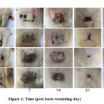 |
Figure 1: Time (post burn wounding day) |
Level of VEGF
VEGF level in plasma of the rats with full-thickness second-degree burn wound have increased for the following days of 7, 14, 21, 28 from the control group. The growth of the VEGF level in plasma varied within research groups.
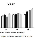 |
Figure 2: Serum level of VEGF in rats |
For instance, group of Silver sulfadiazine’s plasma VEGF level in 7, 14, 21, 28 days were 343.0±14.4, 370.0±18.5, 391.0±20.1, 472.0±19.4 (pg/ml), Calvacin-1 group was 331.2±13.0, 317.0±19.6, 421.0±13.0, 506.0±17.4 (pg/ml); and Calvacin-2 groups result showed 332.0±18.8, 394.6±19.2, 391.0±12.0, 546.0±19.7(pg/ml) respectively. Above mentioned group results showed statistically significant improvement (p=0.0032).
Level of TGF-β1
TGFβ1 is a control of cell growth and differentiation, induces fibrosis and scar formation (the process of wound healing).
Table 4: Serum level of TGFβ1 in rats
| Time (day) | Control (pg/ml) | Silversulfadiazin (pg/ml) | Calvacin-1 (pg/ml) | Calvacin-2 (pg/ml) |
| 7 | 90.3±3.3 | 88.5±3.9 | 79.4±6.0 | 88.5±7.8 |
| 14 | 82.0±4.4 | 77.7±4.2 | 78.6±6.8 | 77.3±3.7 |
| 21 | 81.1±3.4 | 75.0±2.7* | 84.4±4.8 | 80.9±6.6 |
| 28 | 81.1±5.4 | 70.4±2.1* | 81.0±5.2 | 87.3±5.8* |
*p=0.05
In the control group, the level TGFβ1 decreased burn induction. We found significant differences (p=0.05) between the treatment groups. In the present study, the levels of TGF-β1 were increased in all groups and decreased after 14 to 21 days in the SSD treated groups.
Histopathological Examination
The study of the effects of Calvacin gel in the full thickness second degree burns in the experimental rats. The first week after the of the microscopic examination showed that all the groups except the Calvacin 2 were exposed to epithelial edema, epithelial and dermis inflammatory cell infiltration rates. (Figure 3 зураг, A-D).
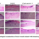 |
Figure 3: Histological examination of burn wounds stained with hematoxylin and eosin |
Scab (1), inflammatory cell infiltration (2), epithelial cell growth (3), hyperplasia of fibroblast (4), blood vessel formation (5 arrow), growth of granular tissue (6). НЕ, х40
After 14 days the burning of the wound, the epidermis layer of the control group and the Calvacin-1 epithelium group was relatively larger than the other groups, and the neutrophils and macrophages observed in the control group were more penetrating than the cells. The presence of over burdishness, the formation of new vessel and collagen fibers in all the groups of experimental and control groups in the dermis layer indicates the wounds in the wound stage 3 or proliferation stage (Fig. 3, E-H). In the 21st days of observation, the epithelial cell regeneration didn’t complete in the control and experimental groups. In the epidermis and the dermis of the skin, neutrophils and macrophages were transverse in the control group of the cells more than in other groups (Fig.3, I, J, K, L). During the study 28 days, the control group epithelium of the regeneration was not fully completed, and the inflammatory cells were observed in epithelial layer, scab not completely removed. The of the Calvacin-2 group skin epithelium were regenerated, in some parts of the very small size of scab, Calvacin-1 group and SSD group of adult tissue epithelium mature but some parts of the scab were observed. In the skin dermis, accumulation of collagen, accumulation of fibroblast cells, and new vascular formation are indicative of phase 4 regeneration or wound healing (Fig. 3, M-P).
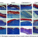 |
Figure 4: Histological examination of burn wounds stained with masson-trichrome |
Scab of wound (1), penetration of inflammation cells (2), growth of epithelial cell (3), hyperplasia of fibroblasts (4), formation of vessel (5), growth of granular tissue (6). Массон трихром, х40, х100.
7 days after observed for the group of calvacin-2 groups, was more likely to have swelling and wound in control, SSD and Calvacin-1 groups. The accumulation of new collagen fibers in the dermis layer of all experimental groups was observed (Figure 4 A-D). On the 14th day of study, experimental and control groups did not disappeared the ruptures of the wound and swell, and the formation of new vessels and collagen fibers in the dermis layer was observed, but the SSD, Calvacin-2, and Calvacin-1 groups were slightly more than the control group (Figure 4 E-H). In the 21st day of the study, wound of redness was reduced to all groups, and the epidermal layer of the regeneration Calvacin-2, Calvacin-1 were better compared to other groups, such as control and SSD groups. In the Dermis layer increased the vascular formation of the true skin layer, were significantly larger compared to other groups in the Calvacin-1 and SSD groups (Figure 4 F-I). After the 28th day of the all group, rupture of the wound, which were scarred, were scavenging, epidermis re-mature, and the newly formed collagen fiber in the dermis layer were significantly observed and were more active in the Calvacin-1, Calvacin-2 and SSD group compared to control (Figure 4 M-P).
Discussion
Calvacin gel was prepared with 5% spor of Calvatia gigantea Batsch. ex Pers. and extract of Gentiana macrophylla Pall. The most suitable for the base of gel former was 1% carbomer. Carbomer aqueous gels are more viscous at pH 6 to 11.14 So we used 1.1% triethanolamine as a neutralizer. Calvacin gel containing 1% propylene glycol had good moisturizing effect and did not change viscosity. Calvacin gel base composition is similar to Cacalia gel and Indian researcher’s gel composition.9 They prepared herbal gel containing Clerodendron in for tunatum leaves extract with 1% carbomer, 0.2% methylparaben 0.1% propylparaben, 5% propylene glycol and 1.2% triethanolamine16. Cutaneous wound healing is an essential physiological process consisting of the collaboration of many cell strains and their products17. Wound healing and tissue repair in burn injuries are considered as a complex process including inflammation, granulation and remodeling of the tissue. We evaluated for the first time the effect of a gel of Calvacin in the full-thickness second-degree burn wound model. The result of this evaluation revealed that treatment with Calvacin gel significantly increased wound healing. Re-epithelialization and increased migration of myofibroblasts, fibroblasts, and macrophages were more prominent in Calvacin -treated groups showing that natural substances played a prominent role in wound healing process after burn injury. Angiogenesis is an important factor in proliferative phase of wound healing. VEGF is one of the most potent proangiogenic growth factors in the skin. In the last stage of wound healing, VEGF plays a role of promoting scar formation.18 These results suggested that Calvacin-1, 2 could heal scald wounds faster and result in less scarring than other treatments by regulating VEGF in the whole wound healing process. TGF-β1 is a key growth factor secreted by several cells and is involved in a number of processes in wound healing, i.e., inflammation, angiogenesis, fibroblast proliferation, collagen synthesis, and remodeling of new extracellular matrix.18 In the control group, the level TGFβ1 decreased burn induction. In the present study, the levels of TGF-β1 were increased in all groups and decreased after 14 to 21 days in the SSD treated groups. But Calvacin believes that all stages of the wound healing process are affected. Many natural substances such as flavonoids,18 iridoids.19,20 geniposide21 and tannin22 have shown the effect of healing experimental induced burn via increasing of re-epithelialization cytokine secretion. Because Calvacin is a composed polyphenol, organic acid, iridoid, tannins. These have high wound healing and antioxidant, anti-inflammatory properties.22.23 Therefore, several publications revealed that a ointment and gel had beneficial effects in animal models of burn disorders. The Mebo has been reported to promote chronic ischemic and neurogenic ulcer healing in patients. MEBO significantly promoted the formation of granulation tissue in cutaneous excisional wounds, shortened the time of wound healing, and increased neovascularization and the number of fibroblasts.26
Arnebia euchroma has exerted a healing effect against standard second degree burn wounds were induced as well as its significant impact on fibroblast proliferation and collagen synthesis.27
These wound healing effects of medicinal plant and ointment related to had significantly stimulatory influence on fibroblast proliferation, collagen bundle synthesis, and revascularization it was similar to our result.
Conclusions
The current study showed that Calvacin has a burn healing activity.
Acknowledgments
We thank the colleagues of the Institute of Traditional Medicine and Technology of Mongolia for their support during this study.
Conflict of interest
No conflict of interest
Funding information
Foundation of science and technology, Mongolia, Project/Award number: Shut/I-2017/27
References
- Health indicators of Mongolia 2017 p.72-77
- DeSanti, Leslie BS, RN. Pathophysiology and Current Management of Burn Injury
- Advances in Skin & Wound Care., 2005;18:333-334
- Baavgai C, Boldsaikhan B.:Mongolian traditional ,pp153-154, State Publishing House, Ulaanbaatar, 1990.
- Tumurbaatar N, Khatanbaatar Z, Tserendagva D.: An introduction to Mongolian traditional medicine., pp25-26 Munkhiin Useg publishing Ulaanbaatar 2006.
- Dejidmaa B, Nyamdemberel Ts,Chimedragchaa Ch,Dagvatseren B. The chemical and pharmacological study of the new drug “calvacin”. Mong Med Sci J., 2011;158(4):74-78
- Badshah H, Ullah F. Pharmacological activities of selected wild mushrooms in South Waziristan (FATA), Pakistan. South African Journal of Botany., 2015;97:107-110
- Glenn S. Bulmer, Everett S. Beneke and J. A. Stevens. Studies on Calvatia gigantea. III. Antitumor Substances Produced by Mycelium from Germinated Spores and Parent Basidiocarps.,1962;6: 621-625
- Ministry of Health. Mongolian Pharmacopoie. pp54-56 Munkhiin Useg publishing Ulaanbaatar 2011.
- Jambaninj D, Syed Azhar Syed Sulaiman, Syed Wasif Gillani et al. Technological study of preparing gel from semi-solid extract of Cacalia hastata,J. Adv. Pharm. Tech. Res. 2012; 3: 25-29.
- Method for definition of mould and fungus. Mongolian National Standard. Dosage form of Drugs, raw materials. MNS-5194-2002. Ulaanbaatar: Center of Standardization and Measurement of Mongolia; 2002. pp.1-5.
- Basic indices of microbiological requirements, method for sampling. Mongolian National Standard. Dosage form of Drugs, raw materials. MNS-5189-2002. Ulaanbaatar: Center of Standardization and Measurement of Mongolia; 2002. pp. 1-7.
- Preparation of samples for microbiological analysis. Mongolian National Standard. Dosage form of Drugs, raw materials. MNS- 5190-2002. Ulaanbaatar: Center of Standardization and Measurement of Mongolia; 2002. pp. 1-6.
- Method for definition of total number of bacterium. Mongolian National Standard. Dosage form of Drugs, raw materials. MNS- 5193-2002. Ulaanbaatar: Center of Standardization and Measurement of Mongolia; 2002. pp. 1-3.
- Rowe RC.:Handbook of pharmaceutical excipients. 6th p 121.London and Chicago: Pharmaceutical Press: (2009).
- Meyer, Tufi Neder and SILVA, Alcino Lázaro da.A standard burn model using rats. Acta Cir. Bras., 1999; 14:.4
- Das S, Haldar P, Pramanik G. Formulation and evaluation of herbal gel containing Clerodendroninfornatum leaves extract. Int J Pharm Tech Res.,2011;3:140–3.
- Shaw TJ, Martin P. Wound repair at a glance. J Cell Sci. 2009;122: 3209–3213.
- Lee K, Lee B, Lee MH, et al. Effect of Ampelopsis Radix on wound healing in scalded rats. BMC Complement Altern Med., 2015;15:213.
- Gouma E , Simos Y. Healing effects of quercetin on full thickness epidermal thermal injury in Wistar rats.International Journal of Phytomedicine., 2016; 8:277-281
- Barreto RS, Albuquerque-Júnior RL, Araújo AA, et al. A systematic review of the wound-healing effects of monoterpenes and iridoid derivatives. Molecules. 2014;19(1):846–862.
- Tanideh N, Haddadi MH, Rokni-Hosseini MH, et al. The healing effect of scrophularia striata on experimental burn wounds infected to pseudomonas aeruginosa in rat. World J Plast Surg. 2015;4(1):16–23.
- Kıvrak İ, Kıvrak Ş, Harmandar M.: Food Chem. 1,158(2014)
- Kıvrak İ, Kıvrak Ş, Harmandar M. Free amino acid profiling in the giant puffball mushroom (Calvatia gigantea) using UPLC-MS/MS. Food Chem. 2014;158:88–92.
- Singleton, V.L., Orthofer, R. and Lamuela-Raventos, R.M. Analysis of Total Phenols and Other Oxidation Substrates and Antioxidants by Means of Folin-Ciocalteu Reagent. Methods in Enzymology., 1999; 299:152-178.
- Tang, Q., Han, S., Feng, J., Di, J., Qin, W., Fu, J., Jiang, Q.”Moist exposed burn ointment promotes cutaneous excisional wound healing in rats involving VEGF and bFGF”. Molecular Medicine Reports., 2014; 9.4:1277-1282.
- Ashkani-Esfahani S, Imanieh MH, Khoshneviszadeh M, et al. The healing effect of arnebia euchroma in second degree burn wounds in rat as an animal model. Iranian Red Crescent Medical Journal., 2012;14(2):70-74.
Abbreviations
| CON | Control |
| SSD | Silver Sulfadiazine |
| Calvacin-1 | Calvatia Gigantea Batsch.
ex Pers was Extracted Gel |
| Calvacin-2 | Calvatia Gigantea Batsch.
ex Pers not Extracted Gel/ |

