
Manuscript accepted on :26-03-2020
Published online on: 11-04-2020
Plagiarism Check: Yes
Reviewed by: Mohammad Alamgeer
Second Review by: Cherry Bansal
Final Approval by: Prof. Juei-Tang Cheng
S. M. Nazia Fathima, R. Tamilselvi* and M. Parisa Beham
Department of ECE, Sethu Institute of Technology, Tamilnadu-626 115.
Corresponding Author E-mail : tamilselvi@sethu.ac.in
DOI : https://dx.doi.org/10.13005/bpj/1931
Abstract
Bone Mineral Density (BMD) trial retrieves the vigor of the bone in the medical arena. Peril of the subjects bone fracture are appraised or estimated by the BMD experiment. Risk condition of the bone in the low or high state is defined as osteopenia or osteoporosis disease respectively. The proclamation identifies the manifestation of this disease which is the most communal bone disease. In the past medical era, the BMD is assessed by conservative x-rays. There are lot of techniques available to measure BMD. One of the newer technique is the DEXA scanning method. Thus it is mandatory to have a regular databank in Dexa images for the young sprout scholars to carry out their research in fracture risk analysis from the Dexa images. The proposed updated version of databank is dubbed as DEXSIT-V2, a timely approach to bid a set of images of Arm, Spine and Femur (Right and Left) bones for osteoporotic images. The subject information such as age, gender and distinct Id are construed in the databank.
Keywords
Arm; BMD; Database; DEXA; Femur Bone
Download this article as:| Copy the following to cite this article: Fathima S. M. N, Tamilselvi R, Beham M. P. DEXSIT-v2: Detection of Fracture Risk Condition. Biomed Pharmacol J 2020;13(2). Available from: 0 |
| Copy the following to cite this URL: Fathima S. M. N, Tamilselvi R, Beham M. P. DEXSIT-v2: Detection of Fracture Risk Condition. Biomed Pharmacol J 2020;13(2). Available from: https://bit.ly/3a6xLYJ |
Introduction
Intrusion of engineering concepts in medical field is an attractive challenge in healthcare. Also engineering sciences algorithms plays a main role in healthcare applications. The biomedical engineering solves a lot of undefined problems and creates an awareness in the medical field research challenges. Lot of research challenges in the medical era need a solution and problem solving capacity from engineering concepts. The gap between engineering and medicine is routed by the Biomedical engineering field in today’s concept. In the present scenario, the researchers find solution to carry out health care conducts such as analysis, rigorous care, prevention of diseases and therapy.
Early challenges in the fracture risk condition are assessed through the X-Ray scan images. But the analysis involves the physician involvement for analyzing the risk of bone condition. In later stage, of of 1963, single-photon absorptiometry (SPA) by Cameron and Sorenson is used for the quantitative analysis of the bone. The results are highly appreciable in terms of accuracy, but have its limitation in the region of measurement. In extension, single energy is replaced with dual energy for improvement in the BMD measurement. Gamma rays joins hand in the part of Dual-photon absorptiometry (DPA) with the vims of the photon [1]. Single and Dual Energy x-ray (SXA) (DEXA) contributed a lot in the medical field for the detection of fracture risk condition [2]. The various scanning methods used for the BMD measurements portrays the risk condition of the bone called as osteoporosis or lower risk condition called as osteopenia. Osteoporosis are noticed more in women in the age after 30 or else affects severe in the women in menopause stage. In men, those who are more than 50 years also typically affected by osteoporosis.
Osteoporosis condition can be visualized in the whole body region, but mainly identified in the hip and femur region. So it is a required one to measure the BMD for the detection of fracture risk condition. The regions which are prone to the risk are assessed frequently for the detection of osteoporosis. Osteoporosis makes the tissue of the bone to become thin and fragile over time and leads to risk of bone fractures [3]. In present day scenario, biomedical is an attractive domain for the medical researchers to find optimal solutions for various challenges and treatment. [4].
The discussed literature deals with the BMD measurement and risk assessment in the Bones. The proposed paper explains the second version database of Dexa images which is initiated as ‘DEXSIT-V2 Database’, for the osteoporotic research. Medical datasets are available for imaging modalities , but osteoporosis images are still a dream in medical world. Even though some survey has discussed the issues of SPA, SXA and DEXA, still now no databases available for osteoporotic research.
The previous version [5] of the database discusses the details of spine, right and left femur images from a GE machine. Also it deals with the T-score and BMD values of a specific subject along with the images. Also the risk condition is discussed along with the physicians treatment. This extended version discusses the images of Spine Femur and arm. The
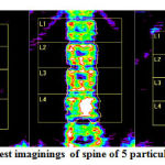 |
Figure 1: Sample DEXA test imaginings of spine of 5 particular specimen – DEXSIT-V2 |
Thus interested by these factors, our main contributions in this exertion are:
Produce a database, which comprises of 200 images of Spine, Femur and Arm bone scan images.
Construes the ID, gender and age of the patients.
DEXSIT-V2
In the bone research, there is no standard dataset for the exact measurement of BMD. Taking in to account of the challenges and attracted by this, a new updated exceptional database is created. This version is chiefly engrossed for the density assessment.
These bone images are collected in real time from a standard centre. The derived dataset comprises of 200 DEXA images poised from diverse specimens. For simplification, the necessary particulars of the subjects are revealed in Table 1 and Table 2. Five Trial sample images of spine, Dexa right & left femur images and Arm scan images are depicted in Figure, figure 2 and Figure 3.The DEXSIT-V2 database is deliberated through subsequent stages:
Arrangement Details
Designing the images
Addendum
The factors are enlightened in fact in the subsequent sections.
Arrangement Details
In the dataset, only Indian population people are considered. More than 500 Spine, 500 Femur and 500 Arm scan images are collected and annotated. Footnote is carried out initially manually and hoarded as png (portable network graphics) format. All the needed things are incorporated along with the image information. DEXSIT-V2-SP, DEXSIT-V2-FE and DEXSIT-V2-AR are the hosted groups in the dataset for Spine, femur and Arm images.
Designing the Images
For better understanding of the database, the annotation is carried out in a very simple way. The documentation of the identification, part of the bone and gender is detailed by means of a footnote. For sample, a DEXA spine scan image is given as DEXSIT-V2_SP_001.png. The notation of the image is understood as spine image of the first subject. Likewise DEXSIT-V2_FE_002.png corresponds to the second subjects’ femur image. Likewise DEXSIT-V2_AR_002.png represents to DEXA Arm image of a subject 002.
Table 1: Construal of Spine images
| Subject Id | Category | Age | Image |
| DEXSIT-V2_SP_001 | F | 40 | Spine |
| DEXSIT-V2_SP_002 | F | 21 | Spine |
| DEXSIT-V2_SP_003 | F | 34 | Spine |
| DEXSIT-V2_SP_004 | F | 52 | Spine |
| DEXSIT-V2_SP_005 | F | 35 | Spine |
Table 2: Construal of femur bone images al of femur
| Subject Id | Category | Stage | Image |
| DEXSIT-V2 _FE_001 | F | 78 | Femur |
| DEXSIT-V2_FE_003 | F | 56 | Femur |
| DEXSIT-V2 _FE_006 | F | 45 | Femur |
| DEXSIT-V2 _FE_009 | F | 35 | Femur |
| DEXSIT-V2 _FE_010 | F | 39 | Femur |
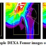 |
Figure 2: Sample DEXA Femur images of 5 subjects |
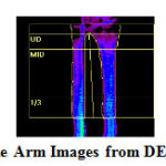 |
Figure 3: Sample Arm Images from DEXA database |
Table 3: Elucidation of Arm Dexa images
| Subject Id | Category | Stage | Image |
| DEXSIT-V2_AR_001 | F | 59 | Arm |
| DEXSIT-V2_AR_002 | F | 24 | Arm |
| DEXSIT-V2_AR 003 | F | 30 | Arm |
| DEXSIT-V2_AR_004 | F | 56 | Arm |
| DEXSIT-V2_AR_005 | F | 36 | Arm |
Table 4: T Score and Risk Condition
| Risk condition | T-score |
| Normal | T-score at or above -1 SD |
| Osteopenia | T-score between -1 and -2.5 SD |
| Osteoporosis | T-score at or below -2.5 SD |
Table 1 shows the health report of spine images for the 5 subjects. Table 2 shows the report of femur bone images for the same subjects. Table 3 shows the report of Arm images. Table 4 illustrates the WHO standard definition for osteoporosis condition based on the T-score value.
Clinical Data Analysis
The risk of bone is called as osteoporosis which is branded by lessening bone mass and structure of the bone. The bone mass decrease leads to the risk of osteoporosis.
From the health report, indication of T-score and Z- score, medication is suggested by the physician. Hormone replacement therapy (HRT), Bisphosphonates, Calcitonin and SERMs are the recommended medicines. If the physician suspects osteoporosis, the patients are advised to have an intake of Vitamin D and calcium supplement daily. By exact study of the Dexa scan images and their analysis, people can be barred from the osteoporosis disease. The database clearly gives the necessary values for predicting the osteoporosis condition. The researchers can authenticate the accuracy of the proposed methods with the clinical reports provided by the physician/experts.
Conclusion
The updated database of DEXA offered a medical image databank called DEXSIT-V2 for bone research. It is established with the meaning of enhancing an ordinary for orthopedic research and allied growth. Database has the principal features of a) spine, femur, and Arm scan images b) Tagging the subject’s natural data. This database offers a solution to various undefined problems in the BME research. The database along with all medical procedures will be openly made handy for research. In order to continue the research or start with the orthopedic research the database can be perceived at the succeeding web address: http://www.sethu.ac.in/ DEXSIT-V2/.
References
- Bonnick,S.L, “Bone Densitometry for Technologists” Thesis Report: Springer, pp1-64, 2006.
- M.Nazia Fathima, R.Tamilselvi and M.Parisa Beham, “Role of Dual-Energy X-ray Absorptiometry in Assessment of Bone Mineral Density – A Review” Proceedings of International Conference on Informatics Computing in Engineering Systems ICICES, 2018.
- Rosa Lorente-Ramos Javier Azpeitia-Armán Araceli Muñoz-Hernández José Manuel arcía- Gómez Patricia Díez-Martínez Miguel rande- Bárez Dual-Energy X-Ray absorptiometry in the Diagnosis of Osteoporosis: A Practical Guide, AJR ; 196:897–904 0361–803X/11/1964–897, 2011.
- K. Garg and Sandeep Kharb ,”Dual energy X-ray absorptiometry: Pitfalls in measurement and interpretation of bone mineral density” Indian Journal of Endrocrinology and metabolism,2013.
- M.Nazia Fathima, R.Tamilselvi and M.Parisa Beham, “XSITRAY: A Database for the Detection of Osteoporosis Condition”, Biomedical and Pharmacology Journal, Vol.12,No.1,2019.

