
Manuscript accepted on :
Published online on: 28-03-2020
Plagiarism Check: Yes
Reviewed by: Łukasz Łopusiewicz
Second Review by: Mihir Kulkarni
Kailas D. Datkhile* , Satish R. Patil, Pratik P Durgavale, Madhavi N. Patil, Nilam J. Jagdale and Vinit N. Deshmukh
Molecular and Genetic Laboratory, Department of Molecular Biology and Genetics, Krishna Institute of Medical Sciences “Deemed to be University”, Taluka-Karad, Dist-Satara, Pin-415 539, Maharashtra, India
Corresponding Author Email: kddatkhile@gmail.com
DOI : https://dx.doi.org/10.13005/bpj/1904
Abstract
Biogenic nanoparticles have emerged as novel contender in biomedicine because of their comprehensive tendency and remarkable potential in therapeutics. This study was intended to synthesize biogenic silver nanoparticles using Nothapodytes foetida leaf extract (Wight) (NFAgNPs). The biosynthesis of silver nanoparticles was continuously monitored by time dependent UV-visible spectrophotometric analysis. The morphology, size and shape of biogenic NFAgNPs were studied by transmission electron microscopy (TEM). In vitro antioxidant potential of biogenic nanoparticles evaluated by assaying percentage inhibition of 2, 2’-diphenyl-1-picrylhydrazyl (DPPH) and 2, 2’-azinobis-(3-ethylbenzothiazoline-6-sulfonic acid (ABTS) radicals. Antimicrobial activity of NFAgNPs was judged by minimum inhibitory concentration (MIC) assay on Gram-positive (Staphylococcus aureus) and Gram- negative (Escherichia coli, Pseudomonas aeruginosa and Klebsiella pneumoniae bacterial strains. The results confirmed that the NFAgNPs exhibited strong antioxidant potential where more than 93.80 % of inhibition in DPPH radicals occurred at 100 µg/mL concentration NFAgNPs whereas comparatively lower ABTS radical scvenging activity (84.59 %) was observed in NFAgNPs. The biogenic NFAgNPs also exhibited strong antibacterial activity against selected gram positive and negative human pathogenic bacteria. The highest percentage of growth inhibition in E. coli i.e (95.03 ± 1.16) followed by P. aeruginosa (94.24 ± 1.27) and K. pneumoniae (91.90 ± 2.69) after exposure to 1mg/mL concentration of NFAgNPs. Similarly, 63.03 ± 0.5 percentage growth inhibitory effects occurred in Gram positive S. aureus. Thus, the studied characteristics of biosynthesized NFAgNPs imply their relevance as a probable antioxidant and antibacterial agent.
Keywords
Notaphodytes Foetida; Silver Nanoparticles; Nfagnps; Antioxidant Activity; Antibacterial Activity
Download this article as:| Copy the following to cite this article: Datkhile K. D, Patil S. R, Durgavale P. P, Patil M. N, Jagdale N. J, Deshmukh V. N. Studies on Antioxidant and Antimicrobial Potential of Biogenic Silver Nanoparticles Synthesized Using Nothapodytes foetida Leaf Extract (Wight) Sleumer. Biomed Pharmacol J 2019; 13(1). |
| Copy the following to cite this URL: Datkhile K. D, Patil S. R, Durgavale P. P, Patil M. N, Jagdale N. J, Deshmukh V. N. Studies on Antioxidant and Antimicrobial Potential of Biogenic Silver Nanoparticles Synthesized Using Nothapodytes foetida Leaf Extract (Wight) Sleumer. Biomed Pharmacol J 2019; 13(1). Available from: https://bit.ly/3bAkJDY |
Introduction
In recent years biogenic nanomaterials have emerged with an immense scope in biomedicine as these nanostructures are proved to combat against deadly communicable and non-communicable diseases. Intensifying application of antibiotics in therapeutics endorsed multidrug resistance (MDR) in numerous microbial strains especially in bacterial pathogens. The abridged efficacy of drugs evidenced that the MDR bacteria could be a major warning to the human wellbeing.1 Therefore, to cope up with the threat of pathogenic executors, pioneered domain of nanotechnology has come up with an increasingly compelling and non-toxic treatment where biogenic nanomaterials are of enormous concern in the field of biomedical research because of their biological origin, mode of action and response. The biomaterials including phytochemicals with natural origin are gifted products of medicinal plants come forward to battle against deadly infectious microorganisms.2-3 Various attempts have been made synthesize different metal nanoparticles using biomaterials and to explore their antioxidant and antimicrobial potential.4-5 Now days, bio-synthesis of nanoparticles has become a keen interest of research for their advanced application in curative treatment to overcome the overall burden of deadly infectious diseases. Biogenic nanoparticles synthesized using extracts of medicinal plants are significantly illustrated for their biological efficacies including antimicrobial and antioxidant potential6-8 but, still there remains paucity in the knowledge on application of biogenic silver nanoparticles synthesized using Nothapodytes foetida.
Nothapodytes foetida (Wight) Sleumer is an important endangered medicinal plant with extensive antioxidant9 and cytotoxic potential10 with high therapeutic value because of abundance in active constituents including camptothecin and its derivatives predominantly used in clinical complications. Earlier, studies on biogenic silver nanopartiles synthesized using Nothapodytes species, reported cytotoxicity potential.11-12 But, the documentary evidences on antioxidant and antimicrobial potential of silver nanoparticles synthesized using any of the Nothapodytes species remained unexplored till date. Therefore in this study we focused our attention at synthesizing silver nanoparticles using crude aqueous leaf extract of N. foetida and subsequently explore for their biological properties including antioxidant and antimicrobial potential.
Materials and Methods
Biosynthesis and purification of silver nanoparticles
The silver nanoparticles were synthesized by adding 10 mL of aqueous plant extract of N. foetida to 90 mL of 1 mili molar (mM) silver nitrate (AgNO3) solution. The preparation was then incubated at 800C in dark and monitored continuously for synthesis of nanoparticles with change in colour of reaction mixture. After formation of NFAgNPs, the mixture was centrifuged at 15000 rpm for 10 minutes and washed several times with sterile deionized distilled water (ddw) to remove unwanted traces of contaminants followed by redispersion of the pellet in sterile ddw for further characterizations.
Visual Observation
The reduction of silver nitrate using aqueous plant extract was monitored for time periods (10, 30, 60, 120 and 180 minutes) and the appearance of dark brown color indicates the formation of silver nanoparticles.
UV-Visible Spectral Analysis
Preliminary characterization of biosynthesized AgNPs was carried out using UV-Visible spectroscopy. The reduction of pure silver ions was monitored by measuring UV visible spectrum of the reaction mixture at a wavelength of 300-800 nm by sampling the aliquots withdrawn from reaction mixture at different time intervals. The measurements were recorded on UV–visible Dual Beam Spectrophotometer (UV-1800 Shimadzu).
Characterization of NFAgNPs
Characterization of biosynthesized NFAgNPs were carried out by Transmission Electron Microscopy (TEM) at Sophisticated Analytical Instrument Facility at Sophisticated Test and Instrumentation Centre, Cochin University, Kerala.
Antioxidant activity of NFAgNPs
The antioxidant activity of NFAgNPs was characterized by DPPH & ABTS assay.
DPPH Radical Scavenging Assay
In vitro reaction of DPPH assay consisted of a mixture of freshly prepared 1 mL DPPH (0.1 mM) solution in methanol with different concentrations (5, 10, 25, 50, 75 and 100 μg/mL of NFAgNPs in final volume of 0.1 mL in distilled water. The mixture was shaken vigorously and allowed to stand at room temperature for 30 min in dark thereafter the absorbance was measured at 517 nm by using a UV-Visible 1800 spectrophotometer (Shimadzu). The percentage of DPPH scavenging activity was calculated using the formula: Percentage inhibition (%) = (A0-A1) / A0) × 100, where: A0 is the Absorbance of control and A1 Absorbance of test. The results were compared with Butylated hydroxy toluene (BHT) as standard
ABTS Radical Scavenging Assay
ABTS radicals are produced by reacting ABTS (7 mM) and potassium persulfate (2.4 mM) and incubating the mixture at room temperature in the dark for 16 hr. Different concentrations of NFAgNPs in final volume of 0.1 mL were added and allowed to react with 1 mL of ABTS for 30 minutes and the absorbance was recorded at 734 nm after incubation, thereafter calculated percent inhibition of ABTS radicals using BHT as standard.
Evaluation of Antimicrobial Properties of NFAgNPs
The antimicrobial activity of NFAgNPs was performed against pathogenic Gram-positive bacteria Staphylococcus aureus (S. aureus) (ATCC® 29213™) and Gram-negative bacteria, Escherichia coli (E. coli) (ATCC® 25922™), Klebsiella pneumoniae (K. pneumoniae) (ATCC® 700603™) and Pseudomonas aeruginosa (P. aeruhinosa) (ATCC® 2617™) by minimum inhibitory concentration (MIC) assay.
Statistical Analysis
All experiments were done in four replicates and then values were expressed as mean ± standard error (SE) of four measurements. Statistical significance was evaluated by one-way analysis of variance (ANOVA) followed by Student’s t-test by SPSS 11 for windows.
Results
Biosynthesis & Characterization of NFAgNPs
Aqueous leaf extract of N. foetida was used for biosynthesis of silver nanoparticles without any other stabilizing agent. After adding faint yellow plant extract to clear solution of AgNO3 solution changed to dark brown color within 3 hours of incubation at 800C (Figure 1A and C) demonstrated bioreduction of silver ions to nanosilver which is accelereted by the N. foetida leaf extract. The change in color of the AgNO3 solution after addition of aqueous leaf extract of N. foetida confirms the formation of silver nanoparticles which may occurred because of phytochemicals present in N. foetida might be responsible for reduction and synthesis of AgNPs.
The primary characterization of NFAgNPs synthesized using aqueous extract of N. foetida was done by UV-visible spectroscopy. The time dependent extinction of surface plasma resonance in biosynthesized NFAgNP solutions was indicated in UV- visible spectra (Figure 1B & D) which exhibited a characteristic absorbance maxima at 440 nm for NFAgNPs at various time points which showed synthesis of nanoparticles initiated after 30 minutes of incubation and the intensity increases progressively without any shift of the peak and completed after 180 minutes of incubation. The morphology and shape of biosynthesized NFAgNPs were determined by TEM which shows that the biosynthesized NFAgNPs are spherical in shape and 20-50 nm size.
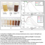 |
Figure 1: A. Biosynthesis of NFAgNPs, bottle containing N. foetida plant extract, sample of 1mM AgNO3 and colloidal solution of NFAgNPs.B. Click here to view Figure |
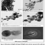 |
Figure 2: TEM analysis of NFAgNPs |
In Vitro DPPH Radical Scavenging Activity
The results of the DPPH and ABTS radical scavenging activity of NFAgNPs are shown in figure 3A & B. As shown in figure 3A, maximum percentage scavenging of DPPH radicals by NFAgNPs was 22.70 ± 1.76, 36.40 ± 1.73, 46.81 ± 1.30, 65.17 ±1.90, 81.80 ± 1.04 and 93.80 ± 1.98 for 5, 10, 25, 50, 75 and 100 µg/mL concentrations respectively.
ABTS Radical Scavenging Activity
As shown in figure 3B, maximum percentage scavenging of ABTS for NFAgNPs was 84.59%, at 100 µg/mL followed by 72.25%, 59.19%, 43.38%, 32.13% and 17.53% at decreasing concentrations of NFAgNPs i.e. 75, 50, 25, 10 and 5 µg/mL respectively. The results showed that the NFAgNPs exhibited strong DPPH radical scavenging ability with 50% inhibitory concentration IC50 22.56 μg/mL than the ABTS radical scavenging activity (IC50 value of 41.47μg/mL).
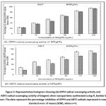 |
Figure 3: Representative histogram showing. |
Antibacterial activity of NFAgNPs.
The antibacterial activity of the NFAgNPs was assayed in vitro by minimum inhibition concentration method against four bacteria species, E. coli P. aeruginosa, K. pneumoniae and S. aureus. Different concentrations of NFAgNPs (0.1, 0.2, 0.4, 0.8 and 1.0 mg/mL) were tested on Muller Hinton media along with amphicillin as positive control. The results of antibacterial activity of biogenic NFAgNPs were represented in Figure 4A-D. The MIC results indicated that higher concentrations of NFAgNPs (1.0 mg/mL) showed greater sensitivity of E.coli P. aeruginosa strains and K. pneumoniae whereas S. aereus required higher concentrations of NFAgNPs to inhibit bacterial cell growth. When we tested antibacterial effect of NFAgNPs, we observed the highest percentage of bacterial growth inhibition in E.coli (95.03 ± 1.16), P. aeruginosa strains (94.24 ± 1.27) and K. pneumoniae (91.90 ± 2.69) when exposed to 1 mg/mL concentration whereas 63.02 + 0.50 % growth inhibition occurred in S. aureus.
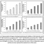 |
Figure 4: Representative histogram showing bacterial growth inhibition of. |
Discussion
Present work explains antioxidant and antibacterial potential of biogenic AgNPs synthesized using N. foetida leaf extract. After addition of aqueous extract of N. foetida, the color of silver nitrate solution turned to dark brown after 3 h of incubation indicating formation of silver nanoparticles. There was no change in color of the reaction observed up to 10 minutes after addition of plant extract to silver nitrate solution which started after 30 minutes of incubation in dark at 800C. In initial UV-visible spectrophotometric analysis, a single surface plasmon resonance peak between 420-450 nm with kmax at 440 nm was recorded in the observations which confirmed the formation of silver nanoparticles thereafter confirmed by TEM. The spherical shaped NFAgNPs were seen in TEM analysis. TEM images clearly demonstrated 20-50 nm size range of NFAgNPs. In addition, TEM micrograph revealed that most of the nanoparticles were spherical in shape. The biosynthesized NFAgNPs were evaluated for their free radical quenching ability by various in vitro assays which exhibited higher DPPH radical scavenging activity as compared to ABTS in response to different concentrations of AgNPs. The free radical scavenging activity of NFAgNPs was found to increase significantly with increasing concentrations similar with observations noted in former some of the studies.13-14 It is demonstrated earlier that the radical scavenging ability of biogenic silver nanoparticles is attributed due to integration of existed functional groups onto the surface of biogenic AgNPs originated from plant extract.15 Earlier studies on in vitro antioxidant potential of biosynthesized silver nanoparticles from Pongamia pinnata showed considerable free radical scavenging potential.16 Also, Kharat and Mendhulkar reported strong antioxidant activity in terms of DPPH radical scavenging by phytosynthesized silver nanoparticles using Elephantopus scaber.17 Similarly silver nanoparticles synthesized using plant extracts are significantly illustrated for their antioxidant potential.6-7
Recently several reports in the literature proved that many of the bacteria developed resistance to multiple antibiotics which ultimately leads necessity of alternate substitute for treatment of pathogenic microbes.18-19 Several researchers developed alternate strategies to overcome the drug resistance. The biologically synthesized nanoparticles have emerged as a combatant to fight against microorganisms and overcome the dilemma of drug resistance. The enhanced antibacterial activity of biogenic nanoparticles is due to the synergistic effect between nanoparticles and natural compounds present in phytochemicals.20 In our results we observed that the NFAgNPs exhibited promising antibacterial activity against booth Gram-positive and Gram negative bacteria. The extent of inhibitory effects on bacterial growth was observed on selected bacterial strains where E.coli found to be more sensitive followed by P. aeruginosa and K. pneumoniae strains. Similar effects of biogenic silver nanoparticles synthesized using Eucalyptus globulus21 and Murraya koeniggi 22 are reported earlier. The biologically synthesized AgNPs using different plant extracts also showed a similar potent bactericidal activity.23-26 Our results reported comparatively lower antibacterial activity of NFAgNPs against methicillin resistant Gram- positive S. aureus bacteria in contrast to the results obtained by Ansari and M. A. Alzohairy27 with the biosynthesized silver nanoparticles using Phoenix dactylifera seed extracts. In conclusion, this is the first study investigate antioxidant and antibacterial effects of biogenic AgNPs synthesized by using N. foetida. Our study also provide possible evidences supporting N. foetida plant extract derived AgNPs exhibited strong antioxidant and antibacterial potential against selected bacterial strains.
Acknowledgement
The authors gratefully acknowledge all the facilities and financial support provided by the Krishna Institute of Medical Sciences “Deemed to be University” Karad, India for experimental work. Authors are thankful to Mr. Santosh Jadhav & Mrs. Ashwini More for technical support.
Conflict of Interest
There is no conflict of interest
References
- Walker B., Barrett S., Polasky S., Galaz V., Folke C., Engstrom G., et al. Environment. Looming global-scale failures and missing institutions. Science. 325:1345-1346 (2009).
- Compean K. L. and Ynalvez R. A. Antimicrobial activity of plant secondary metabolites: A Review. Res J Med Plant. 8: 204-213 (2014).
- Barbieri R., Coppo, E., Marchese, A., Daglia, M., Sobarzo-Sanchez, E., Nabavi, S. M., Nabavi, S. M. Phytochemicals for human disease: An update on plant-derived compounds antibacterial Microbiol Res. 196. 44-68 (2017).
- Prasad T. N. V. K. V., Kambala V. S. R., Naidu R. A. Critical review on biogenic silver nanoparticles and their antimicrobial activity. Curr Nanosci. 7: 541-544 (2011).
- Roy A., Bulut O., Some S., Mandal A. K., Yilmaz M. D. Green synthesis of silver nanoparticles: Biomolecule nanoparticle organizations targeting antimicrobial activity. RSC Advanc. 9: 2673-2702 (2019).
- Nagaich U, Gulati N, Chauhan S. Antioxidant and Antibacterial Potential of Silver Nanoparticles: Biogenic Synthesis Utilizing Apple Extract. J Pharm. 2016: Article ID 7141523, 8 pages http://dx.doi.org10.1155/2016/7141523 (2016).
- Mohanta Y. K., Panda S. K., Jayabalan R., Sharma N., Bastia A. K., Mohanta T. K. Antimicrobial, Antioxidant and Cytotoxic Activity of Silver Nanoparticles Synthesized by Leaf Extract of Erythrina suberosa (Roxb.). Front Mol Biosci. 4:14. doi: 10.3389/fmolb.2017.00014 (2017).
- Singh P., Garg A., Pandit S., Mokkapati V. R. S. S., Mijakovic I. Antimicrobial effects of biogenic nanoparticles. 8: 1009, doi:10.3390/nano8121009 (2018).
- Datkhile K. D., Durgavale P. P., Patil M. N., Vhaval R. D., Khamkar T.S. Evaluation of in vitro Antioxidant and Free Radical Scavenging Properties of Different Parts of Medicinal Plant Nothapodytes foetida (Family: Icacinaceaae). Int J Sci Res. 5: 80-84 (2016).
- Datkhile K. D., Durgavale P. P., Patil M. N. In Vitro evaluation of cytotoxic and genotoxic effects of plant extracts from Nothaphodytes foetida (Wight) Sleumer (Family: Icacinaceae) on Cancer Cells. AsianJ Pharm Clin Res. 11: 284-291 (2018).
- Mahendran G., Ranjitha Kumari B. D. Biological activities of silver nanoparticles from Nothapodytes nimmoniana (Graham) Mabb. Fruit extracts. Food Sci Human Wellness. 5: 207-218 (2016).
- Datkhile K. D., Durgawale P. P. Patil M. N. Biogenic silver nanoparticles from Nothapodytes foetida kills human cancer cells in vitro through inhibition of cell proliferation and induction of apoptosis. J Bionanosci. 11:416-427 (2017).
- Dipankar C., Murugan S. The green synthesis, characterization and evaluation of the biological activities of silver nanoparticles synthesized from Iresine herbstii leaf aqueous extracts. Colloids Surf B Biointerf. 98:12-119 (2012).
- Reddy Nakkala J., Mata R., Bhagat E., Sardas S. R. Green synthesis of silver and gold nanoparticles from Gymnema sylvestre leaf extract: study of antioxidant and anticancer activities. J Nanopart Res. 17:151 (2015). DOI: 10.1007/s11051-015-2957.
- He Y., Wei F., Ma Z., Zhang H., Yang Q., Yao B., et al. Green synthesis of silver nanoparticles using seed extract of Alpinia katsumadai, and their antioxidant, cytotoxicity, and antibacterial activities. RSC Advances. 7(63): 39842–39851 (2017).
- Priya R. S., Geetha D., Ramesh P. S. Antioxidant activity of chemically synthesized AgNPs and biosynthesized Pongamia pinnata leaf extract mediated AgNPs – A comparative study. Ecotoxicol Environ Saf. 134:308–318 (2016).
- Kharat S. N., Mendhulkar V. D. “Synthesis, characterization and studies on antioxidant activity of silver nanoparticles using Elephantopus scaber leaf extract. Mater Sci Eng (C). 62: 719–724 (2016).
- Krychowiak M., Grnchloc M., Banasiuk R., Kraze-Baranowskova M., Gold D., Kawiak A. Combination of silver nanoparticles and Drosera binataextract as a possible alternative for antibiotic treatment of burn wound infections caused by resistant Staphylococcus aureus. PLoS One. 9: 1-20 (2014).
- Ramesh P., Kokila T., Geetha D. Plant mediated green synthesis and antibacterial activity of silver nanoparticles using Emblica officinalis fruit extract. Spectrochimica Acta Part A: Mol and Biomol Spect. 142: 339-343 (2015).
- Duran N., Duran M., deJesus MB., Seabra A. B., Favaro W. J., Nakazato G. Silver nanoparticles: a new view on mechanistic aspects on antimicrobial activity. Nanomed: Nanotech Biol Med. 12 (3): 789–799 (2016).
- Ali K., Ahmed B., Dwivedi S., Saquib Q., Al-Khedhairy A. A., Musarrat J. Microwave accelerated green synthesis of stable silver nanoparticles with Eucalyptus globulus leaf extract and their antibacterial and antibiofilm activity on clinical isolates. PLoS One. 10 (7): Article ID e0131178, 20 pages (2015).
- Qais F. A., Shafiq A., Khan H. M., Husain F J., Khan R A., Alenazi B, Alsalme A, Ahmad I. Antibacterial Effect of Silver Nanoparticles Synthesized Using Murraya koenigii (L.) against Multidrug-Resistant Pathogens. Bioinorg Chem Appl. 2019: Article ID 4649506, 11 pages https://doi.org/10.1155/2019/4649506 (2019).
- Murugan K., Senthilkumar B., Senbagam D., Al-Sohaibani S. Biosynthesis of silver nanoparticles using Acacia leucophloea extract and their antibacterial activity. Int J Nanomed. 9: 2431–2438 (2014)
- Nithya Deva Krupa A., Raghavan V. Biosynthesis of silver nanoparticles using Aegle marmelos (Bael) fruit extract and its application to prevent adhesion of bacteria: a strategy to control microfouling. Bioinorg Chem Appl. 2014: 1–8 (2014)
- Muthukrishnan S., Bhakya S., Kumar TS., Rao M. V. Biosynthesis, characterization and antibacterial effect of plant-mediated silver nanoparticles using Ceropegia thwaitesii—An endemic species. Ind Crops Prod. 63:119-124(2015)
- Bhakya S., Muthukrishnan S., Sukumaran M., Muthukumar M. Biogenic synthesis of silver nanoparticles and their antioxidant and antibacterial activity. Appl Nanosci. 6:755–766 (2016).
- Ansari M. A., Alzohairy M. A. One-pot facile green synthesis of silver nanoparticles using seed extract of Phoenix dactylifera and their bactericidal potential against MRSA. Evid Based Complem Alt Med. 2018: Article ID 1860280, 9 pages (2018).

