
Manuscript accepted on :27-11-2019
Published online on: 28-12-2019
Plagiarism Check: Yes
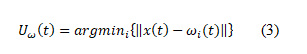
Reviewed by: Dr. Nilesh Bhaskarrao Bahadure
Second Review by: Ram kumar
Final Approval by: Dr Ayush Dogra
Abd El Kader Isselmou1* , Guizhi Xu2 and Shuai Zhang3
1Department of Biomedical Engineering, Hebei University of Technology, Tianjin City, China, 30030
2Dean of School of Electrical Engineering, Hebei University of Technology, Tianjin City, China, 30030
3Vice Dean of School of Electrical Engineering, Hebei University of Technology, Tianjin City, China,30030
Corresponding Author E-mail: Isselmou_kader@yahoo.com
DOI : https://dx.doi.org/10.13005/bpj/1793
Abstract
Medical image processing techniques play an important role in helping doctors and facilities for patient diagnosis, the aim of this paper is comparison between three improved methods to identify the brain tumor using magnetic resonance brain images and analysis of the performance of each method according to different values, accuracy, nJaccard coeff, ndice, sensitivity, specificity, recall and precision values,We used three improved methods the first method improved fuzzy c-means algorithm (IFCM), the second method is improved feed-forward neural network (IFFNN), and the third method is a hybrid self-organizing map with a fuzzy k-means algorithm,the significance of these methods is complementary among them where each one has an advantage in a certain value as shown in the paper results, the three methods gave a very good performance, generally they can identify the tumor area clearly in MR brain image with different performance of the values, each method gave better values than others according to a comparison between the performance value of three methods,Finally, the improved methods allow the development of algorithms to diagnose a tumor more accurately and for a short period of time and each method is distinguished from each other in the performance and value, this gives integrity and strength to this work, these methods can be used in pre and post radio surgical applications
Keywords
IFCM; IFFNN; ISOM-FKM algorithm; MRI; Tumor identification
Download this article as:| Copy the following to cite this article: Isselmou A. E. K, Xu G, Zhang S. Improved Methods for Brain Tumor Detection and Analysis Using MR Brain Images. Biomed Pharmacol J 2019;12(4). |
| Copy the following to cite this URL: Isselmou A. E. K, Xu G, Zhang S. Improved Methods for Brain Tumor Detection and Analysis Using MR Brain Images. Biomed Pharmacol J 2019;12(4). Available from: https://bit.ly/37qPJUT |
Introduction
Brain incorporates a perplexing structure and known as piece part in the body, which is a delicate, light mass of tissue. It very well may be secured by the bones in the skull, three slight layers of tissue (meninges) and a watery liquid (cerebrospinal liquid) that courses through spaces identifying with the meninges and all through spaces (ventricles) inside the mind.
A brain tumor or intracranial neoplasm is where irregular cells structure inside the mind [1], Intracranial tumor are different band of tumor that varies in restriction, side effects, histological creation and the event of certain species dictated by age, there are a few pictures that can be tended to and connected similarly with regards to the cerebrum MR pictures, so as to encourage the viable analysis of the patient, furthermore, there are numerous hidden issues and research in the field of MR cerebrum pictures, this proposal centers around a portion of these issues and medicines.
Tumor detection and analysis using segmentation in multi-directed MR brain images, which are in various different chains-structures axis is the main concern for this research work, compared with conventional image processing means, PCNNs have several significant advantages, which include Immunity against noise, the individuality of geometric variations in input patterns, the capability of linking minor intensity variations in input patterns, etc.
Oritz et al [2] recommend two fully unsupervised methods of MR brain image segmentation using SOM-based strategies, segment T1-weighted images are determined by SOM (Self-Organizing Map) technique, the user of the segmentation algorithm is still restricted to process T1-weighted images, Further, the sensitivity value manufactured by the algorithm requires improvement.
Logeswari and Karnan [3] “reported brain tumor detection using a segmentation process based on Self-Organizing Maps (SOM)” The segmentation process is curbed to segment T2-weighted image sequences.
Logeswari and Karnan [4] “offered brain tumor detection using segmentation process based on Hierarchical Self-Organizing Map (HSOM) technique” were T2 weighted pictures were divided in normal time of 29.9708 seconds, which requires further minimization.
Shen et al [5] projected a novel FCM algorithm named as Improved FCM (IFCM), two significant parameters that address neighborhood pixel fascination are recommended in this work. The principal parameter is worried about the element contrast between neighboring pixels present in the picture and the subsequent parameter is connected to grants the computation of the overall area of the neighboring pixels.
Sahoo et al [6], Yang and Laaksonen [7], Guler et al [8], Ong et al [9], Sun [10] and Fan et al [11] have developed segmentation techniques using ANN classifiers.
Elliott et al [12] have introduced an algorithm, which identifies multiple sclerosis lesions; the algorithm utilizes Bayesian classifier to identify the brain tissues and random-forest for lesion identification. The false detection rate.
Ulagammai et al [13] “used a wavelet-based neural network technique; initially trained by BFO algorithm for load forecasting”.
Yu and Yang [14] prescribed a summed up FCM (GFCM) structure to unite the varieties found in FCM and afterward watched its optimality test utilizing parameter choice. GFCM does not include the optimality test related to the two bunches and enrollment capacities. Alternatively, maybe, GFCM included distinctly in optimality test with the group model.
Sultan Aljahdali et al[15] “suggested an automated fuzzy rule-based algorithms for reliable image segmentation” T1weighted image were fragmented utilizing both fluffy K means and Kernel-based Fuzzy C-Means (FCM) calculations Time utilization required by the calculation plays a noteworthy imperative in portioning the pictures. In addition, the calculation creates a division exactness running somewhere in the range of 59.454 and 80.46, which can further be increased.
Mohamad et al [16] have utilized Improved FCM algorithm for segmenting MR brain images, where Breeding Swarm (BS) optimization values are assigned as the input parameters for FCM algorithm. The usage of this algorithm is narrowed down to segment T1 weighted images.
In this paper, we proposed a comparison between three improved methods, IFCM algorithm, IFFNN algorithm, ISOM-FKM algorithm, the results of this comparison gave a different performance of each method and we improved the values compared with the results of other papers as presented it in the results of this paper.
Materials or data source
Approximately 20 images we obtained from the different patients and different Age groups have been used in this paper, from the Nouakchott Hospital of Oncology, Mauritania details are listed below from patients:
The clinical image of the patient from the age of thirty-seven suffering from used meningioma.
The clinical image of the patient age of thirty-two suffering from and using high- quality astrocytes.
Images of patients with metastatic tumor and bronchial cancer, taken from Nouakchott hospital of oncology and used inventory, figure.2 shown example of different types of MR brain images with the tumor.
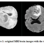 |
Figure 1: original MRI brain images with the tumor
|
Methodology
The methodology of this work include 5 stages of each method, the first stage (input MRI brain image) original image, the second stage (pre-processing) it depends on every method in IFCM algorithm applied morphology operation, IFFNN method applied trilateral filter, ISOM-FKM algorithm applied color quantization, the third stage we started with IFCM algorithm (segment IFCM), after we used IFFNN method, the last applied ISOM-FKM algorithm, the fourth stage identify the tumor area using each method independently, the fifth stage is the performance of each method independently, figure.2 also explain more about the methodology stages.
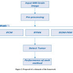 |
Figure 2: Proposed of a schematic of the framework
|
Fuzzy C-means Algorithm
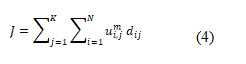
Fluffy C-implies (FCM) calculation is deliberate accumulation made by Dunn, Bezdek that has been upgraded by extra titivated by Mathieu Patitucci voxels gathering (information) through the attractive reverberation picture (MR) mind pictures are arranged and assembled under n number of bunches determined by PSO calculation, (_x^→) (t+1), the refreshed position vector of the molecule (voxel or pixel) inferred with the assistance of PSO calculation goes about as the centroid esteem and dependent on this worth, the number of groups for FCM calculation are additionally doled out utilizing PSO. It is resolved enrolment line focuses and bunch redundancy to restrain the proposed and capacity of the gathering voxels.
𝑁 Describes the number of incoming data points as input to the algorithm (the total number of voxels or voxels in the image). 𝐾 shows the number of iterations to be performed.
Feed Forward Neural Network
The neurons are masterminded in layers and they have unidirectional associations between them, they produce just one lot of the yield esteem, they are alluded to as a static system in light of the fact that inside this the yield esteems planned just relying upon current information. The yield esteems will not be dictated by past info esteem, luckily, they are alluded to as a memory-less system.
This work will manage feed-forward ANNs (Hertez et al., 1991: Haykin, 1994) it comprises of bury associated layers of preparing units or nerve cells, blogging from the loads and inclinations pursues (Hertz et al, 1991) loads contacts layer between the and player symbol image predisposition, information and it alluded to the yield layer tankers by, and more remote, individually. On the head, feed-forward ANNs is an exceptional parameter is a versatile capacity vector, which might be prepared to perform characterization or relapse capacities. A rating bolstering forward ANN plays out the mapping.
Self-Organizing Map Algorithm
The initial step of preparing a SOM calculation begins with instatement, where voxel forces are at first evaluated and displayed with the help of SOM models, this procedure is executed utilizing histogram examination, which goes about as a substitution for blends of likelihood thickness capacities. Histogram based model breaks down the pinnacles and valleys of the information picture and further backings in supporting extraordinary enlightening estimations of the voxel, which can be incredibly utilized in division process [15, 16 and 17]. The SOM calculation is clarified as pursues:
Let represents the diversified input data and is the input vector; the warning unit is calculated in each iterative steps as:
Fuzzy K-Means Algorithm
The Fuzzy k-means algorithm, also known as FKM, the company’s image Behavior competitors, FCM formula, as mentioned by Chin-Tang Chang et al fluorine rubber partition for the given input image is divided into several ‘k’ clusters (divided voxels).
Here, represents the number of data points or voxels within the type MR brain image, Refers to the number of groupings from FKM. Make clear the series and columns of the input image. Defines the membership functions of FKM with fuzziness coefficient (fuzziness co-efficient is obtained from the overlapping of clusters). Is the squared Euclidean distance between pixel and cluster representative cluster center or centroid value.
Accuracy Value
Accuracy, unwavering quality additionally called division exactness; it is utilized to decide the adequacy of the division calculation assessment factors. The precision is indicated in Equation (5).

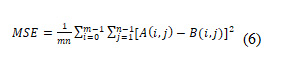
Mean Square Error (MSE)
Mean Square Error (MSE) characterizes squaring the separated amounts [18]. It tends to be communicated in light of the normal of the squares of the mistakes gotten by subtracting the contribution just as the yield esteems. MSE could be the total squared blunder worth including the information image and the segmented image.
Dice Overlap Index (DOI)
It is expressed with the help of the value of the Jaccard index). DOI identifies the purpose of overlap of the input image, as well as the resulting segmented image. DOI mentions calculating in the equation (7).
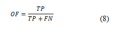
Sensitivity
Overlap Fraction (OF) or sensitivity values alludes back to the best possible division or order in the information picture [19, 20]. Additionally, characterizes the adequacy in precise recognizable proof of tumor locale just as other tissue areas. It is expressed in Equation (8).
Specificity
Specificity defines specific word or algorithms to classify segments of normal tissue that are present in a region in the input image capability, specificity is shown in Equation (9).
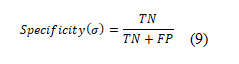
The experiment result, are divided into 3 categories according to the result of every method as described in the following steps.
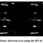 |
Figure 3: Tumor detected area using the IFCM algorithm.
|
 |
Figure 4: Performance of the IFCM algorithm.
|
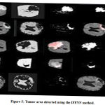 |
Figure 5: Tumor area detected using the IFFNN method.
|
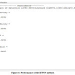 |
Figure 6: Performance of the IFFNN method.
|
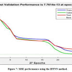 |
Figure 7: MSE performance using the IFFNN method.
|
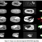 |
Figure 8: Tumor area detected using the ISOM-FKM algorithm.
|
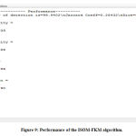 |
Figure 9: Performance of the ISOM-FKM algorithm.
|
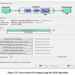 |
Figure 10: Neural network training using the SOM algorithm.
|
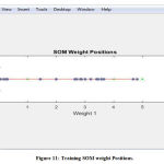 |
Figure 11: Training SOM weight Positions.
|
Discussion
The comparison between, the performance of the three methods gave a difference results and it was generally best results , the three methods can detect the tumor in MR image brain image clearly (Figure3,5 and 8) using 9 MR brain image, the performance of values ISOM-FKM algorithm gave the best result in accuracy value (Figure 9, Table 1), IFCM the best in sensitivity, specificity and ndice values (Figure 4, Table 1), IFFNN method produces a good result in the mean square error (MSE) sensitivity and specificity value as shown in (Figure 6, 7), the ISOM-FKM algorithm produces also a better iterations and SOM training weight values as shown in (Figure, 10,11).
Conclusion
The proposed method based on IFCM algorithm is capable to segment either MR brain images and identify the tumor with high-performance values of sensitivity, specificity, and ndice, IFCM is used to improve the values that we got from previous results.
The proposed method based on IFFNN (Supervised method) is efficient in the terms of MSE, Jaccard Index, DOI, OF, SI values. T1-T2-weighted and FLAIR type MR brain images are segmented while using proposed IFFNN (Supervised method), which affirms that the proposed methodology has the capacity to segment heterogeneous types of tumor in most the axes of MR brain image sequences. The level of tumor infiltration is clearly distinguished from edema portion. The large level of clinical data sets may be analyzed efficiently. Thus, the IFFNN method assists the radiologist in diagnosing the tumor region with a minimized manual operation. ISOM-FKM algorithm, proposed gave the best results in segmentation MR brain image processed, with a best accuracy value 99, 94% and detect the tumor in different location, the objective of this paper to improve the performance the three methods according to the values of accuracy, sensitivity, specificity, ndice value and MSE value and to assist the surgeon in diagnosis within a short period of time with high accuracy.
Acknowledgment
Authors are grateful to the National Natural Science Foundation of Hebei province under grant Number: E2015202292 and Number: E2015202050, high-level talent support project in Hebei province under grant Number: C2015005012, Key research, and development program under grant Number: 15272002 and Number: 15275704 for unlimited help and support to carry out this work.
References
- National Cancer Institute (NCI) Adult Brain Tumors Treatment Publications Locator. 2015-02-13.
CrossRef - Ortiz A, Gorriz J M, Ramirez J, et al. Two fully unsupervised methods for MR brain image segmentation using SOM-based strategies.Applied Soft Computing J. 2013, 13(5):2668-2682.
CrossRef - Logeswari T, Karnan M. An Enhanced Implementation of Brain Tumor Detection Using Segmentation Based on Soft Computing C. International Conference on Signal Acquisition and Processing, Icsap, Bangalore, India, February. DBLP, 2010:243-247.
CrossRef - Logeswari T, Karnan M. An Improved Implementation of Brain Tumor Detection Using Segmentation Based on Hierarchical Self-Organizing Map. Journal of Cancer Research & Experimental Oncology J, 2010, 17(1):591-595.
CrossRef - Shen S, Sandham W, Granat M, et al MRI fuzzy segmentation of brain tissue using neighborhood attraction with neural-network optimization. IEEE Transactions on Information Technology in Biomedicine J. 2005, 9(3):459-67
CrossRef - Sahoo P, Soltani S, A Wong, et al A survey of thresholding techniques. Computer Vision, Graphics, and Image Processing J, 2010, 41(2):233–260.
CrossRef - Yang Z, Laaksonen J. Interactive Retrieval in Facial Image Database Using Self-Organizing Maps. Iapr Conference on Machine Vision Applications C. DBLP, 2005:112-115.
- İnan Güler, Demirhan A, Karakış R. Interpretation of MR images using self-organizing maps and knowledge-based expert systems.Digital Signal Processing J, 2009, 19(4):668-677.
CrossRef - Ong S H, Yeo N C, Lee K H, et al. Segmentation of color images using a two-stage self-organizing network. Image & Vision Computing J, 2002, 20(4):279-289.
CrossRef - Sun W, Wang Y Segmentation Method of MRI Using Fuzzy Gaussian Basis Neural Network. Neural Information Processing Letters & Reviews J, 2005(2):19-24.
- Tian D, Fan L a Brain MR Images Segmentation Method Based on SOM Neural Network. The International Conference on Bioinformatics and Biomedical Engineering C. IEEE Explore, 2007:686-689.
CrossRef - Colm Elliott, Douglas L. Arnold, Louis Collins. D, and Tal Arbel, Temporally Consistent Probabilistic Detection of New Multiple Sclerosis Lesions in Brain MRI, IEEE Transactions on Medical Imaging J, (2013) Vol. 32, No. 8, 1490 – 1503.
CrossRef - L, Venkatesh. P, Kannan. S. P and Padhy. N.P, Application of bacteria foraging technique trained and artificial and wavelet neural networks in load forecasting, Neuro computing J. 2007, 70, 2659–2667.
CrossRef - Yu J, Yang M S. Optimality test for generalized FCM and its application to parameter selection. IEEE Transactions on Fuzzy Systems J, 2005, 13(1):164-176.
CrossRef - Sultan Aljahdali and. Zanaty. E. A, Automatic Fuzzy Algorithms for Reliable Image Segmentation IJCA, (2012). Vol. 19, No. 3.
- Mohamad Forouzanfar, Nosratallah Forghani, and Mohammad Teshnehlab, Parameter optimization of improved fuzzy c-means clustering algorithm for brain MR image segmentation, Engineering Applications of Artificial Intelligence C (2010),23, 160–168.
CrossRef - Chaplot S, Patnaik L M, Jagannathan N R. Classification of magnetic resonance brain images using wavelets as input to support vector machine and neural network. Biomedical Signal Processing & Control J, 2006, 1(1):86-92.
CrossRef - Thomos N, Boulgouris N. V, and Strintzis, M. G, Optimized Transmission of JPEG2000 Streams Over Wireless Channels, IEEE Transactions on Image Processing J. (2006), 15 (1).
CrossRef - Nan Zhang, Su Rua, Stéphane Lebonvallet, Qingmin Liao and Yuemin Zhu, Kernel feature selection to fuse multi-spectral MRI images for brain tumor segmentation, Computer Vision and Image Understanding, (2011) 115 256–269.
CrossRef - Rasoul Khayati, Mansur Vafadusta, Farzad Towhidkhaha, and S. Massood Nabavi, Fully automatic segmentation of multiple sclerosis lesions in brain MRFLAIR images using adaptive mixtures method and Markov random field model, Computers in Biology and Medicine, (2008),38, 379 – 390.
CrossRef

