
Manuscript accepted on :20-Jul-2019
Published online on: 23-07-2019
Plagiarism Check: Yes
Reviewed by: Nicolas Padilla
Second Review by: Hendry Irawan
Final Approval by: Prof. Juei-Tang Cheng
Nida Fathima1 and Sreedevi Dharman2
1Saveetha Dental College, Saveetha Institute of Medical and Technical Science, Saveetha University, Chennai, 600053-India.
2Department of Oral Medicine and Radiology, Saveetha Dental College, Saveetha Institute of Medical and Technical Science, Saveetha University, Chennai, 600053-India.
Corresponding Author E-mail: sanjamrut@gmail.com
DOI : https://dx.doi.org/10.13005/bpj/1753
Abstract
The main objective of this review was to identity the usage of thermography in diagnosis of orofacial pain. Thermography is a method of measurement of skin temperature distribution on the body over a given period of time. Hence using the skin temperature, an advantages result can be obtained in identifying diseased condition in orofacial region. This article highlights the basics of thermography as a diagnostic tool in dentistry in identifying orofacial pain. PUBMED and Google Scholar were searched to identify all the clinical trials which evaluated the use of thermography in diagnosis of orofacial pain. The included studies involved 6 clinical trials. From all the included studies, an advantages result was obtained by using thermography in assessing orofacial disorders between symptomatic and asymtomatic patient’s. All the findings obtained, suggest that thermography can be helpful in evaluating myogenous temperomandibular joint disorder and can be used as a clinical screening method and for improving diagnostic accuracy. In few studies, the sensitivity of thermography in the diagnosis of orofacial disorder is low, but has high specificity which helped in evaluation of orofacial disorder and is therefore applicable to patients with orofacial pain. Thermography benefits by locating the temperature difference between symptomatic and asymtomatic facial region thus indicating its aid in diagnoses of orofacial disorder. However due to liminted evidence and short term studies, it is recommended that more of long term studies with large sample size should be emphasised to use thermography as routine diagnostic tool in identifying orofacial disorder.
Keywords
Asymtomatic Facial; Emphasised; Orofacial Disorder; Thermography
Download this article as:| Copy the following to cite this article: Fathima N, Dharman S. Role of Thermography in Assessment of Myogenous Temperomandibular Disorders. Biomed Pharmacol J 2019;12(3). |
| Copy the following to cite this URL: Fathima N, Dharman S. Role of Thermography in Assessment of Myogenous Temperomandibular Disorders. Biomed Pharmacol J 2019;12(3). Available from: https://bit.ly/2LEid6y |
Introduction
Orofacial pain disorders are highly prevalent and debilitating conditions involving the head, face, and neck. These conditions represent a challenge to the clinician since the orofacial region is complex and therefore, pain can arise from many sources. The clinician needs to have solid knowledge of the pain conditions that arise from these structures for proper diagnosis and a multidisciplinary approach of management is strongly recommended.
Temperomandibular disorder (TMD) defines a number of clinical problems that involve the masticatory musculature, the TMJ, and associated structures.1 TMD is considered to be a subclassification of musculoskeletal disorders2 and is the most prevalent condition for which patients seek treatment.3,4 The careful evaluation of these facial structures in conjunction with clinical symptoms is important in forming a proper differential diagnosis. The patient may present with jaw ache, earache, toothache, facial pain, and/or headache; however, the complaint may be as benign as general facial fullness or pressure. Treatment planning depend on various factors, including the chief complaint, medical history, presenting symptoms, examination, and diagnosis. The therapeutic methods offer patients a wide range of treatment modalities with higher success rates.
Thermology is the study and application of biothermal processes to assess health or disease and the word thermography employs imaging and visual evaluation of those thermal changes from an object to detect, display, and record thermal patterns across the surface of the object.5
The thermography principle is as the amount of blood circulation at different layers of the skin varies, the temperature also changes accordingly. Consequently, disorders that affect the blood flow too result in abnormalities in temperature distribution and these when evaluated will provide valid diagnostic information.6
Based on the method of application, thermography can be classified into the following types7:
Semi‑quantitative contact method – liquid crystal thermography
Quantitative infrared‑detecting noncontact methods which are categorized as follows.
Infrared telethermography.
Dynamic telethermography.
Facial telethermography.
Thus the aim of this systematic review is to analyse the existing clinical trials of thermography role on diagnosis of orofacial pain.
Materials and Method
Search Strategy
Internet sources were used to search for appropriate papers that satisfied the study purpose. The total number of articles obtained are 26. PUBMED and Google Scholar were searched to identify all the clinical trials in which evaluates the use of thermography in diagnosis of orofacial pain. The current review article aims in evaluating the application of thermographs in diagnosis of orofacial pain mostly temperomandibular pain, hence the following inclusion and exclusion criteria was devised to select the appropriate clinical trial. The articles were screened on the basis of title and abstract. Full text was then procured for the relevant articles which fulfilled the inclusion criteria. Only articles published in the English language were considered for this review. In addition, the reference lists of the included articles were screened for additional relevant articles.
Eligibility Criteria
Inclusion Criteria
Randomized controlled clinical trials (RCTs), controlled clinical trials or case.
Papers written in English.
Studies conducted on humans.
Population: patients with symptomatic and Asymtomatic of TMD.
Clinical parameters: evidence in diagnosis of orofacial pain using thermography.
Exclusion Criteria
In-vitro studies, Animal studies, literature reviews. All irrelevant studies were excluded and the reasons for their exclusion were noted.
Outcome Measures
Values obtained by utilising thermography on symptomatic and asymtomatic patients of temperomandibular disorder containing orofacial pain.
Methodological Quality Assessment
Quality assessment was done for all the included articles. All the required data’s collected from the included studies were formulated showing the results and outcome measures. Table (1) contains the included articles used in this review and Table (2) contains the characteristics of thermography in orofacial pain.
Result
This systematic review includes 6 trials in which thermography is used in the diagnosis of orofacial pain mostly patient with TMD. The total number of patients included in all the five studies was 189. The major outcomes measured in these studies vary in temperature between symptomatic and asymtomatic patient’s. The results of these trials are tabulated as follows in Table 2. Krzysztof Woźniak et al [2015], in his trial, he obtained before chewing test showed Tmax=36.01°C, ΔT=0.26°C and after chewing test showed Tmax= 36.31°C, ΔT= 0.52°C. Thus chewing test helped to increase the diagnostic efficiency of thermography in identifying patients with TMD.8 D S Hadda et al [2014] in his trial the result obtained were Absolute mean temperature for asymptomatic patient: anterior temporalis (34.78°C), masseter (33.48°C) and symptomatic patient: anterior temporalis (34.37°C), masseter (32.85°C). Thus temperature of facial cutaneous areas over the masseter and anterior temporalis muscles decreases in the presence of myogenous TMD.9 U. Snekhalatha et al [2018] in her trial obtained mean temperature for the normal group which was calculated was found out to be ≅31°c and that of abnormal group was calculated to be ≅34°c which ≅ 3°C more than normal subjects. Hence the skin surface temperature of the body, that is, the region of interest, is elevated.10 Dong-Joon Han et al [2001] in his trial resulted with mean temperature for TMJ: 28.22°C, Anterior Temporalis: 28.32°C, Masseter anterior: 27.90°C, Masseter inferior: 27.89°C. Thus providing an effective diagnostic aid in evaluation of orofacial pain.11 Ana C. S. Costa et al [2012] in his trial obtained an array of values showing patient’s with TMD: LMA=32.84°C, RMA=32.99°C, LAT=34.09°C, RAT=34.29°C, SHY=32.99°C, LUT= 32.60°c, RUT= 32.85°C. Patients Without TMD: LMA=32.67°C, RMA=32.65°C, LAT=34.22°C, RAT=34.21°C, SHY=33.02°C, LUT= 32.59°C, RUT= 32.57°C. Hence obtaining intra and inter-rater reliability of the temperature measures of the masseter, anterior temporalis, supra-hyoid and upper trapezius muscles symptom were found to be slightly elevated in patient’s with TMD.12
According to all the trials, the thermographic value or the facial temperature of patient’s with orofacial pain was found to be raised than with that of symptomatic patient’s . However this studies gives a weak evidence of the obtained result. The available in literature are short-term studies, hence Long-term studies are required with larger sample size to emphasize the use of thermographs in diagnosis of orofacial pain.
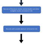 |
Figure 1: Flowchart showing search strategy.
|
Table 1: Article selected for systematic review.
| S.no | Article | Author / year of publication | Journal |
| 1. | Assessment of the Sensitivity, Specificity, and Accuracy of Thermography in Identifying Patients with TMD. | Krzysztof Woźniak et al [2015] | International Medical Journal of Experimental and Clinical Research. |
| 2. | Thermographic characterization of masticatory muscle regions in volunteers with and without myogenous temporomandibular disorder: preliminary results. | D S Hadda et al [2014] | Journal of British institute of radiology. |
| 3. | Facial thermography: a potential complimentary tool for evaluation of dental disorders. | U. Snekhalatha et al [2018] | International Journal of Engineering & Technology |
| 4. | Reliability of infrared thermographic imaging in the orofacial region. | Dong-Joon Han et al [2001] | Journal of oral medicine |
| 5. | Intra and inter-rater reliability of infrared image analysis of masticatory and upper trapezius muscles in women with and without temporomandibular disorder. | Ana C. S. Costa et al [2012] | Brazilian journal of physical therapy. |
| 6. | Infrared Thermography in the Assessment of Temporomandibular Joint Dysorder. | Francis Sahngun Nahm et al [2007] | Korean journal of pain |
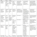 |
Table 2: Characterisitics of included studies.
|
Discussion
Thermography has many advantages – it is easy to use and non-invasive. However, there is a lack of standardized protocol for the temperature measurement of the masticatory muscles using infrared thermography. Gratt et al.13,14 evaluated the orofacial region by establishing 5 measurement areas: small TMJ, large TMJ, mandible, midface, and entire half-face. In another study by Dibai Filho et al.,15 the measurement of skin surface temperature was done in the muscle central point.There is difference in the region of muscle temperature may be explained on the basis of anatomy: the temporalis muscle is thinner than the masseter muscle and is influenced by the superficial path of the temporalis artery, which makes the region more hyper-radiant.16
Currently thermal imaging techniques were used for the evaluation of various dental disorders by qualitative and quantitative methods. This technique is also used as research tool in the field of dentistry for identifying the root canal inflammation, cavitations and in the development of abscess. Asymptomatic dental issues can be diagnosed using thermography, as seen in the study with patients showing no apparent problems or complaints about pain or discomfort. In a study similar to findings by Merla et al wherein they distinguished between healthy patients and patients suffering from myofascial pain using functional infrared imaging.17-19
The evaluation by the infrared thermography has some limitations, as the amount of subcutaneous fat, since this interferes in the skin surface temperature.20 Furthermore, it is an indirect measure of the blood flow21 and of the activity of the autonomic nervous system.22 Gratt and his colleagues classified the patients with chronic orofacial pain as normal, hot or cold when the temperature in a selected anatomic area (∆T) fell between certain assigned ranges.23
The symmetry of temperature distribution in the face and neck has been analyzed as a local prognostic factor in many studies. Based on 4000 measurements of thermograms of 100 healthy adults, Weinstein24 described the cut-off point for identifying pathologies associated with pain in the head as the absolute value of the temperature differences between corresponding homonymous structures in excess of 0.5°C. This value was determined using spot measurements. Normal temporomandibular joint (TMJ) examination using thermography had showed symmetrical thermal patterns with a mean ΔT value of 0.1°C.25 On the other hand, patients affected with internal derangement and TMJ osteoarthritis showed ΔT values of + 0.4°C.12 Beth and Gratt in 1996 conducted a double‑blinded clinical.26
Krzysztof Woźniak et al [2015], the study confirmed its limited diagnostic efficiency in identifying subjects with TMD – 95.5% specificity in identifying patients with no symptoms of dysfunctions according to Di when the absolute difference in temperature between the right and left side (ΔT) zones was lower than 0.26°C with sensitivity 44.3% and accuracy 52.4% before chewing test and 0.52°C with sensitivity 46.4% and accuracy 56.3% after test. The chewing test was a major factor that increased the efficiency of thermography in identifying patients with TMD. Further research is needed on the use of thermography in the diagnosis of TMD.8
In D S Hadda et al [2014], infrared imaging revealed a difference of 1.4°C between mean anterior temporalis and masseter muscle temperatures and a difference of 0.09 between the corresponding normalized temperatures in asymptomatic subjects, indicating that the temporalis was significantly more hyper-radiant than the masseter (p < 0.05). The results of sensitivity and specificity of the thermographic assessment for masticatory muscle regions suggest greater diagnostic accuracy when absolute mean temperature (×T) is used, even when translated to dimensionless temperature (×θ). These results suggest that thermal values, if used in conjunction with physical. assessment, can serve as a means of screening and improved diagnostic accuracy in myogenous TMDs.9
In U. Snekhalatha et al [2018] study has shown the value of infrared thermography as a technique which can be used for obtaining additional relevant information of patients with dental disorders, both symptomatic, and asymptomatic. In this study all abnormal subjects showed elevated temperature over the affected region, which is mostly due to the in- creased muscular activity and therefore a reciprocal increase in blood supply.10
Conclusion
This systematic review has proven the demand for future clinical trials to assess diagnosis of orofacial pain by using thermography. Hence it can be concluded that from analyzing the above 6 articles which consist of both patient’s with symptomatic and asymtomatic conditions of orofacial pain, indicates a positive result in which the thermographic value or the facial temperature of patient’s with orofacial pain was found to be raised than with that of asymtomatic patient’s. Even though the results in some of the studies were positive and promising, it has certain limitations pertaining sensitivity of orofacial disorders, which was found to be low compared to that of specificity. Hence more of a long term evidence based studies should be performed pertaining the use thermography as routine diagnostic tool in orofacial disorders.
References
- McNeill C. Temporomandibular Disorders: Guidelines for Classification, Assesment, and Management. 2nd ed. Chicago, IL: Quintessence Publishing Co, Inc; 1993.
- Okeson JP. Bell’s Orofacial Pains. The Clinical Management of Orofacial Pain. 6th ed. Carol Stream, IL: Quintessence Publishing Co, Inc; 2005.
- Dworkin SF. Temporomandibular disorder (TMD) pain-related disability found related to depression, nonspecific physical symptoms, and pain duration at 3 international sites. J Evid Based Dent Pract. 2011;11(3):143–144.
- Dworkin SF, Huggins KH, LeResche L, et al. Epidemiology of signs and symptoms in temporomandibular disorders: clinical signs in cases and controls. J Am Dent Assoc. 1990;120(3):273–281.
- Sudhakar S, Bina kayshap, Sridhar Reddy P. Thermography in dentistry‑revisited. Int J Biol Med Res. 2011; 2(1):461‑465.
- Komoriyama M, Nomoto R, Tanaka R, Hosoya N, Gomi K, Iino F, et al. Application of thermography in dentistry – Visualization of temperature distribution on oral tissues. Dent Mater J 2003;22:436‑43.
- Mehrotra A, Aggarwal A. Thermography: A review. J Dent Sci Oral Rehabil 2013; 4‑8.
- Krzysztof Woźniak, Liliana Szyszka-Sommerfeld and Dagmara Piątkowska. Assessment of the Sensitivity, Specificity, and Accuracy of Thermography in Identifying Patients with TMD. Med Sci Monit. 2015; 21: 1485–1493.
- D S Haddad, M L Brioschi,R Vardasca, M Weber, E M Crosato, and E S Arita. Thermographic characterization of masticatory muscle regions in volunteers with and without myogenous temporomandibular disorder: preliminary results. Dentomaxillofacial Radiology, December 2015, Vol. 43:, Issue. 8.
- U. Snekhalatha, Nida Mir, Mehvish Khan, Parimal Raj, Vimaladhithan, Yeshi Choden. Facial thermography: a potential complimentary tool for evaluation of dental disorders. International Journal of Engineering & Technology, 7 (2.8) (2018) 175-181.
- Dong-Joon Han, Ki-Suk Kim. Reliability of infrared thermographic imaging in the orofacial region. JournalOMP, 2001,vol.26, no.4.
- Ana C. S. Costa; Almir V. Dibai Filho; Amanda C. Packer; Delaine Rodrigues-Bigaton. Intra and inter-rater reliability of infrared image analysis of masticatory and upper trapezius muscles in women with and without temporomandibular disorder. Braz. J. Phys. Ther, November 2, 2012, vol.17 no.1.
- Gratt BM, Sickles EA, Ross JB, et al. Thermographic assessment of craniomandibular disorders: diagnostic interpretation versus temperature measurement analysis. J Orofac Pain. 1994;8:278–88.
- Gratt BM, Sickles EA. Thermographic characterization of the asymptomatic temporomandibular joint. J Orofac Pain. 1993;7:7–14.
- Dibai Filho AV, Packer AC, Costa AC, Rodrigues-Bigaton D. Accuracy of infrared thermography of the masticatory muscles for the diagnosis of myogenous temporomandibular disorder. J Manipulative Physiol Ther. 2013;36:245-52.
- Haddad DS, Brioschi ML, Arita ES. Thermographic and clinical correlation of myofascial trigger points in the masticatory muscles. Dentomaxillofac Radiol 2012; 41: 621–9.
- Merla A, Ciuffolo F, D’Attilio M, Tecco S, Festa F, De Michele G, Tangherlini A, Romani GL; Functional Infrared Imaging in the Diagnosis of the Myofascial Pain.
- Moinuddin Hassan, Tyuichi Kimura, Jcakimasa Asai, Akihiro Shi- mase,Masakazu Fukuoka and Ttatsuo Togawa; Imaging Of Skin Thermal Properties By Changing Ambient Radiation Temperature-An Electrical Control System For Stepwise Change In Ambient Radiation temperature; Engineering in Medicine and Biology Society,1-3;1998.
- Sarbani Deb Sikdar, Anshul Khandelwal, Savita Ghom, Rajkumar Diwan, FM Debta: Thermography: A New Diagnostic Tool in Dentistry. Journal of Indian Academy of Oral Medicine and Radiology 22(4):206-210, October-December 2010.
- Savastano DM, Gorbach AM, Eden HS, Brady SM, Reynolds JC, Yanovski JA. Adiposity and human regional body temperature. Am J Clin Nutr. 2009;90(5):1124-31.
- Seifalian AM, Stansby G, Jackson A, Howell K, Hamilton G. Comparison of laser Doppler perfusion imaging, laser Doppler flowmetry, and thermographic imaging for assessment of blood flow in human skin. Eur J Vasc Surg. 1994;8(1):65-9.
- Holey LA, Dixon J, Selfe J. An exploratory thermographic investigation of the effects of connective tissue massage on autonomic function. J Manipulative Physiol Ther. 2011;34(7):457-62.
- Gratt BM, Sickles EA, Ross JB. Thermographic characterization of an internal derangement of the temporomandibular joint. Journal of Orofacial Pain 8: 197-206, 1994.
- Weinstein SA, Weinstein G, Weinstein EL, Gelb M. Facial thermography, basis, protocol, and clinical value. Cranio. 1991;9:201–11.
- Anbar M. Diagnostic thermal imaging: A historical technological perspective. In: Quantitative Dynamic Telethermography in Medical Diagnosis. Boca Raton: CRC Press; 1994. p. 1‑9.
- Gratt BM, Graff‑Radford SB, Shetty V, Solberg WK, Sickles EA. A 6‑year clinical assessment of electronic facial thermography. Dentomaxillofac Radiol 1996;25:247‑55.

