
Manuscript accepted on :16 Aug 2019
Published online on: 14-09-2019
Plagiarism Check: Yes
Reviewed by: Nour Rano
Second Review by: Grigorios Kyriakopoulos
Final Approval by: Juei Tuei Cheng
Nevin E. Sharaf1 , Asmaa F. Galal2*, Mohamed S. El-Sawy3, Aziza B. Shalby4, Alaa H. Sayed4,5 and Hanaa H. Ahmed4
1Department of Environmental and Occupational Medicine, National Research Centre, Giza, Egypt.
2Narcotics, Ergogenics and Poisons Department, National Research Centre, Giza, Egypt.
3Department of Architecture, Faculty of engineering, Misr University, Giza, Egypt.
4Department of Hormones, Medical Research Division, National Research Centre, Giza, Egypt.
5Applied Medical Sciences Department, Community College in AlQurayyat Al-Jouf, Saudi Arabia
Corresponding Author E-mail: asma.galal.79@gmail.comDOI : https://dx.doi.org/10.13005/bpj/1751
Abstract
This study investigated the impact of Wi-Fi signals exposure on cognitive function and its relevant brain biomarkers and the possible role of designed Bio-Geometrical forms in restoring the neurobehavioral alterations resulting from the exposure to the emerging radiation.Rats were assigned into 3 groups; Gp I control group (away from exposure to radiation); Gp II, III were exposed to wireless router signals for 24 h for 6 months and Gp III was protected by a set of designed BioGeometrical shapes. Animals were tested for spatial memory, anxiety and emotionality in addition to the related neurotransmitters (dopamine, serotonin and acetylcholine) in different brain areas. Melatonin, Heat Shock Protein (HSP-70) and acetylcholine esterase (AchE) were also measured in various brain regions and histopathological examination was carried out as well. Wi-Fi radiation exposed group showed elevated anxiety level and impaired spatial memory. Moreover, significant decline in dopamine, serotonin and acetylcholine levels in the investigated brain areas has been recorded. Melatonin levels were decreased in the cortex, striatum and hippocampus while HSP-70 was depleted in the cortex only. Using Bio-Geometrical forms along with Wi-Fi exposure could combat the burden of Wi-Fi radiation. This was evidenced by the recovery of the anxiety level and the improvement of memory task. In addition, the presence of Bio-Geometrical shapes could retrieve dopamine, serotonin and acetylcholine as well as melatonin and HSP-70 levels This study provides solid foundation for the potential use of Bio-Geometrical shapes to modify the insult of Wi-Fi radiation on brain function and structure.
Keywords
Bio-Geometrical Shapes; Brain Regions; Memory-Anxiety; Wi-Fi Radiation
Download this article as:| Copy the following to cite this article: Sharaf N. E, Galal A. F, El-Sawy M. S, Shalby A. B, Sayed A. H, Ahmed H. H. Role of designed Bio-Geometrical forms in antagonizing neurobehavioral burden of Wi-Fi radiation: Evidence-based experimental study. Biomed Pharmacol J 2019;12(3). |
| Copy the following to cite this URL: Sharaf N. E, Galal A. F, El-Sawy M. S, Shalby A. B, Sayed A. H, Ahmed H. H. Role of designed Bio-Geometrical forms in antagonizing neurobehavioral burden of Wi-Fi radiation: Evidence-based experimental study. Biomed Pharmacol J 2019;12(3). Available from: http://biomedpharmajournal.org/?p=28451 |
Introduction
The ever-increasing exposure to electromagnetic radiation (EMR) owing to usage of wireless technologies has drawn much attention on the social and scientific levels.1 As internet has become indispensible in modern life, people everywhere, at home, workplace, public areas, and schools are exposed to electromagnetic fields (EMF) from multiple sources such as smart phones, office devices connected to Wi-Fi, laptops, and desktops.2 The EMF is a type of non-ionizing radiofrequency, with a large frequency band pattern and can interact with information structures of living cells; lipids, proteins and nucleic acids.1 Exposure to EMF radiation has been proposed to promote various neurological insults, including headaches, changes in sleep pattern, imbalance in the neurotransmitters, anxiety, learning disability, and alteration in the neuronal electrical activity.3, 4 Chronic cumulative EMF exposures in children may negatively influence their growth and capacity for normal development, learning and judgement.5
EMFs affects the CNS through various mechanisms including modifications of biomolecules, disturbing membrane potential and ion currents, which lead to disturbed cellular signaling cascades. EMFs also induce altered transportation and release of neurotransmitters such as serotonin (5-HT), and dopamine (DA) which play crucial roles in learning, and memory functions.6 Furthermore, neurotransmitter perturbation and imbalance can lead to anxiety in rodents. Alterations in hippocampal 5-HT level have been positively correlated to deficits in spatial and non-spatial functions.7, 8 In addition; decreased 5-HT transmission in prefrontal cortex (PFC) is connected with anxiety alteration.6
Also, the hippocampal DA has been shown to be implicated in synaptic plasticity and is involved in the encoding of spatial information at the behavioral level.9 Ach, a cholinergic neurotransmitter, plays a key role in learning, memory, locomotor functions as well as cerebral blood flow.10-12AchE converts Ach to choline and acetate in the synaptic cleft. AchE activity is implicated in cell proliferation and neurite outgrowth.12The inhibition of AchE has been involved in the neurodegenerative disorders. A strong relationship between EMR and cholinergic system has been proposed.10,12
Melatonin, the pineal gland neurohormone, represents the potent antioxidant that scavenges reactive oxygen species (ROS). It has been demonstrated that melatonin has a modulator role against 2.45 GHz-induced oxidative damage in dorsal root ganglion neurones of rats.13 Previous studies demonstrated that microwave radiation disrupt the melatonin/serotonin cycle in the brain.14
Heat shock proteins (HSPs) are known as stress proteins as they are upregulated in response to various kinds of stress. The cytoprotective actions of HSPs are greatly explained by their anti-apoptotic ability as they can interact with various key apoptotic proteins to block all apoptotic cascades. HSP70 is a glucocorticoid receptor chaperone that manipulates inflammatory reactions.15 French et al.16 hypothsized that repeated exposure to RF radiation may act as recurring stressors leading to continuous overexpression of HSPs in exposed cells.
Bio-Geometry is a recent science that utilizes the energy principles of shapes, colors, sounds and motions to amplify natural energy, balance biological energy systems and achieve harmony between inner and outer environments and thereby giving the body the power to heal itself.17 Bio-Geometry has been developed and patented by the Egyptian architect Ibrahim Karim as a response to the hazards of modern technology.17 Bio-Geometry energy quality harmonizing applications are being utilized in architecture, industrial design and telecom networks.18 A recent work of our research team provided evidence that the use of Bio-Geometrical shapes can abrogate the neurodegenerative effects induced by EMR through intensifying the antioxidant defense mechanism.19
The rational of the present approach was to gain better understanding of the devastating effects of the extreme exposure to Wi-Fi radiation on the learning and memory functions with their reliable biochemical indices in various brain areas of rats. Also, the study was intended to appraise the mechanisms underlying the protective outcomes of using Bio-Geometrical shapes against neurobehavioral aberration and histoarchitectural deformation of rat brain regions induced by radiofrequency radiations.
Materials and Methods
Wireless Devices with Internet Access
Two commercially available Linksys wireless-N300 (WAP300N-EE), operating at 802.11.g standards were applied in this research work. The output power was 95 mW average, as equivalent iso-tropically radiated output power. The maximum specific absorption rate (SAR) of the wireless gateways in the conformity assessment test was 0.091 W/kg.
Designed Bio-Geometrical Shapes
Five Bio-Geometrical shapes were used in this study, as previously described in Sharaf et al.19, the first one called Hamburg acrylic stands form shown in Fig. 1. This form was designed by Ibrahim Karim and put in the front of Wi-Fi router to harmonize the waves and lessen the harmful impacts. The second shape was paper form as shown in Fig. 2 which has been designed by Mohamed Elsawy and put on Wi-Fi router antenna to reduce the harmful effects of waves. The third shape was Bio-Geometric acrylic corner stand as shown in Fig. 3 which has been designed by Ibrahim Karim to amplify the energy quality of space. The fourth shape called Bio-Geometric acrylic cube as shown in Fig. 4 which has been designed by Ibrahim Karim and put beside the animal cage to elevate the energy quality of space. The fifth shape was plastic form as shown in Fig. 5 which has been designed by Mohamed Elsawy and put on Wi-Fi router antenna to accommodate the harmful insults of the waves.
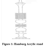 |
Figure 1: Hamburg Acrylic stand |
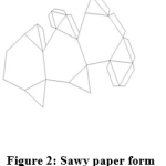 |
Figure 2: Sawy paper form |
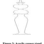 |
Figure 3: Acrylic corner stand |
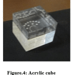 |
Figure 4: Acrylic cube |
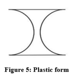 |
Figure 5: Plastic form |
Animal Handling and Experimental Study
Adult female Wistar rats of the same age (3 months), and weight (120 ± 5 g), were procured from the Animal Facility of the National Research Centre, Giza, Egypt. The rats were kept in a ventilated animal house with a 12 h light/dark cycle at ambient temperature of 22-25 ̊ C throughout the experimental period. The rats were allowed to acclimatize to their environment for at least 10 days before the initiation of the experiment. The rats were fed with standard rodent chow and water ad libitum. The experimental design and procedures were approved by Institutional Ethics Committee of the National Research Centre, Giza, Egypt and the experiment was performed as per guidelines of Ethical Committee for Medical Research at the National Research Centre (registration number: 15-175).
In this study, rats were randomized into three equal groups as follows: Group I; (control group) in which the rats were housed in the ordinary plastic cage in a separate room away from any electromagnetic waves contamination, Group II; (exposed group) ) in which the rats were kept in a similar cage placed in another room mounted with one wireless device. These rats were exposed for 24 h over a period of 6 months to the electromagnetic radiation emitted from the Wi-Fi device and Group III; (protected group) in which rats were maintained in a third similar cage placed in a third room mounted with the other wireless device, over the above mentioned period but the rats in this group were protected by a set of Bio-Geometrical shapes.
The two applied Wi-Fi devices were placed at a distance of 25 cm from each cage; the rooms housing the cages are identical to each other in shape and dimensions but located away from each other by a distance not less than 30 m (Fig. 6).
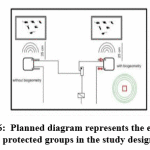 |
Figure 6: Planned diagram represents the exposed and protected groups in the study design |
Behavioral Assays
Open Field Test
The open field test was carried out in a square wooden arena (80 cm x 80 cm x 40 cm) with white smooth polished floors divided by black lines into 16 equal squares.20The test was performed under dim light in a quiet room. Each rat placed at the center square, and observed during 5 min period. Ambulation, rearing and grooming frequencies were measured.
Elevated Plus Maze (EPM)
The elevated plus maze was constructed of varnished wood and consisted of four arms of the same size (50 cm×10 cm) arranged in such a way as to form a cross. All arms extended from a central square (10 cm×10 cm) and were raised 50 cm above the floor. Two opposed arms were surrounded only by a short wall of 1 cm (open arms), while the other two opposed arms were surrounded by 40 cm high walls, except for the entrance (enclosed arms). Each animal was placed on the central square of the maze facing an enclosed arm and was allowed to explore the maze for 5 min.21
Morris Water Maze (MWM)
Spatial learning was evaluated using Morris water maze consisted of a circular water pool of 180 cm diameter and 50 cm depth divided into four equal quadrants designated A, B, C, and D. A platform of 10 cm diameter was fixed in one quadrant, lying 1.5 cm below the water surface. Over a period of four days, rats were trained to find the platform, and their latencies (time required to reach the platform) were recorded in each trial; rats were rescued if they failed to find the platform within 120 sec. On the fifth day, the platform was removed and the time spent in target quadrant was recorded.22
Episodic-Like Memory Test
This test evaluates the recollection of a unique past experience in terms of what happened, and where and when it happened. Each animal received two training trials and one test trial. There was a delay of 50 min between each trial. The exploration time allocated for each trial was 6 min. On the first training trial four black blocks known as old familiar objects were placed symmetrically one in each corner of the open-field. On the second training trial four grey cylinders known as recent familiar objects were placed in a T-shaped configuration. On the test trial, two old familiar objects were placed in the same locations as in the first training trial, one in the northwest corner and one in the southeast corner. Two recent familiar objects were placed one in the northeast corner and one in the southwest corner. Thus, one recent familiar object was displaced, whereas the other recent familiar object was stationary. The time spent exploring the old versus the new familiar objects is measured in the test trial.23
Sample Collection and preparation
After the experimental period was over, all animals were sacrificed by decapitation and each brain was quickly excised, washed in ice-cold saline, blotted dry and stored immediately at -80°C. Frozen brain was then dissected to cortex, striarum and hippocampus according to the method described by Glowinski and Iverson.24 Three brains were fixed in formalin-saline for histopathological investigation. The rest of brains were prepared for assessment of biochemical measurements. Half of the chosen samples from each group were homogenized immediately to give 10% (w/v) homogenate in ice-cold medium containing 50 mM Tris-Hcl (pH 7.4) and 300 mM sucrose. The homogenate was centrifuged at 1800 xg for 10 min at 4 ºC. The supernatant (10%) was stored in aliquots in -20°C for biochemical analysis.
Neurochemical and Biochemical Analyses
Serotonin (5-HT) and dopamine (DA)
5-HT and DA were analyzed in the other half of the selected areas of each group using High Performance Liquid Chromatography (HPLC) according to the method of Pagel et al. 25 Prior to analysis, each brain area was homogenized in 1/5 w/v of 75% aqueous methanol to obtain homogenate. Each homogenate was centrifuged at 1800xg (4 oC) for 10 min. The supernatant was injected onto AQUA column C18 (150×4.6 mm I.D., 5µm). Samples were injected in the HPLC system under the following conditions: mobile phase 97/3 20 mM potassium phosphate buffer pH 3.0/methanol (v/v), at flow rate of 1.0 ml/min. The detection wavelength was 270 nm and the injection volume was 40 µl.
Acetylcholine (Ach)
Quantitative determination of acetylcholine in brain regions was done using rat acetylcholine ELISA kit obtained from Glory Science Co., Ltd, Veterans Blud, Suite, USA, under guidance of the manufacturer.
Acteylcholinesterase (AchE) Activity
Acetylcholinesterase activity was measured in brain regions as described by Gorun et al.26 The assay is based on measurement of thiocholine produced when acetylcholine is hydrolyzed; the color was read immediately at 412 nm using spectrophotometer.
Melatonin
Assessment of melatonin in brain regions was done using rat melatonin ELISA kit (WKEA MED supplies crop Co., Ltd, China) according to the manufacturer’s manual.
Heat shock protein 70 (HSP-70) level
HSP-70 was quantified using rat HSP-70 ELISA kit (WKEA MED supplies crop Co., Ltd, China) according to the standard protocol provided by the manufacturer.
Histopathological examination
After fixation of the chosen brain regions from each group in buffered formalin (10%) for twenty four hours, washing was done in the tap water, and then serial dilutions of alcohol (methyl, ethyl and absolute ethyl) were used for dehydration. Specimens were cleared in xylene and embedded in paraffin at 56oC in hot air oven for twenty four hours. Paraffin bees wax tissue blocks were cut into 4 µm thick slices using slidge microtome. The obtained tissue sections were collected on glass slides, deparaffinized, stained by heamatoxylin and eosin stain for examination with a light electric microscope.27
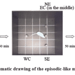 |
Figure 7: Schematic drawing of the episodic-like memory test. |
Statistical Analysis
Statistical analysis was performed using GraphPad Prism version 7. Statistical significance of difference was determined using analysis of variance (one way ANOVA). A p value < 0.05 was considered as significant. Percent of change in comparison with the corresponding control group was calculated according to the following formula:
Difference (%) = Treated value – Control value/ Control value X 100
Results
Testing of cognitive function and general behavioral screening
Elevated Plus Maze (EPM)
Rats exposed to Wi-Fi radiation showed preference to spend more time in closed arms than open arms and center area, where time spent in closed arms increased by 335% and time spent in open arms decreased by 90 %, versus the control counterparts (Fig.8a). Also, Wi-Fi radiation exposure increased number of entries in closed arms (129 % relative to control rats) as shown in Fig. 8b. These results indicate that exposure to Wi-Fi radiation increases anxiety and fear-related behaviors.
On the other side, rats protected by Bio-Geometrical shapes displayed preference to explore open arms and center area than closed arms (710 % compared to Wi-Fi- exposed rats) and experienced an increased number of entries in open arms (80 % compared to Wi-Fi- exposed rats) reflecting the normal level of fear and anxiety behavior of these rats (Fig.8 a and b).
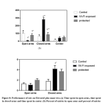 |
Figure 8: Performance of rats on Elevated plus maze test, (a) Time spent in open arms, time spent in closed arms and time spent in center. (b) Percent of entries in open arms and percent of entries in closed arms. |
Open Field Test (OFT)
Rats exposed to Wi-Fi radiation were highly thigmotactic or wall-seeking spending most of their time in the corners and along the walls, and performing stretch attends behavior that can be considered to represent different aspects of fear and avoidance of danger. Latencies to move in OFT were prolonged by 940 % in respect with the controls. Two behaviors involved in locomotion; explore and rear showed an overall increase (146%, 700 % respectively versus the controls) in the outer ring and along the walls indicating an overall increase in spontaneous activity (Fig.9).
In contrast, rats protected by Bio-Geometrical shapes exhibited normal exploratory behavior in the center and the inner ring of OFT representing normal fear-mediated response and normal anxiety level. Locomotor activity dropped by 27 % in comparison with Wi-Fi exposed rats. While, latency to move was lowered by 81.9 % relative to Wi-Fi exposed rats. Grooming frequency did not differ significantly between all the tested groups (Fig.9).
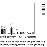 |
Figure 9: Performance of rats in Open field test; ambulation, rearing, latency, sit and grooming. |
Morris Water Maze (MWM)
Exposure to Wi-Fi radiation significantly prolonged the escape latency in MWM by 189 % compared to the controls indicating deficits in learning and memory task performance. However, protection by BioGeometrical shapes significantly reduced escape latency by 49.6 % versus Wi-Fi- exposed rats indicating improved rats’ performance in learning and memory task in MWM. Wi-Fi radiation exposure decreased target quadrant occupancy (TQO) by 40% compared to control rats. Whereas, TQO decreased in the protected rats by Bio-Geometrical shapes by 18.75% compared to the control group (Fig.10)
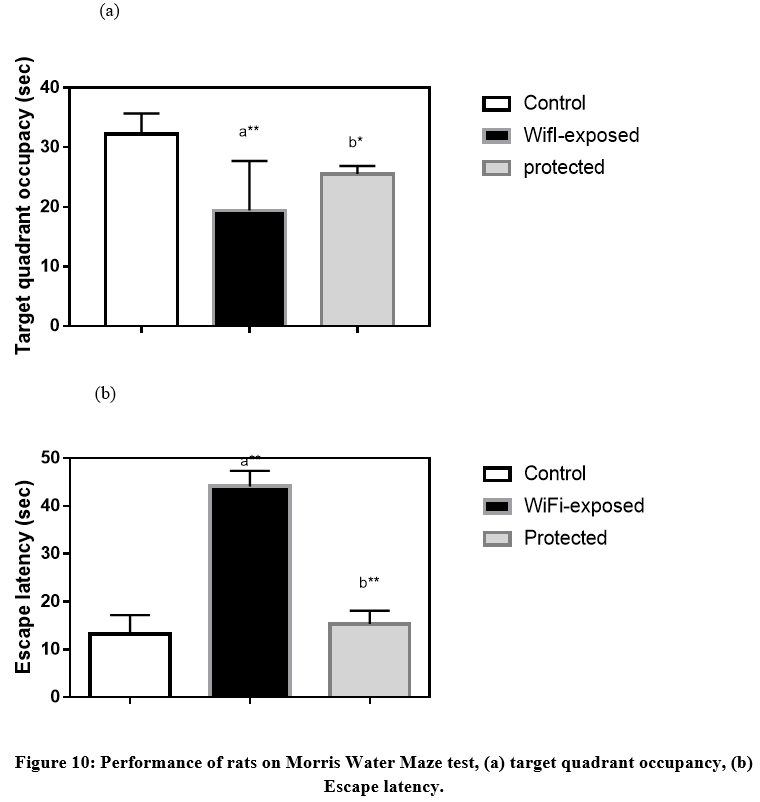 |
Figure 10: Performance of rats on Morris Water Maze test, (a) target quadrant occupancy, (b) Escape latency. |
Episodic-like memory
In this test, all groups did not show any significant difference in the mean exploration time for old familiar and recent familiar objects (Fig.11). Also, time spent exploring stationary and displaced objects did not differ significantly between all the tested groups.
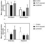 |
Figure 11: Performance of rats in Episodic-like memory test, (a) Mean object exploration of old and recent objects, (b) Mean object exploration of stationary and displaced objects. |
Neurochemical findings
Serotonin (5-HT)
Levels of 5-HT were decreased in the cortex (58%, p<0.001), striatum (48%, p<0.05) and hippocampus (50%, p<0.001) in Wi-Fi radiation exposed rats as compared to the control rats. On the other hand, protected group by Bio-Geometrical shapes showed recovery in 5-HT levels in all the investigated brain areas (94%, 42%, and 66%, respectively) in comparison with the Wi-Fi radiation exposed group (Table 1).
Dopamine (DA)
Exposure of rats to Wi-Fi radiation decreased DA levels in the cortex (60%, p<0.001), striatum (41%, p<0.001) and hippocampus (47%, p<0.001) versus the control counterparts. The protection of rats with Bio-Geometrical shapes normalized DA in the cortex (127%, p<0.001) and striatum (90%, p<0.001) while the hippocampal DA showed depression in comparison with Wi-Fi radiation exposed rats (Table 1).
Acetylcholine (ACh)
Exposure to Wi-Fi radiation led to a reduction in ACh levels in the cortex (68%, p<0.05), and hippocampus (10.5%, p<0.05) relative to the controls. The protection with Bio-Geometrical shapes did not modify cortical ACh levels in comparison with Wi-Fi radiation exposed group (Table 1).
Table 1: Serotonin, dopamine, Ach levels in cortex, striatum and hippocampus of rats of the three studied groups
| Control | Wi-Fi exposed | Protected | |
| Serotonin
Cortex Striatum Hippocampus |
|||
| 16 ± 0.35 | 6.7a** ± 0.94
(-58.12%) |
13b** ± 0.73
(94.03%) |
|
| 15 ± 1.1 | 7.7a* ± 1.3 (-48.6%) | 11 ± 1.6 (42.8%) | |
| 12 ± 0.19 | 6a** ± 0.37 (-50%) | 10b** ± 0.45 (66.66%) | |
|
Dopamine Cortex Striatum Hippocampus |
|||
| 28 ± 0.79 | 11a** ± 0.6 (-60%) | 25b** ± 1.4 (127.27%) | |
| 17 ± 1.3 | 10a** ± 1.2 (-41%) | 19b** ± 0.12 (90%) | |
|
19 ± 1.4 |
10a** ± 1 (-47%) |
12 ± 0.63 (20%) |
|
| Ach
Cortex Striatum Hippocampus |
|||
| 4.1 ± 0.031 | 1.3a* ± 0.064 (-68.3%) | 1.3a* ± 0.017 (0%) | |
| 2.805 ± 0.14 | 2.6 ± 0.11 (-7.14%) | 2.6 ± 0.29 (0%) | |
| 1.9 ± 0.014 | 1.7a*± 0.023 (-10.52%) | 1.7 ± 0.077 (0%) | |
a* Significant difference at p< 0.05 compared to control group.
a** Significant difference at p< 0.001 compared to control group.
b* Significant difference at p< 0.05 compared to Wi-Fi exposed group.
b** Significant difference at p< 0.001 compared to Wi-Fi exposed group.
% Percent of change in comparison with the corresponding group.
Acetylcholinesterase
AChE activity was increased in cortex (31%, p<0.05), and hippocampus (58%, p<0.05) of Wi-Fi radiation exposed animals while protected animals with Bio-Geometrical shapes showed normal levels of AChE (Table 2).
Biochemical outcomes
Melatonin
Melatonin level showed reduction in the cortex (33%, p<0.001), striatum (86%, p<0.001) and hippocampus (77%, p<0.001) of Wi-Fi radiation exposed animals as compared to the control animals. However, protection by Bio-Geometrical shapes caused enhancement in melatonin level in all the investigated brain areas relative to the Wi-Fi radiation exposed animals (Table 2).
HSP-70
Exposure to Wi-Fi radiation increased HSP-70 in the cortex (44%, p<0.05) only as compared to the controls while protected animals with Bio-Geometrical shapes revealed normal level of HSP-70 in all the investigated brain areas (Table 2).
Table 2: AchE, melatonin and HSP-70 levels in cortex, striatum and hippocampus of rats of the three studied groups
| Control | Wi-Fi exposed | Protected | |
| AchE
Cortex Striatum Hippocampus |
|||
| 0.66 ± 0.027 | 0.87 a* ± 0.055 (31.8%) | 0.66 ± 0.014 (-24.14%) | |
| 1.3 ± 0.22 | 1.6 ± 0.0374 (23.07%) | 0.88 ± 0.21 (-45%) | |
| 0.82 ± 0.061 | 1.3a* ± 0.97 (58.5%) | 0.72 ± 0.088 (-44.6%) | |
| Melatonin
Cortex Striatum Hippocampus |
|||
| 21 ± 0.2 | 14a** ± 0.84 (-33.33%) | 18 a,b** ± 0.31 (28.6%) | |
| 24 ± 0.41 | 3.3a** ± 0.13 (-86.25%) | 16 a,b** ± 0.31 (384.8%) | |
| 24 ± 0.33 | 5.4a** ± 0.29 (-77.5%) | 19 a,b** ± 0.16 (251.8%) | |
| HSP-70
Cortex Striatum Hippocampus |
|||
| 2.5 ± 0.32 | 3.6* ± 0.13 (44%) | 2.9 b*± 0.1 (19.4%) | |
| 3.2 ± 0.017 | 3.2 ± 0.11 (0%) | 3.1 ± 0.065 (3.1%) | |
| 2.9 ± 0.082 | 3.0 ± 0.096 (3.45 %) | 2.8 ± 0.041 (6.6%) | |
a* Significant difference at p< 0.05 compared to control group.
a** Significant difference at p< 0.001 compared to control group.
b* Significant difference at p< 0.05 compared to Wi-Fi exposed group.
b** Significant difference at p< 0.001 compared to Wi-Fi exposed group.
% Percent of change in comparison with the corresponding group.
Histopathological Description
Histological examination of sections of brains of control group revealed normal histological structure of the cerebral cortex, hippocampus (subiculum, fascia dentate &hilus ) , striatum and cerebellum (Fig.12 a-e). Exposure to Wi-Fi radiation caused nuclear pyknosis and degeneration in the cerebral cortex (Fig.13a) associated with focal gliosis (Fig.13b). The subiculumm, fascia dentate and hilus in the hippocampus revealed nuclear pyknosis and degeneration in the neurons (Fig.13c and 13d). The striatum showed nuclear pyknosis and degeneration in the neurons with diffuse gliosis in between (Fig.13e).
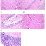 |
Figure 12: H&E staining of brain sections of control group. (a) Cerebral cortex, (b) hippocampus (subiculum), (c) hippocampus (fascia dentate), (d) hippocampus (hilus), (e) Striatum and cerebellum. |
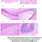 |
Figure 13: H&E staining of brain sections of Wi-Fi exposed group. |
Protection with Bio-Geometrical shapes resulted in normal histological feature of the cerebral cortex (Fig.14a), subiculum of the hippocampus (Fig.14b). The fascia dentate in the hippocampus showed nuclear pyknosis and degeneration in some neurons (Fig.14c). The striatum showed intracellular oedema in the neurons (Fig.14d).
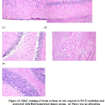 |
Figure 14: H&E staining of brain sections of rats exposed to Wi-Fi radiation and protected with BioGeometrical shapes group. |
Discussion
In the present study, we adopted two classic behavioral paradigms, OFT, EPM for anxiety testing in addition to MWM and episodic memory tests for memory evaluation. The current findings revealed that the exposure to Wi-Fi radiation for a long period of time altered anxiety and emotion in EPM test as indicated by the decline in open arm entries, and open arm exploration time. This test assesses the exploratory activity in EPM as a measure of anxiety and fear induced by high and open spaces. Upon exposure to open arms of EPM, the rodent experiences more anxiety behavior derived by the conflict between natural exploratory behavior of novel area and the avoidance of high and open spaces. Both open and closed arms evoke similar exploratory motivation; hence, avoiding open arms is linked to high levels of fear.28 These results corporate previous studies that demonstrated that exposure of rats to 3 different frequencies of microwave radiation increased closed arm entries in EPM test.29 Exposure to mobile phone radiation (900 MHz) also decreased percentage of entries into the open arm, percentage of time spent on the open arm and distance travelled on the open arm.28 Similarly, Liu et al.30 showed that exposure to low frequency magnetic field MF 4h/day for 25 days increased the anxiety-like behaviors in rats in EPM.
Locomotor activity after long term Wi-Fi radiation exposure was examined in open field test. This test evaluates various features of rodents’ behavior and its underlying drives including changes in emotion created by exposure to a novel arena. Being a major reaction to exposure to novel and open areas, thigmotaxis is considered as a reliable index in measuring emotion of rodents.31 The increase in thigmotaxis in Wi-Fi radiation exposed group implied that animals are highly anxious to stress induced by unfamiliar and open central part. The frequency of locomotion and rearing is generally affected in anxiety-like behavior.30 The observed increase in rearing frequency is a measure of the excitability in CNS and positively correlated with exploratory behavior in rodents.19 Liu 2008 showed that chronic exposure to ELF MF increased thigmotaxis and grooming without altering locomotor activity. In contrast, Salunke et al.32 proved that exposure of mice to HF-EMF up to 120 days did not produce any alteration in OFT.
Testing the performance of animals in MWM learning task, which aims to assess spatial memory, revealed that Wi-Fi radiation exposure affected the acquirement of learned reactions in the MWM test. In trial sessions, the Wi-Fi radiation exposed group showed longer latencies than the control group to reach the hidden platform. In memory retention test, the Wi-Fi radiation-exposed animals spent shorter time in the target quadrant indicating that animals were unable to recall the correct location of the hidden platform. This in turn points to the reduced spatial navigation capability as well as the object-place configurations of the Wi-Fi radiation exposed animals.33 Our findings are in line with findings of Li et al.34 reported that 6 weeks exposure to 2.45 GHz microwaves induced deficits in spatial learning and memory. Also, Deshmukh et al.29 demonstrated decline in rats’ performance in MWM after 90 days-exposure to 900, 1800, and 2450 MHz microwave.
On the contrary, Wi-Fi radiation exposure did not affect working memory in episodic-like memory test. The episodic-like memory task estimates the relative recency of two remembered objects, the old familiar one and the recent familiar one.23 Wi-Fi radiation exposed animals were able to recognize previously explored objects indicating that rats had the ability to discriminate object location based on the novelty.33
These alterations in behavioral assays could be a result of neurochemical alterations. In this study, analysis of monoamines in brain regions (cortex, striatum and hippocampus) revealed a detrimental effect of Wi-Fi radiation on the CNS. Wi-Fi radiation exposure induced a decrease in DA and 5-HT levels in cortex, striatum and hippocampus. This effect can be attributed to the oxidizing capability of the low-intensity radiofrequency radiation and the ability to cross the blood brain barrier (BBB) resulting in neurodegeneration.35 Previous works indicated that electromagnetic fields influence the biophysical roles of nerve cells and induce alterations of neurotransmitter levels. It has been demonstrated that 30 min radiation on mature nerve cells may generally suppress the synthesis of most monoamines and/or accelerate their degradation. EMRs have been proposed to act through vast cellular responses, through downstream activation of voltage-gated calcium channels (VGCCs), such as increasing intracellular calcium, oversignaling of calcium and nitric oxide and also excessive free radicals and peroxynitrite.36, 37 The presence of high densities of various VGCCs in neurons throughout the CNS implicates that CNS is highly sensitive to EMR.9 In addition, dopaminergic neurons are susceptible to oxidative response owing to their reduced antioxidant capacity and the high contents of DA, iron and lipids.38 The metabolism of DA generates superoxide radicals, hydrogen peroxide, and auto-oxidation quinone products of DA.7
Exposure to EMR induces alterations in Ach level39 and release40, choline uptake41 and acetylcholine esterase activity.42,43 Mahdavi and colleagues 39 mentioned that Ach is altered following exposure to EMR (1 and 5 Hz radiation) in male rats through activation of AchE gene expression level in the brain which ascertained cholinergic/inflammatory reactions and molecular injury in the brain cells. These changes can explain the observed alterations in AchE activity in the cortex and hippocampus with consequent induction of anxiety found in the exposed rats in the present study.
Melatonin is the main secretory neuro-hormone of the pineal gland, derived from serotonin at night that regulates the daily sleep/wake cycle, blood pressure, heart and hormone production. The pineal gland is located at the end of the visual system and connected to the eyes by retinal neuronal cell bodies. Through this neural connection with the eyes, the production of melatonin is regulated by the 1ight: dark cycle.45 Melatonin’s synthesis in the pineal gland is always low during the day and increases greatly at night.46 Since light is EMR in the visible electromagnetic spectrum, exposure of humans or animals to EMR in the visible range rapidly inhibits the capacity of the pineal gland to either produce or secrete melatonin.47 Previous studies reported that the exposure of animals to an altered geomagnetic field environment, altered the synthetic ability of the pineal gland.48
In the current study, HSP-70 was elevated in the cortex of Wi-Fi radiation exposed rats. These findings corporate previous studies that recorded significant increases in HSP-90 and HSP-70 in cerebellar hemispheres following exposure to EMF at 900 and 2450 MHz.49 Also, Yang et al.50 observed that exposure of adult rats to 2.45 GHz (6 W/kg) induces a stress reaction in hippocampus through upregulating the expression of HSP-27 and HSP-70. On the opposite side, some research investigators found no response of Wi-Fi exposure on HSP expression in their experiments. Ding et al.51 noticed that RF radiation field (1950 continuous wave for one hour) has no apparent impact on HSP-27 and HSP-70.
In the present study, Wi-Fi radiation exposure induced nuclear pyknosis and degeneration in cerebral cortex, striatum and hippocampus. Furthermore, focal gliosis was observed in cerebral cortex and diffuse gliosis in striatum which is indicative of neuroinflammation. These results are in line with the previous findings of Ibitayo et al.52 who showed that chronic Wi-Fi exposure induces vascular congestion and abnormal architecture. Both neurons and glia interact dynamically to facilitate information processing and behavior. The observed behavioral deficits in the current study can be attributed to the detrimental effect of EMR on neurons and glial cells, thus, altering the neuronal activity in the rat hippocampus. Long term exposure to microwave radiation has been previously shown to nuclear pyknosis, capillary congestion53, 54 in addition to irregular neuronal arrangement with evidence of Karyopyknosis especially in hippocampus.55
In the present study, the designed BioGeometrical shapes could modulate the deleterious effects of EMF emitted from Wi-Fi router. The protected group displayed normal behavioral pattern; normal anxiety and fear levels, and normal exploratory behavior in concomitant with restoration of dopamine, and serotonin concentrations. We propose that these forms by some way could mitigate negative effects of energy induced by EMR and combat the insults of EMR in the protected group, through supporting of self-healing. The histological features of the tissue of brain areas constitute the direct evidence for the neuroprotective influence of the BioGeometrical shapes.
The human body has an energy field around it, which has its own north-south axis. As we move around, the angle formed between our individual axis and that of the earth is constantly changing, and this in turn either strengthens or weakens our energy field. 17
BioGeometry uses the subtle energy principles of geometric form to introduce natural balance to the different energy qualities found in any living system. The natural function of this energy is to provide balance, or centering to the different energy qualities or effects within any living system. In addition, the space quality is affected by air quality, ventilation and geometric forms in outer environment. Each geometric form resonates with a specific resonance frequency that yields a change in brain waves that guide consciousness to a particular status.52 For example, the pyramid affect delta waves that guide the users’ consciousness to relaxation and comfort status, while the cube and cylinders affect alpha waves that help in mental stability, learning and improve general mood. Accordingly, the BioGeometrical forms made remarkable achievement in reversing the devastating effects of Wi-Fi radiation on rat brain.56
Conclusively, the present investigation illuminate the mechanisms underlying the detrimental effects of Wi-Fi routers on the chemistry and structural organization of rat brain which lead to aberration in the anxiety behavior and memory deficits. Also, the present approach provides clues for the potent power of using well designed BioGeometrical forms in minimizing these deleterious impacts via maintaining the energy structures and motivating a process of energy-quality equilibrium of the living system.
Recommendation
The favorable neuroprotective effects of the utilized Bio-Geometrical forms open a line of thought on designing these forms in small size to be inserted in the Wi-Fi devices in order to protect the users from the harmful insult of the emitted radiation.
Acknowledgement
The authors express sincere appreciation to Prof. Adel Bakeer Kholoussy, Professor of Pathology, Faculty of Veterinary Medicine, Cairo University, for his kind cooperation in Conducting Histological Examination In This Study.
Conflict of Interest
The authors declare no conflict of interests.
References
- Akdag MZ, Dasdag S, Canturk F, Karabulut D, Caner Y, Adalier N. Does prolonged radiofrequency radiation emitted from Wi-Fi devices induce DNA damage in various tissues of rats? J Chem Neuroanat 2016; 75 (Pt B): 116–22. http://dx.doi.org/10.1016/j.jchemneu.2016.01.003.
- Hardell L. World Health Organization, radiofrequency radiation and health – a hard nut to crack (Review) INT J ONCOL 2017; 51: 405-413. https://doi.org/10.3892/ijo.2017.4046.
- Sage C, Carpenter DO. Public health implications of wireless technologies. Pathophysiol 2009; 16 (2-3):233-46. https://doi.org/1016/j.pathophys.2009.01.011.
- Çelik Ö, Kahya MC, Nazıroğlu M. Oxidative stress of brain and liver is increased by Wi-Fi (2.45 GHz) exposure of rats during pregnancy and the development of newborns. J ChemNeuro anat 2016, 75: 134-9. https://doi.org/10.1016/j.jchemneu.2015.10.005.
- Hardell L, Sage C. Biological effects from electromagnetic field exposure and public exposure standards. Biomed Pharmacother 2007; 62(2):104-9. https://doi.org/10.1016/j.biopha.2007.12.004.
- Maaroufi K, Had-Aissouni L, Melon C, Sakly M, Abdelmelek H, Poucet B, et al. Spatial learning, monoamines and oxidative stress in rats exposed to 900 MHz electromagnetic field in combination with iron overload. Behav Brain Res 2014; 258:80-9. https://doi.org/10.1016/j.bbr.2013.10.016.
- Lotharius J, Brundin P (2002) Pathogenesis of Parkinson’s disease: dopamine, vesicles and α-synuclein. Nat Rev Neurosci 2002; 3: 932-42. https://doi.org/1038/nrn983.
- Haider S, Naqvi F, Batool Z, Tabassum S, Perveen T, Saleem S, Haleem DJ. Decreased Hippocampal 5-HT and DA Levels Following Sub-Chronic Exposure to Noise Stress: Impairment in both Spatial and Recognition Memory in Male Rats. Sci Pharm 2012; 80(4):1001-11. https://doi.org/ 10.3797/scipharm.1207-15.
- Thurm F, Schuck NW, Fauser M, Doeller CF, Stankevich Y, Evens R, Riedel O, Storch A, Lueken U, Li SC. Dopamine modulation of spatial navigation memory in Parkinson’s disease. Neurobiol Aging 2016; 38:93-103. doi: 10.1016/j.neurobiolaging.2015.10.019.
- Deiana S, Platt B, Riedel G. The cholinergic system and spatial learning. Behav Brain Res 2011; 221: 389–411. https://doi.org/1016/j.bbr.2010.11.036.
- Klinkenberg I, Sambeth A, Blokland A. Acetylcholine and attention. Behav Brain Res 2011; 221: 430–42. https://doi.org/1016/j.bbr.2010.11.033.
- Goncalves JF, Nicoloso FT, daCosta P, Farias JG, Carvalho FB, daRosa MM, et al. Behaviour and brain enzymatic changes after long term intoxication with cadmium salt or contaminated potatoes. Food Chem Toxicol 2012; 50: 3709–18. https://doi.org/1016/j.fct.2012.07.016.
- Tan DX, Manchester LC, Terron MP, Flores LJ, Reiter RJ. One molecule, many derivatives: A never-ending interaction of melatonin with reactive oxygen and nitrogen species?. J Pineal Res 2007; 42. 28–42. DOI:1111/j.1600-079X.2006.00407.x
- Henshaw DL, Reiter RJ. Do magnetic fields cause increased risk of childhood leukaemia via melatonin disruption? Bioelectromagnetics 2005; 7: S86–S97. DOI:1002/bem.20135
- Beck IM, Drebert ZJ, Hoya-Arias R, Bahar AA, Devos M, Clarisse D, et al. Compound A, a selective glucocorticoid receptor modulator, enhances heat shock protein Hsp70 gene promoter activation. PLoS One 2013; 8(7):e69115. https://doi.org/ 10.1371/journal.pone.0069115.
- French PW, Penny R, Laurence JA, McKenzie DR. Mobile phones, heat shock protein and cancer. Differentiation 2001; 67: 93–97. DOI:1046/j.1432-0436.2001.670401.x
- Karim I. Back to a Future for Mankind. Florida: CreateSpace Independent Publisher; 2010. ISBN: 9781449963958.
- Sharaf NE, El-Sawy MS, Metwally FM, El-Khayat Z, Abdel-Razik F. Protective Role of BioGeometry against Indoor Pollutants of Some Egyptian Building Materials in Adult Male Rats. World J Med Sci 2014; 10: 337-46. https://doi.org/10.5829/idosi.wjms.2014.10.3.1142.
- Sharaf NE, El-Sawy MS, Ahmed HH, Metwally FM, Hegazy NM, El-Mishad AM. Bio-geometrical shapes: a new option for protection against neurodegenerative insult of Wi-Fi radiation. Biosci Res 2018; 15(3): 2481-8.
- Pruus K, Vaarmann A, Rudissaar R, Allikmets L, Matto V. Role of 5-HT1A receptors in the mediation of acute citalopram effects: a 8-OH-DPAT challenge study. Prog Neuropsychopharmacol Biol Psychiatry 2002; 26(2):227-32. PMID: 11817498.
- Pellow S. Anxiolytic and anxiogenic drug effects in a novel test of anxiety: are exploratory models of anxiety in rodents valid? Methods Find Exp Clin Pharmacol 1986; 8(9): 557-65. PMID: 2877126.
- Morris R. Developments of a water-maze procedure for studying spatial learning in the rat. J Neurosci Methods 1984; 11: 47–60. PMID: 6471907.
- Nittby H, Grafström G, Tian DP, Malmgren L, Brun A, Persson BR, Salford LG, Eberhardt J. Cognitive impairment in rats after long-term exposure to GSM-900 mobile phone radiation. Bioelectromagnetics 2008; 29(3):219-32. https://doi.org/1002/bem.20386.
- Glowinski J, Iversen L. Regional studies of catecholamines in the rat brain. 3. Subcellullar distribution of endogenous and exogenous catecholamines in various brain regions. Biochem Pharmacol 1966; 15(7):977-87. PMID: 5967909.
- Pagel P, Blome J, Wolf HU. High-performance liquid chromatographic separation and measurement of various biogenic compounds possibly involved in the pathomechanism of Parkinson’s disease. J Chromatogr B Biomed Sci Appl 2000; 746(2): 297-304. PMID: 11076082.
- Gorun V, Proinov I, Baltescu V, Balaban G, Barzu O. Modified Ellman procedure for assay of cholinesterases in crude enzymatic preparations. Anal Biochem 1978; 86(1):324-6. PMID:655393.
- Banchroft JD, Stevens A, Turner DR. Theory and practice of histological techniques. 4th ed. Churchill Livingstone, NewYork, London, San Francisco, Tokyo, 1996, 744. http://trove.nla.gov.au/version/45634524.
- Narayanan SN, Kumar RS, Paval J, Kedage V, Bhat MS, Nayak S, Bhat PG. Analysis of emotionality and locomotion in radio-frequency electromagnetic radiation exposed rats. Neurol Sci 2013; 34(7):1117-24. https://doi.org/10.1007/s10072-012-1189-4.
- Deshmukh PS, Megha K, Nasare N, Banerjee BD, Ahmed RS, Abegaonkar MP, Tripathi AK, Mediratta PK. Effect of Low Level Subchronic Microwave Radiation on Rat Brain. Biomed Environ Sci 2016; 29(12):858-867. doi: 10.3967/bes2016.115.
- Liu T, Wang S, He L, Ye K. Anxiogenic effect of chronic exposure to extremely low frequency magnetic field in adult rats. Neurosci Lett 2008; 434 (1):12-7. https://doi.org/1016/j.neulet.2008.01.019.
- Choleris E, Thomas AW, Kavaliers M, Prato FS. A detailed ethological analysis of the mouse open field test: effects of diazepam, chlordiazepoxide and an extremely low frequency pulsed magnetic field. Neurosci Biobehav Rev 2001; 25(3):235-60. PMID: 11378179.
- Salunke BP, Umathe SN, Chavan JG. Behavioral in-effectiveness of high frequency electromagnetic field in mice. Physiol Behav 2015; 140: 32-7. doi: 10.1016/j.physbeh.2014.12.019.
- Dere E, Huston JP, De Souza Silva MA. Episodic-like memory in mice: simultaneous assessment of object, place and temporal order memory. Brain Res Brain Res Protoc 2005; 16(1-3):10-9. https://doi.org/1016/j.brainresprot.2005.08.001.
- Li HJ, Peng RY, Wang CZ, Qiao SM, Yong Z, Gao YB, Xu XP, Wang SX, Dong J, Zuo HY, Li Z, Zhou HM, Wang LF, Hu XJ. Alterations of cognitive function and 5-HT system in rats after long term microwave exposure. Physiol Behav 2015;140:236-46. doi: 10.1016/j.physbeh.2014.12.039
- Pall ML. Microwave frequency electromagnetic fields (EMFs) produce widespread neuropsychiatric effects including depression. J Chem Neuroanat 2016; 75(Pt B):43-51. https://doi.org/10.1016/j.jchemneu.2015.08.001.
- Pall ML. Electromagnetic fields act via activation of voltage-gated calcium channels to produce beneficial or adverse effects. J Cell Mol Med 2013; 17(8): 958-65. https://doi.org/10.1111/jcmm.12088.
- Pall ML. Electromagnetic field activation of voltage-gated calcium channels: role in therapeutic effects. Electromagn Biol Med 2014; 33(4): 251. doi: 10.3109/15368378.2014.906447.
- Greenamyre JT, Hastings TG. Parkinson’s: divergent causes, convergent mechanisms. Science 2004; 304: 1120-2. https://doi.org/1126/science.1098966.
- Obajuluwa AO, Akinyemi AJ, Afolabi OB, Adekoya K, Sanya JO, Ishola AO. Exposure to radio-frequency electromagnetic waves alters acetylcholinesterase gene expression, exploratory and motor coordination-linked behaviour in male rats. Toxicol Rep 2017; 4: 530-4. https://doi.org/10.1016/j.toxrep.2017.09.007.
- Modak AT, Stavinoha WB, Deam AP. Effect of short electromagnetic pulses on brain acetylcholine content and spontaneous motor activity of mice. Bioelectromagnetics 1981; 2(1):89-92. PMID: 7284047.
- Testylier G, Tonduli L, Malabiau R, Debouzy JC. Effects of exposure to low level radiofrequency fields on acetylcholine release in hippocampus of freely moving rats. Bioelectromagnetics 2002; 23(4):249-55. PMID: 11948603.
- Lai H, Carino MA, Horita A, Guy AW. Low-level microwave irradiation and central cholinergic activity: a dose-esponse study. Bioelectromagnetics 1989; 10(2):203-8. PMID: 2712849.
- Dutta SK, Das K, Ghosh B, Blackman CF. Dose dependence of acetylcholinesterase activity in neuroblastoma cells exposed to modulated radio-frequency electromagnetic radiation. Bioelectromagnetics 1992; 13(4):317-22. PMID: 1510740.
- Mahdavi SM, Sahraei H, Yaghmaei P, Tavakoli H. Effects of electromagnetic radiation exposure on stress-related behaviors and stress hormones in male wistar rats. Biomol Ther (Seoul) 2014; 22(6):570-6. https://doi.org/10.4062/biomolther.2014.054.
- Reiter RJJ. Static and extremely low frequency electromagnetic field exposure: reported effects on the circadian production of melatonin. J Cell Biochem 1993; 5 I, 394. PMID:8098713
- McIntyre IM, Norman TR, Burrows GD, Armstrong SM. Human melatonin suppression by light is intensity dependent. J Pineul Res 1989; 6, 149. PMID: 2915324.
- Reiter RJ. Action spectra, dose-response re- lationships, and temporal aspects of light’s effects on the pineal gland. Ann NY Acad Sci 1985; 453, 215. PMID: 3907458.
- Olcese J. The neurobiology of magnetic field detection in rodents. Prog Neurobiol 1990; 35, 325. PMID: 2281140.
- López-Furelos A, Leiro-Vidal JM, Salas-Sánchez AÁ, Ares-Pena FJ, López-Martín ME.Evidence of cellular stress and caspase3 resulting from a combined two-frequency signal in the cerebrum and cerebellum of sprague-dawley rats. Oncotarget 2016, 7: 64674-89. https://doi.org/10.18632/oncotarget.11753.
- Yang XS, Hao YT, Xiao Y, Chen CH, Zhang GB, Yu ZP. Exposure to 2.45 GHz electromagnetic fields elicits a HSP-related stress response in rat hippocampus. Brain Res bulletin 2012, 88: 371-8. https://doi.org/10.1016/j.brainresbull.2012.04.002.
- Ding GR, Wang XW, Li KC, Qiu LB, Xu SL, Tan J, Guo GZ. Comparison of Hsps expression after radio-frequency field exposure in three human glioma cell lines. Biomed Environ Sci 2009; 22(5):374-80. https://doi.org/10.1016/S0895-3988(10)60014-1.
- Ibitayo AO, Afolabi OB, Akinyemi AJ, Ojiezeh TI, Adekoya KO, Ojewunmi OO. RAPD Profiling, DNA Fragmentation, and Histomorphometric Examination in Brains of Wistar Rats Exposed to Indoor 2.5 Ghz Wi-Fi Devices Radiation. Biomed Res Int 2017; 2017:8653286. https://doi.org/ 10.1155/2017/8653286.
- Narayanan SN, Kumar RS, Potu BK, Nayak S, Bhat PG, Mailankot M. Effect of radio-frequency electromagnetic radiations (RF-EMR) on passive avoidance behaviour and hippocampal morphology in Wistar rats. Ups J Med Sci 2010; 115(2):91-6. https://doi.org/10.3109/03009730903552661.
- Wei-Jia Zhi, Li-Feng Wang, Xiang-Jun Hu. Recent advances in the effects of microwave radiation on brains. Military Medical Research 2017; 4:29. https://doi.org/10.1186/s40779-017-0139-0.
- Zhao L, Peng RY, Wang SM, Wang LF, Gao YB, Dong J, Li X, Su ZT. Relationship between cognition function and hippocampus structure after long-term microwave exposure. Biomed Environ Sci 2012; 25(2):182-8. https://doi.org/3967/0895-3988.2012.02.009.
- Eslam Elbaiuomy, Ibrahim Hegazy, Sherif Sheta. The impact of architectural spaces’ geometric forms and construction materials on the users’ brainwaves and consciousness status. Int J Low-Carbon Tech 2018, 13(1): 43–51. https://doi.org/10.1093/ijlct/ctx018.

