
Manuscript accepted on :22-Aug-19
Published online on: 30-09-2019
Plagiarism Check: Yes
Reviewed by: Md.Sarwar Hossain
Second Review by: Fatima Grace
Final Approval by: Dr Pallav Sengupta
Putu Yuliawati1*, Cynthia Dewi M1, A A A Sukartini Djelantik1, Putu Budhiastra1, N K Niti Susila1, Cok Istri Dewiyani P2, I Gede Raka W3 and I G K Nyoman Arijana4
1Department of Ophthalmology, Faculty of Medicine, Udayana University-Sanglah Hospital Denpasar Bali.
2Department of Ophthalmology, Faculty of Medicine, Udayana University-Bali Mandara Eye Hospital Denpasar Bali.
3Department of Internal Medicine, Faculty of Medicine, Udayana University-Sanglah Hospital Denpasar Bali.
4Department of Histology, Faculty of Medicine, Udayana University Denpasar Bali.
Corresponding Author E-mail: putu.yulia@gmail.com
DOI : https://dx.doi.org/10.13005/bpj/1773
Abstract
Pterygium is an eye disease with multifactorial etiopathogenesis. Molecular factors such as cell proliferation and inflammatory mediators are associated with increased calcium mobilization and activation of nuclear factor kβ mediated by histamine-1 receptors (H1R). This study aims to determine whether the expression of H1R primary pterygium tissue is higher than normal conjunctival tissue and the expression of H1R based on pterygium grades. This study was an analytic observational study with a case-control study approach at Sanglah General Hospital, Bali Mandara Eye Hospital, and Mangusada Hospital. The study was conducted from November 2017 to April 2018. The pterygium and conjunctival tissues obtained from 28 subjects in the same eye and examined for H1R expression by immunohistochemistry. The results of this study obtained 64.3% of women with a mean age of 54.2 ± 7.8 years. There was no difference in mean H1R expression between pterygium grades in the final score (P = 0.759). There was a mean difference of H1R between primary pterygium (42.50) and normal conjunctival tissue (14.50) with P <0.001. Only tissue types affected the expression of H1R in the final score (B = 4.893; 95% CI 4.363-5.423; P <0.001). It was concluded that the expression of H1R primary pterygium tissue was higher in primary pterygium than normal conjunctival tissue.
Keywords
Conjunctival Tissue; Histamine-1 receptors; Pterygium Tissue
Download this article as:| Copy the following to cite this article: Yuliawati P, Dewi M. C, Djelantik A, A, A, S, Budhiastra P, Susila N. K. N, Dewiyani P. C. I, Raka W. I. G, Arijana I. G. K. N. Histamine-1 Receptors Expression in Primary Pterygium Tissue is Higher than Normal Conjunctival Tissue. Biomed Pharmacol J 2019;12(3). |
| Copy the following to cite this URL: Yuliawati P, Dewi M. C, Djelantik A, A, A, S, Budhiastra P, Susila N. K. N, Dewiyani P. C. I, Raka W. I. G, Arijana I. G. K. N. Histamine-1 Receptors Expression in Primary Pterygium Tissue is Higher than Normal Conjunctival Tissue. Biomed Pharmacol J 2019;12(3). Available from: http://biomedpharmajournal.org/?p=28690 |
Introduction
Pterygium is a disorder that is often seen in the eye. Pterygium is a disorder characterized by the growth of fibrovascular tissue and conjunctival folds that resemble wings, usually in the nasal area with a horizontal direction that invades the corneal surface.1,2 Pterygium has the potential to cause blindness, in the later stages requires complex surgery for visual rehabilitation.3
Pterygium occurs throughout the world with prevalence rates that vary greatly in age, sex, race, and geography. Starting from 1% in Japan to 26% in the Samoan Islands, as well as an incidence above 33% elder (≥ 50 years) in Asia.4,5 Based on the Basic Health Research Indonesia in 2013, the prevalence rate of pterygium in Indonesia was 8.3%, of which the highest in Bali was 25.2%.6
Etiology and pathogenesis of pterygium is unclear. Environmental factors such as ultraviolet (UV) radiation, low humidity, and dust are related to pterygium pathogenesis. There are various other pathogenic factors that are thought to be associated with pterygium such as viral infection, epigenic aberration, epithelial-mesenchymal transition, anti-apoptotic and immunological mechanisms, lymphangiogenic and angiogenic stimulation, deregulation of extracellular matrix modulators and growth factors, inflammatory mediators, cell proliferation factors, cell collection progenitors and bone-marrow-derived stem cells, as well as modification of cholesterol metabolism.7,8,9
Histamine is the main mediator in allergy and inflammatory reactions. Histamine is not only in the process of allergies but also in the modulation of cell proliferation and migration.10,11 The pleiotrophic effects of histamine are mediated by several different pharmacological receptors such as histamine-1, histamine-2, histamine-3 and histamine-4 receptors (HR) which are all G protein-couple receptors (GPCR).12 Histamine in the conjunctiva acts by stimulating H1R and H2R, causing hyperemia, fibroblast cell proliferation, cytokine secretion, and increased microvascular cell permeability. Histamine via H1R in conjunctival epithelial cells paired with inositol phosphate increases intracellular calcium (Ca2+), which will be associated with the process of secretion and flow of inflammatory cytokines. Intracellular Ca2+ mobilization is used to assess the functional activity of histamine receptors in pterygium fibroblasts. The pterygium tissue is characterized by excessive cell proliferation, trans-differentiation and angiogenesis associated with Ca2+ signaling activity. Histamine-1 receptors can activate nuclear beta factor kappa (NF-kβ) which is a promising transcription factor in inflammation that regulates pro cytokine pro inflammation production and molecular adhesion.9,11,13,14 More than 40 H1R antagonists are available worldwide, where H1R ligands are the most widely used in all treatments. Both health professionals and consumers assess that all H1R antagonists who have been approved show efficacy and safety.15
Fang (2013) based on gene microarray analysis, found four histamine receptors (H1R, H2R, H3R, and H4R) in pterygium fibroblasts.9 Siak et al (2011) study that NF-κβ is found in human pterygium tissue, where the level is higher than normal conjunctival tissue.16 Qin et al (2016) proved the first time that histamine has an important role in pterygium proliferation through H1R, where H1R has the highest expression level and H4R with the lowest expression level. In normal conjunctival tissue also found H1R, H2R, and H4R, where the expression level of H1R conjunctival tissue is only half of the pterygium tissue.10
This research was conducted because of the high prevalence of pterygium in Indonesia and Bali in particular, where Indonesia is in the equatorial region. Until now, as far as the author’s knowledge, research to determine the expression of H1R in primary pterygium compared to normal conjunctival tissue is still limited. Based on the above, this study was conducted to determine whether the expression of H1R primary pterygium tissue is higher than normal conjunctival tissue.
Materials and Methods
This research is an analytic observational study with a case control study approach. 28 study subjects with primary pterygium aged ≥ 40 years and signed informed consent. The study was conducted at Sanglah Hospital, Bali Mandara Eye Hospital, and Mangusada Hospital from November 2017 to April 2018. Sample was all patients with consecutive primary pterygium who met the inclusion and exclusion criteria. Exclusion criteria were the use of topical / systemic anti-allergy drugs in the last two weeks, recurrent pterygium, surgical history / conjunctival trauma, and ocular infection. Ethical clearance was approved by the Research Ethics Commission of the Udayana University Medical Faculty / Sanglah Hospital Denpasar (2018.02.1.0194).
Research Protocol
The preparation stage was carried out interviews, eye examination, and eye documentation by the researcher. Criteria for pterygium grading correspond to Seid and Bejiga (2000) based on the size of the pterygium covering the cornea, ie grade I if the pterygium covers less than 1.5 mm, grade II if the pterygium is less than half the radius of the cornea, grade III if the pterygium is more than half the radius of the cornea, and grade IV if it hits and passes through the edge of the pupil, can be seen in Figure 1.17
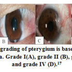 |
Figure 1: The grading of pterygium is based on extension to the cornea. Grade I(A), grade II (B), grade III (C), and grade IV (D).17 |
Sampling of the primary and normal conjunctival pterygium tissue is performed on the same eye of the study subject during surgery. The conjunctival tissue is taken in the normal superotemporal conjunctiva (furthest from the pterygium) during graft removal. The sample size of more than 2×2 mm stored separately in a container containing buffer formaline 10%. Immunohistochemical (IHC) examination of H1R expression was performed at the Histology Laboratory of Udayana University, Denpasar. The H1R expression was obtained from the semiquantitative final score shown in table 1.
Table 1: Assesment Score System18
| % positive cell (A) | Intensity reaction (B) | Final score |
| 0 = 0% | 0 = no reaction | A x B = score
(0-9) |
| 1 = < 30 % | 1 = weak | |
| 2 = 30-60% | 2 = mild | |
| 3 = > 60 % | 3 = strong |
Statistics
Data processing using the SPSS. Categorical scale descriptive data in the form of frequencies and percentages, while the data are numerically scaled in the form of mean and standard deviation. An unpaired t test to examine the differences in expression of H1R between the primary pterygium tissue and the normal conjunctiva. The difference in mean H1R expression between pterygium degrees was analyzed by one way anova test. Generalized linear model for analyzing control variables on different expressions of H1R. The level of significance at p <0.05.
Results
Characteristics of 28 research subjects can be seen in table 2.
Table 2: Characteristics of subjects
| Variable | n (%) | Mean ± SD |
| Age(year) | 54.2 ± 7.8 | |
| Gender
Male Female |
10 (35.7%) 18 (64.3%) |
|
| Outdoor activities (hours/day)
≤ 5 > 5 |
18 (64.3%) 10 (35.7%) |
|
| Onset (year)
<40 ≥40 |
6 (21.4%) 22 (78.6%) |
|
| Smoking status
Yes No |
6 (21.4%) 22 (78.6%) |
|
| Ocular allergy
Yes No |
7 (25.0%) 21 (75.0%) |
|
| Pterygium grading
I II III IV |
– 16 (57.1%) 11 (39.3%) 1 (3.6%) |
The proportion of female was 18 people (64.3%) more than males as many as 10 people (35.7%) with a mean age of 54.2 ± 7.8 years and the age range of 42-70 years. Most subjects (64.3%) performed outdoor activities 5 hours / day with an average length of 4.6 hours / day. Smoking was found in 6 people (21.4%) and the presence of ocular allergy was found in 7 people (25.0%). The highest grade of pterygium is 16 in grade II (57.1%) without any grade I in the subject. The pterygium tissue with 16 right eye samples (57.1%).
Description of tissue with IHC examination on pterygium tissue (A) shows H1R expression (brown colour) more than conjunctival tissue (B). The red arrow indicates the cell expressing H1R. The black arrow shows cells that do not express H1R. In normal conjunctival tissue, there is almost no H1R expression (Figure 2).
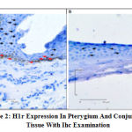 |
Figure 2: H1R Expression in Pterygium and Conjunctival Tissue with IHC Examination |
The expression of H1R primary pterygium tissue based on its grade was assessed from the percentage of positive cells, reaction intensity, and final score in the IHC examination can be seen in table 3. Based on the results of the table, there was no difference in mean H1R expression between pterygium grades.
Table 3: H1R Expression based on Pterygium Grading
| Pterygium Grading | H1R Expression | P Value | |
| % Positive cell
(A) |
II
III IV |
14.63
14.23 15.50 |
0.926 |
| Intensity reaction
(B) |
II
III IV |
14.63
13.91 19.00 |
0.760 |
| Final score
(AxB) |
II
III IV |
14.66
13.86 19.00 |
0.759 |
Expression of H1R pterygium and conjunctival tissue based on the percentage of positive cells, reaction intensity, and the final score on the IHC examination can be seen in Table 4.
Table 4: Differences in the expression of H1R pterygium and conjunctival tissue
| H1R Expression | P value | ||
| Pterygium | Conjunctiva | ||
| % Positive cell (A) | 42.14 | 14.86 | 0.000 |
| Intensity reaction (B) | 42.50 | 14.50 | 0.000 |
| Final score (AxB) | 42.50 | 14.50 | 0.000 |
Based on the results of the table above, there was a difference in the mean expression of H1R between the primary pterygium tissue and the normal conjunctiva, where the mean rank expression of H1R primary pterygium tissue was almost three times higher than the normal conjunctival tissue.
According to the results of table 5, only the type of tissue (primary pterygium tissue versus normal conjunctival tissue) affects the expression of H1R, while other variables do not affect.
 |
Table 5: Differences in H1R expression after controlling gender, age, ocular allergy, outdoor activity, smoking status, onset, pterygium grading, and type of tissue. |
Discussion
The relationship between sex and age with the development of pterygium is still debateable. Wu et al (2002) found prevalence rates above 33% in older age (≥ 50 years) in China.5 Study in the Central India by Nangia et al (2013) found the mean age of subjects with pterygium 56.6 ± 13 years and age range of 30-85 years.2 Nemet et al (2014) in Israel stated the mean age of pterygium patients was 58.4 ± 14 years.4 Total of 28 subjects were included in this study with an average age of 54.21 ± 7.81 years and an age range of 42-70 years. Age as a significant risk factor for pterygium is associated with increased susceptibility to UV exposure in older people with increasing age and spending most of the outdoor activities resulting in accumulation of UV damage. The results obtained may differ from other studies, due to differences in age ranges in collecting study samples.
Some literature shows a higher prevalence in women than men as in studies conducted in Dali populations in China, Doumen County in China, populations in the Barbados Islands, and Kathmandu in Nepal.5,19,20,21 In this study, the percentage of female subjects (64.3%) was more than that of men (35.7%). Research subjects were patients with pterygium who underwent surgery at the hospital. Women also pay more attention to cosmetic reasons so that they perform eye examinations for surgery.
The tropical climate as a risk factor for pterygium is strongly associated with outdoor activity and UV exposure.4 Research in Indonesia has a higher prevalence of pterygium in groups with outdoor activity 10 years earlier.22 Study in Sumatra (Indonesia) by Gazzard et al found a history of outdoor activities > 5 hours / day for the past 10 years associated with doubled risk for pterygium compared without the history.3 In this study obtained outdoor activities > 5 hours / day in the last 5 years amounting to 21.4% of the subjects. Bali is located at 8º south latitude which is included in the “pterygium belt” region with high UV exposure.
Lee et al (2017) in Korea stated that current smoking history was significantly lower in the pterygium group than without pterygium, according to previous studies that found that smoking protects against pterygium.23 Conversely Pyo et al (2016) reported that pterygium in Korea is associated with a lifestyle without smoking.24 Other studies also state that pterygium is not associated with smoking status.3,20 In this study found 21.4% of subjects with smoking and 78.6% without smoking. The mechanism of protective smoking is still unclear.
Histamine through H1R can cause immunomodulatory effects, itching, swelling, erythema, increased vascular permeability, and pain.25 Ocular symptoms can occur in 40-60% of allergy patients.26 In this study 28.6% of the subjects were found with ocular allergy. These results were obtained based on interviews with research subjects, where ocular allergy can occur unnoticed by the subject so that further examination is needed for diagnosis. Feelings that are felt, especially watery eyes.
Study by Shrestha and Kaiti (2016) obtained 58% of pterygium with grade I followed by grade II at 41%.19 Study by Tan et al (2006) in the Riau Islands of Indonesia obtained the highest 59.3% pterygium grade I.22 In this study, 57.1% pterygium II grade was obtained without any grade I. The subjects in this study were only pterygium which caused complaints and performed surgery, so that pterygium grade I was not found because it was generally asymptomatic and did not carry out examinations to health services. The varying degrees of pterygium division between different studies causes difficulties in comparing the severity of the inter-study.
The relationship between H1R and primary pterygium tissue has been demonstrated in this study and other previous studies. This study is the first study to assess RH-1 expression based on pterygium grading. Based on the grade of pterygium, the mean rank of expression of H1R pterygium grade II was 14.66, grade III 13.86, and grade IV 19.0 in the final score which did not have a significant difference between pterygium grades with P = 0.759. Pterygium I grade was not found in this study. The results showed no relationship between pterygium grading and H1R expression.
Histamine-1 receptors paired with Gq/11 proteins activate intracellular Ca2+ signalling, cGMP, phospholipase D, phospholipase A2, and NFκβ.27 Active HR and epidermal growth factors have been found in pterygium tissue. The histamine-1 receptors is the dominant receptor and is known to increase intracellular Ca2+ levels.9 According to Qin et al (2016), the highest H1R expression and H4R were lowest in pterygium fibroblasts, where the proliferative effect of histamine works mainly through H1R. Expression of H1R on pterygium fibroblasts twice from conjunctival fibroblasts with RT-qPCR and DNA agarose gel electrophoresis.10
In this study, the mean rank of H1R primary pterygium tissue was 42.14 and normal conjunctival tissue was 14.86 in the final score with P < 0.001 so that there was a significant difference in H1R expression between pterygium and the conjunctiva tissue. Mean rank expression of H1R primary pterygium tissue is almost three times higher than normal conjunctival tissue. Histamine-1 receptors included in GPCR can increase intracellular Ca2+ levels and activate NFκβ which is a prominently inflammatory transcription factor.9,13,14 Calcium signalling activity is associated with excess cell proliferation, trans-differentiation, and angiogenesis. Active NF-κβ in pterygium, contributing to several biological effects such as collecting inflammatory cells in pterygium tissue, increasing cell motility and invasion, influencing the expression of pro apoptotic and anti-apoptotic genes and cell cycle proteins.7
Conclusion
Based on the results of the study it was concluded that H1R expression primary pterygium tissue was higher than normal conjunctival tissue, and without mean differences between pterygium grades. This finding shows cell proliferation and inflammatory factors are molecular factors associated with the development of pterygium. Further biomolecular study is needed to investigate the susceptibility of H1R expression.
Acknowledgement
This research did not receive any specific grant from funding agencies in the public, commercial, or not-for-profit sectors. We thank our colleagues from Ophthalmology Department, Faculty of Medicine Udayana University who provided insight and expertise that greatly assisted the research.
Conflict of Interest
The authors declare that there is no conflict of interest.
References
- American Academy of Ophthalmology and staff. External Eye Disease and Cornea. United State of America: American Academy of Ophthalmology.2015-2016;324-325.
- Nangia, V., Jonas, J.B., Nair, D., Saini, N., Nangia, P., Panda-jonas, S. Prevalence and Associated Factors for Pterygium in Rural Agrarian Central India. The Central India Eye and Medical Study. PLoS ONE.2013;8(2):e82439.
- Gazzard, G., Saw, S.M., Farook, M., Koh, D., Widjaja, D., Hong, C.Y., Tan, D.T.H. Pterygium in Indonesia: Prevalence, Severity and Risk Factors. Br J Ophthalmol.2002;86(12):1341-1346.
- Nemet, A.Y., Vinker, S., Segal, O., Mimouni, M., Kiserman, Igor. Epidemiology and Associated Morbidity of Pterygium: A Large, Community-Based Case-Control Study. Seminars in Ophthalmology.2014:1-6.
- Wu, K., He, M., Xu, J., Li, S. Pterygium in Aged Population in Doumen Country, China. Yan Ke Xue Bao.2002;18:181-4.
- Kementerian Kesehatan RI. 2013. Riset Kesehatan Dasar Tahun 2013. Tersedia dalam: URL:http/www.depkes.go.id/resources/download.
- Cardenas-Cantu, E., Zavala, J., Valenxuela, J., Valdez-Garcia, J.E. Molecular Basis of Pterygium Development. Seminars in Ophthalmology.2014:1-17.
- Alqahtani, J.M. The Prevalence of Pterygium in Al-Khobar: A Hospital-Based Study. Journal of Family and Community Medicine.2013;20(3):159-161.
- Fang, C. “Elucidating the Regulatory Mechanisms of Pterygium” (Thesis). School of Biological Sciences, University of East Anglia, Norwich, UK.2013.
- Qin, Z., Fu, Q., Zhang, L., Yin, H., Jin, X., Tang, Q., Lyu, D., Yao, K. Proliferative Effects of Histamine on Primary Human Pterygium Fibroblasts. Mediators of Inflammation.2016;ID9862496.
- Bielory, L., Ghafoor, S. Histamine Receptors and The Conjunctiva. Curr Opin Allergy Clin Immunol.2005;5:437-440.
- Jemima, E.A., Prema, A., Thangam, E.B. Functional Characterization of Histamine H4 Receptor on Human. Molecular Immunology.2014;62:19-28.
- Panula, P., Chazot, P.L., Cowart, M., Gutzmer, R., Leurs, R., Liu, W.L.S., Stark, H., Thurmond, R.L., Haas, H.L. International Union of Basic and Clinical Pharmacology.XCVIII. Histamine Receptors. Pharmacol Rev.2015;67:601-655.
- Criado, P.R., Maruta, C.W., Criado, R.F.J., Filho, C.A.M. Histamine, Histamine Receptors and Antihistamines: New Concepts. An Bras Dermatol.2010;85(2):195-210.
- Tripathi, T., Shahid, M., Khan, H.M., Khan, R.A., Siddiqui, M., Mahdi, A.A. The Influence of Histamine H1-Receptor on Liver Functions in Immunized Rabbits. Saudi Journal of Biological Sciences.2011;18:411-418.
- Siak, J.J., Ng, S.L., Seet, L.F., Beuerman, R.W., Tong, L. The Nuclear-Factor kappaB Pathway is Activated in Pterygium. Invest Ophthalmol Vis Sci.2011;52(1):230-236.
- Seid, A., Bejiga, A. Free Conjunctival Autograft in the Management of Advanced Primary and Recurrent Pterygia. East African Medical Journal.2000;77:588-91.
- Klein, M., Vignaud, J.M., Hennequin, V., Toussaint, B., Bresler, L., Plenat, F., Leclere, J., Duprez, A., Weryha, G. Increased Expression of the Vascular Endothelial Growth Factor is a Pejorative Prognosis Marker in Papillary Thyroid Carcinoma. J Clin Endocrinol Metab.2001;86:656-658.
- Shrestha, P., Kaiti, R. A Hospital Based Study of Pterygium in Tertiary Care Hospital of Nepal. Kathmandu Univ Med J.2016;14(3):192-7.
- Zhong, H., Cha, X., Wei, T., Lin, X., Li, X., Li, J., Cai, N., Li, J., Su, X., Yang, Y., Yu, M., Yuan, Y. Prevalence of and Risk Factors for Pterygium in Rural Adult Chinese Populations of the Bai Nationality in Dali: The Yunnan Minority Eye Study. Invest Ophthalmol Vis Sci.2012;53(10):6617-6621.
- Luthra, R., Nemesure, B.B., Wu, S.Y., Xie, S.H., Leske, M.C. Frequency and Risk Factors for Pterygium in the Barbados Eye Study. Arch Ophthalmology.2001;119:1827-1832.
- Tan, C.S.H., Lim, T.H., Koh, W.P.; Liew, G.C., Hoh, S.T. Epidemiology of Pterygium on A Tropical Island in the Riau Archipelago. Eye.2006;20(8):908-12.
- Lee, Y.B., Kim, S.Y., Park, Y.G., Han, K.D., Kim, J.W., Chae, H.S., Lee, Y.C. Evaluation of Sosioeconomic Status as A Risk Factor of Pterygium Using the Korean National Health and Nutrition Examination Survei 2010 to 2011. Medicine.2017;96:11.
- Pyo, E.Y., Mun, G.H., Yoon, K.C. The prevalence and risk factors for pterygium in South Korea: the Korea National Health and Nutrition Examination Survey (KNHANES) 2009-2010.EpidemiolHealth.2016;38:e2016015.
- Wade, L., Bielory, L., Rudner, S. Ophthalmic Antihistamines and H1-H4 Receptors. Curr Opin Allergy Clin Immunol.2012;12:510-516.
- Leonardi, A., Bogacka, E., Fauquert, J.L., Kowalski, M.L., Groblewska, A., Jedrzejczak-Czechowicz, M., Doan, S., Marmouz, F., Demoly, P., Delqado, L. Ocular Allergy: Recognizing and Diagnosing Hypersensitivity Disorders of the Ocular Surface. Allergy.2012;67:1327-1337.
- Jutel, M., Blaser, K., Akdis, C.A. Histamine in Chronic Allergic Responses. J Invest Allergy Clin Immunol.2005;15:1-8.

