
Manuscript accepted on :November 24, 2017
Published online on: --
Plagiarism Check: Yes
Abbas Razzaq Abed and Ibtisam Mohammed Hussein
Al-Furat Al-Awsat Technical University /Technical institute of Babylon.
Corresponding Author E-mail: dr_abas2008@yahoo.com
DOI : https://dx.doi.org/10.13005/bpj/1340
Abstract
The results of this study were detect the action of common anti-fungal used against to Rhodotorula mucilaginosa, these agents were divided into two groups, first group was named specific antifungal agent which included Ketoconazole, Clotrimazole, Itraconazole , Fluconazole and Nystatin, while second group was named specific antifungal agent which included 1% Iodine (I), 3% Sodium bicarbonate (NaHCO3) , 3% Hydrogen peroxide (H2O2), 2% Acetic acid (CH3COOH)and 10% Potassium permanganate (KMnO4). The parameters were used in this study included isolation and identification of Rhodotorula mucilaginosa, antifungal susceptibility testing and minimum inhibitor concentration. All Rhodotorula mucilaginosa isolates were isolated from the fingers of the feet of athletes. Antifungal susceptibility testing showed Rhodotorula mucilaginosa sensitive to Ketoconazole, Nystatin, Clotrimazole, Acetic acid and H2O2 and resistance to Itraconazole, Ketoconazole, Iodine ,Sodium bicarbonate and Potassium permanganate. Acetic acid was recorded lowest MIC value in comparison with other non-specific antifungal agents , while Ketoconazole was recoded lowest MIC value in comparison with other specific antifungal agents.
Keywords
Athletes foot Rhodotorula Mucilaginosa; Specific; Non-Specific Antifungal;
Download this article as:| Copy the following to cite this article: Abed A. R, Hussein I. M. A Comparative Study Between Specific and Non-Specific Antifungal Agents to Treat the Rhodotorula Mucilaginosa Athletes Foot. Biomed Pharmacol J 2017;10(4). |
| Copy the following to cite this URL: Abed A. R, Hussein I. M. A Comparative Study Between Specific and Non-Specific Antifungal Agents to Treat the Rhodotorula Mucilaginosa Athletes Foot. Biomed Pharmacol J 2017;10(4). Available from: http://biomedpharmajournal.org/?p=18295 |
Introduction
An antifungal drug is a pharmaceutical (fungicide or fungistatic) used to treat and prevent fungal infection as athlete’s foot and thrush (Baginski and Czub 2009) . Athlete’s foot, is a skin disease of the feet caused by fungus . Signs and symptoms often include itching, rough skin, and congestion . In chronic cases the skin may blister. Athlete’s foot disease may infect any part of the foot, but most often grows between the toes (Bell-Syer et al. 2012). Rhodotorula is an environmental yeast that is found in air, soil, lakes, water, milk, and fruit juice. Rhodotorula species, part of the Basidiomycota phylum, colonies plants, humans, and other mammals (Larone 1995). Rhodotorula produces pink to red colonies and blastoconidia that are single cell lacking pseudohyphae and hyphae. Several studies have isolated Rhodotorula in different ecosystems and environments as well as described infections in Mammals. Rhodotorula spp. have been recognized as emerging yeast pathogens in humans in the last two decades (Hagan et al.1995). Among the few sources to the pathogenicity of Rhodotorula spp. in animals, there are several studies of an outbreak of skin infections in birds and sea animals and pulmonary infections and otomycosis in ruminants (Fernanda, 2012). This study aimed to select best antifungal agent to treat Rhodotorula mucilaginosa athletes foot.
Materials and Methods
Antifungal Agents
In this study used two types of antifungal agents include Specific and non- specific antifungal agents. Specific antifungal agents were used to treat skin mycological infection between foot toes include Ketoconazole, Clotrimazole ,Itraconazole, Fluconazole and Nystatin, While non-specific antifungal agents are using to treat skin mycological infection between foot toes include 1% Iodine, 3% Sodium bicarbonate, 3% H2O2, 2% Acetic acid and 10% Potassium permanganate. All non-specific antifungal agents were prepared from chemical agents stock solution (10% Iodine, 50% Sodium bicarbonate, 50% H2O2, 50% Potassium permanganate and 98% Acetic acid and use distilled water as a dilute of these substances}. The required concentration has been prepared by V1C1=V2C2 equation (Mary, 2005).
Microbe’s Isolates
Rhodotorula mucilaginosa were isolate from toes skin (Hagan et al.1995 ; Galan-Sanchez et al.,1999) of sport man by cut the small piece of infected skin between their toes figure (1) and kept in the cool container till trans to lab of Microbiology/the Technical institute of Babylon than this small piece cultivated in the Sabouraud Dextrose Agar (SDA) media for 48 hours at 27ºC (Paweł and Anna 2010).
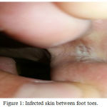 |
Figure 1: Infected skin between foot toes.
|
Lab Diagnosis of Rhodotorula Mucilaginosa
Macroscopic Appearance
Rhodotorula spp. are pigmented basidiomycetous yeasts in the family Sporidiobolaceae (Fell et al. 2000) Rhodotorula spp. produce colonies that are pink to red in color but can also be orange to red on Sabouraud Dextrose agar due to the presence of carotenoid colours . Colony shape has been appeared as thin , smooth, and sometimes mucoid. Rhodotorula spp. are nutritionally non-fastidious, grow easily on common media, and are characterized by a rapid growth rate(Larone 2002).
Microscopic Appearance
A small portion of Rhodotorula mucilaginosa colony cultivated in SDA media after incubation 48 hours at 27ºC taken by bacteriological loop and it place on the clean surface of class slide and mixed with one drop distilled water and one drop of Lactophenol-cotton blue dye. A cover slide was gradually applied with slowly pressure to expulsion air bubbles. The slide was then observed under microscope {Power zoom X10 , X40 , 100X objective lenses respectively} (Cumitech , 1980). Rhodotorula mucilaginosa was appeared as Spherical to elongate cells and budding yeast cells . In some cases, rudimentary pseudomycelium can be observed (Reference Method for Broth Dilution Antifungal Susceptibility Testing of Yeasts 2008) figure (2).
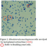 |
Figure 2: Rhodotorula mucilaginosa cells are dyed by lactophenol cotton blue (100X) Ο Refer to Budding yeast cells.
|
Biochemical Test
Rhodotorula mucilaginosa production of urease; and inability to assimilate inositol or to ferment sugars (Reference Method for Broth Dilution Antifungal Susceptibility Testing of Yeasts 2008).
Rhodotorula Mucilaginosa Cellular Counts
In this study the method of Rhodotorula mucilaginosa cells count same calculation method of Candida albicans cell count include:
Rhodotorula mucilaginosa cells suspension was prepared by adding five ml of distilled water to fresh SDA media contains Rhodotorula mucilaginosa (colonies aged 48 hour at 27ºC). Its turbidity was adjusted accordance to the absorbance of 0.08-0.10 at 625nm corresponding to 5 x 10⁶ CFU/ml (Ricardo and Edeltrudes 2013).
Determination of Minimum Inhibitory Concentration (MIC)
MIC of the effective specific and non-specific antifungal agents were determined by tube dilution Method (Cruickshank, 1975)., Ten test tubes with 8 ml of Sabouraud Dextrose Broth (SDB) in each were taken and autoclaved. To the first tube, 2 ml of the each concentration (50% H2O2,10% Iodine and 98% Acetic acid) was added and serial double fold dilution was done up to the 10 tube and from the 10 tube, 2 ml of the mixture was discarded. To each tube 100μl of inoculums Rhodotorula mucilaginosa suspension (5 x 10⁶ CFU/ml) were added and mixed well . The tubes were incubated for 48 hours at 27ºC. The least concentration of each one specific and non-specific antifungal agents capable of inhibiting the Rhodotorula mucilaginosa growth was considered MIC.
Antifungal Susceptibility Testing
Specific Antifungal Susceptibility Testing
A standard antifungal disc diffusion susceptibility testing method (Clinical Laboratory Standards Institute 2009) were used by commercially available discs preloaded with Ketoconazole (10 µg) , Clotrimazole (50 µg), Nystatine (100 I.U.) , Itraconazole (25 µg) and Fluconazole (50 µg) were using to determine the inhibition zone against Rhodotorula mucilaginosa in SDA media.
Non-Specific Antifungal Susceptibility Testing
Ager well diffusion method (Magaldi et al. 2004) were used to Non-Specific antifungal Susceptibility Testing.
One hundred µl of Rhodotorula mucilaginosa suspension were spread uniformly over SDA medium by using the class spreader, then left for one hour to dry of yeast cells on the media surface. By using cork borer, one central well (digs) was worked on the SDA media. One hundred microliter was taken from each non-specific antifungal agents (1% Iodine , 3% Sodium bicarbonate , 3% H2O2 , 2% Acetic acid and 10% Potassium permanganate) that has been prepared previously and put in these wells. Same previous steps were used again for distilled water which considered as control group. Number of petri-dish for each agent repeated five times.
Inhibition activities of the non-specific antifungal agents were determined by measuring the zones inhibition formed around the well in millimeter. The plates were observed for presence of zones of inhibition around the well after 48 hours at 27ºC (Mohit 2013).
Statistical Analysis
Data are presented as M±SE. For the statistical analysis, it was used one-way analysis of variance (ANOVA) using SPSS 13.0. Variances were considered significant if p < 0.05 (Joda 2008).
Results and Discussion
The results of the present study showed a difference in the effectiveness of antifungal agents (Specific and Non-specific) against Rhodotorula mucilaginosa.
Specific Antifungal Agents
In this study Rhodotorula mucilaginosa isolates appeared more susceptible (sensitive) to Ketoconazole, Nystatin and Clotrimazole respectively whereas other azole antifungal drugs (Itraconazole and Fluconazole) did not show any effectiveness (Resistance), as figure (3) and table (1).
The lowest MIC value (mg/ml) of different specific antifungal agents used in this study against Rhodotorula mucilaginosa showed in Ketoconazole in comparison with other agents used in this study, table (2).
Table 1: Zone inhibition diameter of different specific antifungal agents against Rhodotorula mucilaginosa in the SDA media in comparison with distilled water. The age of colonies 48 hours at 27ºC.
| Antifungal
µg or I.U./Disc |
Inhibition Zone
M±SE |
| Ketoconazole (10 µg) | 2.07±0.04 A |
| Clotrimazole (50 µg) | 1.32±0.04 B |
| Nystatine (100 I.U.) | 1.80±0.05 A |
| Itraconazole(25 µg) | 0.00±0.00 C |
| Fluconazole (50 µg) | 0.00±0.00 C |
| Control | 0.00±0.00 C |
M±SE =Mean ±Stranded error
Variant capital letters refer to significant values (P<0.05) between groups.
Table 2: MIC value of different specific antifungal agents against Rhodotorula mucilaginosa in the SDA broth for 48 hour at 27ºC.
| Antifungal | MIC value |
| Ketoconazole | 0.190 µg/ml |
| Clotrimazole | 39.06 µg/ml |
| Nystatine | 79.65 I.U./ml |
| Itraconazole | 1.6 mg/ml |
| Fluconazole | 2.4 mg/ml |
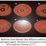 |
Figure 3: Inhibition Zone diameter (disc-diffusion method) of specific antifungal agents against Rhodotorula mucilaginosa in the SDA media for 48 hours at 27°C.
|
Ke= Ketoconazole (10 µg), Cl= Clotrimazole (50 µg), Ny= Nystatin (100 I.U.),
Fl=Fluconazole (25 µg), It= Itraconazole (50 µg), C=Control group.
The difference of antifungal drugs activity dependent on type and mechanism of action.
The Mechanism of Action of azole antifungal group (Ketoconazole, Clotrimazole, Itraconazole and Fluconazole) act by inhibit CYP P450 14 α- demethylase in mould and yeast. CYP P450 14 α- demethylase enzyme is necessary to convert of lanosterol to ergosterol , While mechanism of action of Nystatin act by Linked to ergosterol in mycological membrane causing membrane to become leaky (Myers 2006).
The use of antifungal drugs in the therapy of fungal diseases can lead to the development of antifungal resistance.
The resistance of Rhodotorula mucilaginosa to Ketoconazole and Clotrimazole may be result from low intracellular antifungal concentration by stimulation of efflux pathway or decreased of antifungal penetration, modification of the specific active sites, up regulation of the specific enzyme and development of bypass pathways (Pemán 2009).
Non-Specific Antifungal Agents
In this study Rhodotorula mucilaginosa isolates appeared more susceptible (Inhibition zone) to Acetic acid and H2O2 respectively whereas other non-specific antifungal agents (Iodine ,Sodium bicarbonate and Potassium permanganate) did not show any effectiveness (Resistance) , as figure (4) and table (3).
The lowest MIC value (mg/ml) of different non-specific antifungal agents used in this study against Rhodotorula mucilaginosa showed in acetic acid in comparison with other agents, table (4).
Table 3: Zone inhibition diameter of different non-specific antifungal agents against Rhodotorula mucilaginosa in the SDA media in comparison with distilled water. The age of colonies 48 hours at 27ºC.
| Antifungal | Inhibition Zone
M±SE |
| Acetic acid (2%) | 2.83±0.11 A |
| H2O2 (3%) | 0.71±0.03 B |
| Sodium bicarbonate (3%) | 0.00±0.00 C |
| Iodine (1%) | 0.00±0.00 D |
| Potassium permanganate (10%) | 0.00±0.00 D |
| Control | 0.00±0.00 D |
M±SE =Mean ±Stranded error
Variant capital letters refer to significant values (P<0.05) between groups.
Table 4: MIC value of different non- specific antifungal agents against Rhodotorula mucilaginosa in the SDA broth for 48 hour at 27ºC.
| Antifungal | MIC value |
| Acetic acid | 0.62 mg/ml |
| H2O2 | 20 mg/ml |
| Sodium bicarbonate | 120 mg/ml |
| Iodine | 8 mg/ml |
| Potassium permanganate | 40 mg/ml |
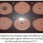 |
Figure 4: Inhibition Zone diameter (Agar well diffusion method) of specific antifungal agents against Rhodotorula mucilaginosa in the SDA media for 48 hours at 27°C.
|
A=Acetic acid (2%) , N=Sodium bicarbonate (3%) , H=Hydrogen peroxide (3%) , P=Potassium permanganate (10%) , I=Iodine (1%) C=Control.
Some non-specific agents used in this study have antifungal effect but variety in cellular yeast damage depended on type, concentration and mechanism of action of antifungal agent used .
CH3COOH has been mostly used in medical fields for more than 6000 years for the disinfection of wounds infections and especially as an antiseptic agent in the treatment and prophylaxis of deferent microorganisms . The antimicrobial effect of acetic acid, even at concentrations as low as 5%, has been attributed to its ability to decrease pH both in intra- and extracellular conditions and therefore to altering the cell membrane’s transportation and integrity, as well as enzymatic activity, and even precipitating cytoplasmic proteins(Ryssel 2009) , Whereas antimicrobial activity of H2O2 acts as an oxidant by producing •OH which linked with macromolecules of cell example lipids, proteins, and DNA. It has been proposed that exposed sulfhydryl groups and double bonds are particularly targeted (Block. and Peroxygen 1991).
NaHCO3 is used as an alkalinizing material for glutaraldehyde sterilization of medical apparatus . In watery solution, it ionizes to form sodium ions and bicarbonate ions ions. The dissociation of HCO−3 ions from NaHCO3 increases the pH of a solution (Enfors and Molin 1975).
I ions acts by lowering the O2 concentration in aerobic microbes cell . I ions interacts with the respiratory chain of the microbes by blocking the transport of electrons through electrophilic reactions with the respiratory chain enzymes. I also interacts high affinity with the proteins of the cytoplasm membrane in a form with a positive (H2O +I) or neutral (I2 or HOI) charge (Maris ,1995).
KMnO4 is a mild antiseptic with astringent properties. It is used in dermatology to treat weeping skin conditions (Anderson 2003). KMnO4 oxidation of organic pollution also may induce biodegradability and/or toxicity to microbes (Bowers 1992), Also KMnO4 act on the cell membrane phospholipids, containing unsaturated fatty acids, may be sensitive to MnO–4 oxidation at C=C bonds that led to defect of cellular components (Bui and Cotton 2002).
All non-specific antifungal agents which resistance to Rhodotorula mucilaginosa may be resulted from:-
First cause
Fungal resistance to chemical biocides is very limited. One commonly accepted theory about the mechanism of fungal resistance to biocides involves natural (intrinsic) resistance. A fungal cell may have an innate ability to present a permeability barrier to one or more biocides, or to inactivate a biocide due to the presence of existing enzymes (McDonnell and Russell 1999).
Second cause
The defiance mechanism of Rhodotorula mucilaginosa to Potassium permanganate, Iodine and Sodium bicarbonate may be resulted from lowest concentrations were used to treatment of Rhodotorula mucilaginosa for that may be Increasing the concentration of these substances may increase their effectiveness against this yeast.
Conclusion
Specific antifungal drugs
Rhodotorula mucilaginosa were highly sensitive to Clotrimazole and Ketoconazole and Nystatin and resistance to Fluconazole and Itraconazole.
Non-Specific antifungal drugs
Rhodotorula mucilaginosa were sensitive to Acetic acid and H2O2 and resistance to Sodium bicarbonate, Iodine and Potassium permanganate.
Recommendation
Molecular study of cellular defect of Rhodotorula mucilaginosa after specific and non-specific antifungal agents treated.
Studying Rhodotorula mucilaginosa resistance to specific and non-specific antifungal agents in this study.
Studying the direct use of antifungal agents (Clotrimazole, Fluconazole, Nystatin, 2% Acetic acid and 3% H2O2) in a sample of patients infected by Rhodotorula mucilaginosa athletes’ foot.
Acknowledgments
The authors are acknowledge to the Al-Furat Al-Awsat Technical University /Technical institute of Babylon in helping us to provide the providing the requirements of research.
References
- Baginski M., Czub B. Amphotericin B and its new derivatives. Current Drug Metabolism. 2009;10(5):459–69.
CrossRef - Bell-Syer S. E., Khan S. M., Torgerson D. J., Bell-Syer., Sally E. M., ed. Oral treatments for fungal infections of the skin of the foot. The Cochrane database of systematic reviews. 2009;10.
- Larone D. H . Medically Important Fungi – A Guide to Identification, American Society for Microbiology, Washington, DC, USA, 3rd edition. 1995.
- Hagan M. E., Klotz S. A., Bartholomew W., Potter L and Nelson M. A pseudoepidemic of Rhodotorula rubra: a marker for microbial contamination of the bronchoscope. Infection Control and Hospital Epidemiology. 1995;16(12):727-728.
CrossRef - Fernanda W and GoldaniL. Z. Epidemiology of Rhodotorula. An Emerging Pathogen. 2012;2012:7. Article ID 465717.
- Mary A. H., Robert L. K and John C. P. Mathematics Exercises in Biotechnology. National Science Foundation .NSF Award : DUE 0003065. 2005.
- Galan-Sanchez F., Garcia-Martos P., Rodriguez-Ramos C., Marin-Casanova P and Mira-Gutierrez J. Microbiological characteristics and susceptibility patterns of strains of Rhodotorula isolated from clinical samples. Mycopathologia. 1999;145:109-112.
CrossRef - Paweł K and Anna B. M . Drug susceptibility of 64 strains of Rhodotorula sp. Wiadomoœci Parazytologiczne. 2010;56(2):167-170.
- Fell J. W., Boekhout T., Fonseca A., Scorzetti G and Statzell-Tallman A. Biodiversity and systematics of basidiomycetous yeasts as determined by large-subunit rDNA D1/D2 domain sequence analysis. Int. J. Syst. Evol. Microbiol. 2000;50(3):1351-1371.
CrossRef - Larone D. H. Medically important fungi. 4th ed. Washington, D.C.: ASM Press. 2002.
- Cumitech. Practical Methods for Culture and Identification of Fungi in the Clinical Microbiology Laboratory. American Society for Microbiology, Washington, D.C. 1980.
- Reference Method for Broth Dilution Antifungal Susceptibility Testing of Yeast. 2008.
- Ricardo D and Edeltrudes O. Anti-Candida Activity and Chemical Composition of Cinnamomum zeylanicum Blume Essential Oil .Br Arch. Biol. Technol. 2013;56 (5):749-755.
CrossRef - Cruickshank R., Duguid J. P and Marmion. Tests for sensitivity of antimicrobial agents. Med. Microbiol. 1975;190-208.
- Clinical Laboratory Standards Institute. CLSI Document M44-A2. Reference Method for Antifungal Disk Diffusion Susceptibility Testing of Yeasts, Approved Guideline, 2nd ed.; CLSI: Wayne, PA, USA. 2009.
- Magaldi S., Mata-Essayag C., de Capriles C., Perez M., Colella T., Carolina O and Yudith O. Well diffusion for antifungal susceptibility testing International Journal of Infectious Diseases. 2004;8:39-45.
CrossRef - Mohit K., Mohammed F., Satyapal S., Anwar S and Ashok K. B. Antifungal activityof the Eucalyptus australe important medicinal plant. International Journal of Engineering Science Invention. 2013;2(1):30-27.
- Joda M.The progressive statistical analysis by using SPSS. (1sted.) Churchill livingstone .Edinburgh. 2008.
- Myers R. S. Immunizing and Antimicrobial Agents. MEDCH. 2006;401.
- Pemán J., Cantón E and Espinel-Ingroff A. Antifungal drug resistance mechanisms. Expert. Rev .Anti Infect. Ther. 2009;7(4):453-60.
CrossRef - Ryssel H., Kloeters O., Germann G., Shafer T. H., Wiedemann G and Oehlbauer M. The antimicrobial effect of acetic acid-an alternative to common local antiseptics. Burns. 2009;35(5):695-700.
CrossRef - Block S and Peroxygen S. compounds. In: Block S S, editor. Disinfection, sterilization, and preservation. 4th ed. Philadelphia, Pa: Lea and Febiger. 1991;167–181.
- Enfors S. O and Molin G .,ed. Gerhardt P., Sastilow R. N and Sadoff H. L. Inhibition of germination in Bacillus cereus spore by high gas pressure. In Spores VI Ann Arbor, MI. American Society for Microbiology. 1975;506–512.
- Maris Modes of action of disinfectants. Rev. sci. tech. Off. int. Epiz. 1995;14(1):47-55.
- Anderson . Should potassium permanganate be used in wound care? Times. 2003;5-11;99(31):61.
- Bowers A. R., Cho S. H and Singh A. Symposium W.W. Eckenfelder A. R., Bowers and Roth J. A (eds.). Chemical oxidation of aromatic compounds: comparison of H2O2, KMnO4, and O3 for toxicity reduction and improvements in biodegradability. In Chemical Oxidation, Technologies for the Nineties. Proceedings of the First International Technomic Publishing Co., Inc., Lancaster, Penn. 1992.
- Bui C. T and Cotton R. G. H. Comparative study of permanganate oxidation reactions of nucleotide bases by spectroscopy. Bioorganic Chemistry. 2002;30:133–137.
CrossRef - McDonnell G and Russell A. D. Antiseptics and disinfectants: activity action and resistance. Clin. Microbiol. Rev. 1999;12:147-149.

