Rational In Silico Design of a Multi-Epitope Peptide Vaccine Targeting SPP1 for Non-Small Cell Lung Cancer
 , Murugesh Easwaran2, Prabhu Dhamodharan3, Shanmughavel Piramanayagam4* and Luke Elizabeth Hanna5
1Central Research Laboratory for Biomedical Research, Vinayaka Mission’s Medical College and Hospital, Vinayaka Mission’s Research Foundation (VMRF-DU), Karaikal, India
2Department of Bioinformatics, Bharathiar University, Coimbatore, India
3Department of Biotechnology, Center for Drug Discovery, Karpagam Academy of Higher Education, Coimbatore, India
4Department of Botany, Bharathiar University, Coimbatore, India
5Department of Virology and Biotechnology, ICMR-National Institute for Research in Tuberculosis, Chennai, India
Corresponding Author E-mail:shanmughavel@buc.edu.com
Download this article as:
ABSTRACT:Lung cancer is defined as the exponential growth of mutated cells. Significantly, It is noteworthy because it is the second most prevalent kind of cancer in both men and women. Eighty-five percent of lung cancer fatalities are caused by non-small cell lung cancer (NSCLC). Immunotherapy has been shown to improve patient health, including survival rate, progression-free survival, and percentage of response. Peptides are more effective than antibodies at penetrating tissue and internalizing cells, thereby facilitating more effective delivery of immunomodulators to tumors. A multi-epitope peptide vaccine was designed to function as an antigen and elicit an immune response against NSCLC in response to natural immunity. The immunization with the peptide is expected to be more specific and efficient as the target protein is highly expressed in the worldwide NSCLC patient samples. We employed a set of computational tools and software, such as the IEDB database for epitope mapping, ToxinPred for Toxicity, AllerTop for allergenicity and vaxijen for antigenicity, Cluspro for molecular docking, C-ImmSim for molecular dynamics, and for the construction and validation of peptides. As a result, the generated multi-epitope peptide vaccine with favorable weighted score (-1168.7 kcal/mol) with TLR4. Immune simulation analysis indicates that the immunization response enhances the population of dendritic cells, NK cells, memory cells, B-cell differentiation, and immunoglobulin accumulation. In-silico analysis on cloning and expression of the 11-epitope vaccine construct with pET-28a(+) vector in Escherichia coli (Strain K12) showed high potency with a Codon Adaptation Index value of 1.0. This study presents the in-silico design of a peptide vaccine with 11-epitopes, incorporating CTL (4 epitopes), HTL (1 epitope), and B-cell (2 epitopes) complemented by T-cell epitopes (4 epitopes), targeting the SPP1 antigen for NSCLC immunotherapy. The vaccine construct demonstrated favorable antigenicity (probable antigen, VaxiJen score >0.4) with non-allergenic, and non-toxic properties.
KEYWORDS:Epitope; Immunotherapy; In-Silico cloning; NSCLC; Peptide; Vaccine construct
Introduction
Worldwide, Lung cancer is the second most common in men and women, which is an uncommon histologic alteration of lung structure due to mutation, with the symptoms like coughing, shortness of breath, chest discomfort, headaches, and fatigue, as well as more serious ones like unexpected weight loss, blood in the cough, wheezing, and bone pain.1 As per the latest statistics released by the National Cancer Institute American Cancer Society, 2.5 million new lung cancer cases were recorded in the year 2024, and 2.3 million cases are expected in the year of 2025. Totally 1.8 million deaths were recorded due to lung cancer, and 1.2 million are expected in the year of 2025. The morbidity incidence is observed as one among eight, and the death index is one among five.2,3 Unlike the last decade, women (age >65 years) were more susceptible to lung cancer morbidity than men.4 In India, the second most frequent malignancy in both men and women is lung cancer with 81748 cases; among 58 970 were male.5 One in nine Indians is susceptible to lung cancer morbidity, and one in 29 women and one in 67 men. In the US, lung cancer is consistently behind cancer-related mortality at 1 in 5 persons. The major reason behind this is habitual smoking, which leads to a high risk of lung cancer. Prolonged and heavy consumption is common. Thus, it contains chemicals that can directly damage the DNA, cause mutation, and lead to cancer.6,7 The American Cancer Society reported 85% of lung cancer deaths in the year 2025 were directly attributed to smoking (>1 million deaths), which is higher than the deaths caused by colorectal cancer. The three common types of lung cancer were non-small cell lung cancer, small cell lung cancer, and mesothelioma. Until the last decade, 85% of lung cancer deaths happened due to NSCLC, but in 2025 it rose to 87%.8 Even though NSCLC is slowly growing compared to SCLC, it creates a high risk, as 40% of cases had metastasis before diagnosis.9,10
There are numerous anti-cancer treatments in practice, such as radiation, chemotherapy, immunotherapy, hormonal therapy, and surgery. Chemotherapy alters the immune system to relieve symptoms, prepare for further treatment, destroy cancer cells, and prevent spread. Nausea, vomiting, weakened immune system, palpitations, and mucositis are some notable disadvantages. Temozolomide (TMZ) chemotherapy dramatically lowers T lymphocytes in the immune system.11 Radiation therapy possibly reduces and destroys cancer cells with the side effects of fatigue, hair loss, nausea, vomiting, diarrhea, reproductive troubles, changes in sensation, and harm to healthy tissues. All these therapy approaches have a limited impact.12
On another spectrum, Immunotherapy is trusted to be a successful way for treatment, prevention and overcoming the disadvantages of cancer treatment. Despite its complexity and unreliabilityDespite its complexity and unreliability, it reduces overall survival (OS) and progression-free survival (PFS) among outpatients.13 The effectiveness of immunotherapy is entirely dependent on the sensitivity of adaptive immune cells, including Cytotoxic T-cells, T-helper cells, and B-lymphocytes. During inflammation and antigen recognition, there is a high concentration of tissue-resident memory T-lymphocytes (Trm) in peripheral blood. CD4(+) and CD8(+) receptors of T-cells remember the antigenic substance and start the synthesis of B-cell (plasma cell) antibodies.14 Antigen evasion triggers a humoral and cellular immune response, which in turn activates Th, cytotoxic T-cells and B-cells. After cytotoxicity encounters an existing antigen, T-helpers release chemical messengers to activate B-cells. Ultimately, particular antibodies are secreted, leaving memory B-cells with an antibody impression.15 The host immune system and infectious antigen maintain a reciprocal balance during vaccine delivery, and cross-elimination, equilibrium, and escape phases are used to encourage immunization.16
A major part of immunotherapy, peptides were used to induce successful immunization as they are very specific to hormone receptors, surface receptors, immune receptors, and direct killing of tumor cells.17 Polypeptides have similar features to antibodies when compared to other targeted ligands like polysaccharides, MAbs, aptamers, and protein ligands. Antigenic targets, immune monitoring, and immune system checkpoints are essential for significant advancements in tumor immunology. Immunotherapy has been extensively studied as a therapeutic modality for lung cancer. Antigen recognition is pivotal in the therapeutic efficacy of active immunotherapy, which can be categorized as either non-specific or specific. Based on literatures, L-BLP25 is an immunotherapy targeting the mucin 1 (MUC1) antigen that stimulates a T-cell response against MUC1 in both a preclinical lung cancer mouse model with MUC1 transgene and in human patients. L-BLP25 is a liposome-based vaccine that contains a synthetic 25-amino acid lipopeptide derived from the tandem repeat region of MUC1, combined with the nonspecific adjuvant monophosphoryl lipid A and three different lipids.18 Designing such peptides is challenging because protein-protein and protein-peptide interactions govern most biological processes, and historically, targeting these interactions with small molecules has proven difficult.
Advanced immunotherapy leads to the design of preventive vaccines based on the genome or sequence of infectious agents, to enhance target precision during immunization, resulting in successive and low allergenicity.19 The antigenic properties of pathogens played a wide role in the preparation of vaccines. Consequently, we developed a peptide as a vaccination candidate for NSCLC derived from antigenic regions of SPP1 protein, which is significantly overexpressed in lung cancer. Epitope recognization mechanism of T-cells is basically different from B-cells and antibodies. T-cells identify antigen-presenting cells using MHC proteins in a fragmented state. MHC proteins, encoded by the major histocompatibility gene complex, are integral membrane glycoproteins that bind peptides and display them as T-cell epitopes for recognition by T-cell receptors. Thus, our peptide is designed to induce antigenicity and highly recognized by the immune cells. In this study, we focused on SPP1-based vaccine preparation. Significantly, multi-epitope peptides are designed from the antigenic sites of SPP1. Further, the peptides were analyzed for antigenicity and allergenicity to play a promising role.
Materials and Methods
Physicochemical properties of target protein (SPP1)
A protein sequence [Osteopontin] was obtained from the UniProtKB database [P10451 · OSTP_HUMAN]. A significant non-collagenous bone protein that adheres strongly to hydroxyapatite, seemingly constituting an essential component of the mineralized matrix. It acted as a cytokine crucial to type I immunity, enhancing the synthesis of interleukin-12 and interferon-gamma while diminishing interleukin-10 production. Deep TMHMM was a hybrid model that forecasts the topology of alpha-helical transmembrane proteins (αTMPs) by integrating deep learning neural networks and a Hidden Markov Model.20 It can predict and record the transmembrane helices available in protein structure. Then the physiochemical characters of protein sequences were studies with Protparam tool (https://web.expasy.org/protparam/) of Expasy, including molecular mass, calculated isoelectric point (pI), elemental composition, protein half-life, stability score, aliphatic content, degree of hydrophobicity, and hydropathy index.
B – cell epitope prediction
All the possible B-cell epitopes were mapped with IEDB’s B-cell epitope prediction tool (http://tools.iedb.org/bcell/) tailored as a semi-empirical technique as Bepipred Linear Epitope Prediction 2.0 algorithm combined with the Kolaskar-Tongaonkar antigenicity scale (threshold: 1.0).21
T-cell epitope prediction
MHC-I epitopes were identified with MHC-I binding prediction tool (http://tools.iedb.org/mhci), whereas MHC-II epitopes were delineated by the consensus technique of the IEDB database based on the target sequences. The NetMHCIIpan 4.1 EL algorithm (Version 2023.09) is specifically utilized for the prediction of T-cell epitopes in human, focusing on the HLA-A*01:01 allele for MHC-I epitope representation of the HLA-A gene. All other settings and parameters were kept at their default values, with XHTML tables selected as the output format. The MHC-II prediction integrates the Neural Network alignment algorithm (NN-align) with matrix alignment techniques to estimate peptide binding affinity. HLA-DP option selected for species/locus pair, with reference allele designated as DPA1*01:03/DPB*01:01, All other settings and parameters were kept at their default values, with XHTML tables selected as the output format.22 The protein sequence for the T-cell receptor was analyzed using Neural Network-based prediction with the netCTL program. The prediction algorithm identifies antigenic sites utilizing a neural network using a threshold value of 0.5 and a C-terminal cleavage cutoff of 0.15.
Multistage epitope screening (antigenicity, allergenicity, toxicity)
All predicted epitopes were evaluated for antigenicity, allergenicity, and toxicity to select the most suitable ones. The VaxiJen 2.0 server (http://www.ddg-pharmfac.net/vaxijen) was used to predict antigenicity of T-cell and B-cell epitopes based on protein physicochemical properties, applying a threshold of 0.4.23 AllerTOP (https://www.ddg-pharmfac.net/AllerTOP) assessed the allergenicity of these epitopes. Toxicity was analyzed using the ToxinPred2 server (https://webs.iiitd.edu.in/raghava/toxinpred2/).24
Vaccine construct design (linkers, adjuvant)
The multi-epitope vaccine was formulated using poly-ICLC, a compound consisting of polyinosinic-polycytidylic acid stabilized by poly-L-lysine and carboxymethylcellulose, followed by the incorporation of all accessible epitopes for vaccine production.25 The EAAAK is a linker employed to conjugate B-cell epitope with an adjuvant, followed by coupling of B-cell’s epitopes with a series of T-cell’s epitopes, which include both cytotoxic T-lymphocytes and helper T-lymphocytes, utilizing AAY and GPGPG linkers and a Toll-like receptor 3 (TLR3) agonist that enhances both innate and acquired immune responses, particularly promoting dendritic cell activation and responses from cytotoxic T-cells.26
Construct level Validation
VaxiJen 2.0 (http://www.ddg-pharmfac.net/vaxijen) was utilized to evaluate the antigenicity of the designed multi-epitope vaccine, while AllerTOP (https://www.ddg-pharmfac.net/AllerTOP) predicted its allergenicity. Toxicity was assessed using the ToxinPred server (https://webs.iiitd.edu.in/raghava/txnpred/).23,24
Tertiary structure modeling and refinement (I-TASSER, GalaxyRefine)
The solubility of the multi-epitope construct was evaluated by introducing the construct into bacterial system and assessing the overexpression was conducted using SOLUproT (http://scratch.proteomics.ics.uci.edu/), Protparam tool from ExPASy was accessed to (https://web.expasy.org/protparam/protparam-doc.html) investigate the physicochemical properties.27,28
Three-Dimensional Structure Prediction and Structural Refinement
The tertiary structures of the multi-epitope construct were predicted using structure prediction methods, primarily the I-TASSER tool (https://zhanggroup.org/ITASSER/). I-TASSER analyzes key structural elements such as major helices, sheets, strands, beta turns, gamma turns, and disulfide bonds by generating full-length protein models through the excision of continuous segments from threading alignments, followed by reassembly via replica-exchange Monte Carlo simulations. The SPICKER algorithm clusters low-temperature replicas generated during the simulation and selects the top five cluster centroids to build detailed atomic models. The predicted tertiary structure of the vaccine was then refined by repacking and side-chain rebuilding using the GalaxyRefine server (https://galaxy.seoklab.org/) to enhance model accuracy.29
Molecular docking against TLR4
MolProbity (http://molprobity.biochem.duke.edu/) was utilized to validate the quality of the three-dimensional structure. ClusPro 2.0 (https://cluspro.bu.edu/) was configured to analyze interactions between the TLR4 receptor’s ligand-binding domain (PDB: 2Z63) and the vaccine construct.30 Cluspro 2.0 was employed to dock multiple-epitope peptides with the target receptor and to assess the efficacy of synthesis. It generates docking models ranked by weighted energy scores that integrate contributions from Van der Waals forces, electrostatic interactions, and desolvation energy. The most favorable (most negative) weighted score indicates the best predicted binding orientation.
Immune Simulation (C-ImmSim)
C-Immsim Server is a computational counterpart of celada- seidan model, used to evaluate the immune response (like cell-cell co-operation, homeostasis of immune cells, clonal selection by antigen affinity, MHC restriction, diversity of immune elements as a result of immunization) developed by the constructed peptide vaccine. Immunization efficacy was assessed with a position-specific scoring matrix.31 The C-ImmSim server mimics natural immunological responses to antigens. It forecasts both cellular and humoral immune reactions based on antigen-specific interactions.
Codon Optimization and In-Silico Cloning
Codon adaptation was performed on the proposed vaccine to optimize its codon usage in the prokaryotic bacterium Escherichia coli. Codon was optimized with Java Codon Adaptation Tool (http://www.jcat.de/).32 The E. coli K12 strain was selected to improve expression efficiency of the final vaccine protein. Prokaryotic ribosome binding sites, rho-independent transcription terminators, and restriction enzyme sites were carefully removed to ensure precise translation of the vaccine gene. Restriction sites of XhoI and BamHI were appended to the vaccine’s N-terminus and C-terminus, respectively. Subsequently, it was positioned between the XhoI and BamHI sites in the pET-28a (+) vector.
Results
The SPP1 gene, which encodes osteopontin, serves as a biomarker for non-small cell lung cancer (NSCLC) and shows high expression across global NSCLC samples.33 Its upregulation drives cancer progression and indicates poor prognosis in lung cancer patients.34
The SPP1 amino acid sequence with 314 length was retrieved from the Uniprot Database as follows,
P10451 · OSTP_HUMAN
MRIAVICFCLLGITCAIPVKQADSGSSEEKQLYNKYPDAVATWLNPDPSQKQNLLAPQNAVSSEE
TNDFKQETLPSKSNESHDHMDDMDDEDDDDHVDSQDSIDSNDSDDVDDTDDSHQSDESHHS DESDELVTDFPTDLPATEVFTPVVPTVDTYDGRGDSVVYGLRSKSKKFRRPDIQYPDATDEDITSH MESEELNGAYKAIPVAQDLNAPSDWDSRGKDSYETSQLDDQSAETHSHKQSRLYKRKANDESNE HSDVIDSQELSKVSREFHSHEFHSHEDMLVVDPKSKEEDKHLKFRISHELDSASSEVN
SPP1 significantly contributes to the prognosis and advancement of multiple cancer types. High SPP1 expression is linked to tumor progression, metastasis, and reduced patient survival across various malignancies such as breast, ovarian, Liver, Colon, and Prostate. Thus, SPP1 was believed to be a promising gene to elute vaccine candidates using the reverse vaccinology approach.
Physicochemical Properties of SPP1
As helices play crucial roles related to stability and binding affinity in structure-based studies, the presence and the quantity of alpha-helical and beta-barrel result in no violation in topology analysis Figure 1. The posterior probabilities of the protein stretch in a narrow line with signal; thus the sequence behaviour is narrow outside and from the first amino acid till the last one behaves neutrally as 1.0.
The hydropathicity of SPP1 is -1.100. Protein-protein interactions can be altered by hydropathicity, which can also affect regulatory systems and signalling pathways. Protein misfolding and aggregation linked to neurodegenerative diseases are examples of how changes in hydropathicity might aid in the onset and progression of disease. A higher percentage of aliphatic side chains in proteins were indicated by a higher aliphatic index (AI), which could lead to enhanced thermostability. It is projected that proteins with an AI greater than 40 will remain stable, as SPP1 possesses 57.42. Proteins that include cysteine (Cys) residues may experience reduction, due to the transformation of disulfide bonds (S-S) into thiol groups (-SH), but SPP1 cysteine residues were evaluated a 0.647. The half-life of a protein describes the stability and turnover in a protein’s protein pool shrinking to half its original size. The target protein had 30 hours as a half-life period (mammalian reticulocytes, in vitro). SPP1 protein demonstrated favorable physicochemical properties for vaccine development, including appropriate hydropathicity, half-life period, thermostability, and aliphatic index.
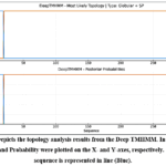 |
Figure 1: Depicts the topology analysis results from the DeepTMHMM. In this graph, sequence and Probability were plotted on the X- and Y-axes, respectively. The query sequence is represented in line (Blue). |
B-Cell Epitope Mapping
As per Kolaskar-Tongaonkar method, antigenic site prediction the hydrophobic residues as Leu, Cys, and Val, code for major antigenic sites. There are 11 epitopes (listed in Table 1) that were identified from the query sequence in the range 7-19 in length. Average score with a threshold value of 1.00, minimum of 0.85, and maximum of 1.27 as depicted in Figure 2.
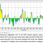 |
Figure 2: Position-based antigenicity score of the SPP1 protein sequence using the Kolaskar-Tongaonkar method. The X-axis represents amino acid residues (positions 0-320), and the antigenicity score is represented along the Y-axis. |
Table 1: B-cell epitopes Mapping from the SPP1
|
S.No |
Peptide | Length | Antigenicity | Allergenicity | Toxicity |
| 1 | AVICFCLLGITCAIPVKQA | 19 | Non-Antigen | Allergen |
Non-Toxin |
|
2 |
PDAVATW | 7 | Antigen | Non-Allergen | Non-Toxin |
| 3 | KQNLLAPQNAV | 11 | Non-Antigen | Non-Allergen |
Non-Toxin |
|
4 |
DELVTDF | 7 | Non-Antigen | Non-Allergen | Non-Toxin |
| 5 | ATEVFTPVVPTVD | 13 | Non-Antigen | Allergen |
Non-Toxin |
|
6 |
DSVVYGLR | 8 | Antigen | Allergen | Non-Toxin |
| 7 | KAIPVAQDLN | 10 | Antigen | Allergen |
Non-Toxin |
|
8 |
HKQSRL | 6 | Antigen | Non-Allergen | Non-Toxin |
| 9 | SDVIDSQ | 7 | Antigen | Allergen |
Non-Toxin |
|
10 |
LSKVSREF | 8 | Non-Antigen | Allergen | Non-Toxin |
| 11 | DMLVVDP | 7 | Non-Antigen | Allergen |
Non-Toxin |
Table 1- The predicted B-cell epitopes from the SPP1 protein sequence, using the IEDB database, are summarized with details on the total number of peptides, their positions, and lengths. The table contains the output data form the IEDB database, AllerTop, ToxinPred.
T-cell Epitope Mapping
All lengths of MHC-I epitopes of T-cell were predicted between 0.0 (Rank 100)- 0.9 (Rank 1). Totally 2128 epitopes were retrieved. Based on the score (>0.5) and Rank (1 and above), 12 MHC-I epitopes were predicted between the length of 9-13, were tabulated in Table 2.
In MHC-II epitope analysis, HLA-DP is a non-classical MHC gene present as a cell-surface receptor in immune recognition and antigen presentation. MHC-II epitopes (total no. of 600) were identified with score range of 0.0029-0.5965 and ranked between 0.03-100. The scrutiny was based on the score (>0.3) and rank (>1.5), and 2 epitopes were selected for further studies showed in Table 3. In both MHC-I and MHC-II, high scores represent a good binder.
There are 307 antigenic sites identified from the sequence by the netCTL tool. Based on the top-down score (>1), 8 peptides were taken for further analysis, listed in Table 4. In total, 27 T-cell epitopic regions were chosen for multi-epitope construction.
Table 2: T-cell epitopes of MHC-I molecule derived from SPP1
|
S.No |
Peptide | Length | Score | Rank | Antigenicity | Allergenicity |
Toxicity |
|
1 |
SSEEKQLYNKY | 11 | 0.94711 | 0.02 | Non-Antigen | Allergen | Non-Toxin |
| 2 | QADSGSSEEKQLY | 13 | 0.92114 | 0.03 | Antigen | Allergen |
Non-Toxin |
|
3 |
ESEELNGAY | 9 | 0.875545 | 0.04 | Antigen | Allergen | Non-Toxin |
| 4 | GSSEEKQLY | 9 | 0.796305 | 0.07 | Antigen | Non-Allergen |
Non-Toxin |
|
5 |
MESEELNGAY | 10 | 0.733674 | 0.09 | Non-Antigen | Non-Allergen | Non-Toxin |
| 6 | SGSSEEKQLY | 10 | 0.713499 | 0.09 | Antigen | Non-Allergen |
Non-Toxin |
|
7 |
SSEEKQLY | 8 | 0.699811 | 0.1 | Antigen | Non-Allergen | Non-Toxin |
| 8 | HMESEELNGAY | 11 | 0.673376 | 0.1 | Non-Antigen | Non-Allergen |
Non-Toxin |
|
9 |
SAETHSHKQSRLY | 13 | 0.613084 | 0.13 | Antigen | Allergen | Non-Toxin |
| 10 | FTPVVPTVDTY | 11 | 0.529123 | 0.18 | Antigen | Allergen |
Non-Toxin |
|
11 |
HSHKQSRLY | 9 | 0.51324 | 0.18 | Antigen | Non-Allergen | Non-Toxin |
| 12 | SHMESEELNGAY | 14 | 0.511991 | 0.18 | Non-Antigen | Allergen |
Non-Toxin |
Table.2- Listed the T-cell epitopes (MHC-I molecule) derived from the SPP1 protein sequence from the IEBD database. The table contains the output data form the IEDB database, AllerTop, and ToxinPred.
Table 3: T-cell epitopes of MHC-II molecule derived from SPP1
|
S.No |
Start | End | Length | Peptide | Score | Rank | Antigenicity |
Allergenicity |
Toxicity |
|
1. |
269 | 283 | 15 | FHSHEFHSH | 0.5965 | 0.03 | Non- Antigen |
Non- Allergen |
Non-Toxin |
| 2. | 199 | 213 | 15 | YKAIPVAQD | 0.3863 | 0.71 | Antigen | Non- Allergen |
Non-Toxin |
Table 3- Listed the T-cell epitopes (MHC-II molecule) derived from the SPP1 protein sequence using the IEBD database. The data presented in the table were the output from the IEDB database, AllerTop, and ToxinPred.
Table 4: T-cell epitopes predicted by netCTL
|
S.No |
Length | Peptide Sequence |
Predictions score |
Antigenicity |
Allergenicity |
Toxicity |
|
1. |
9 | ESEELNGAY | 4.7139 | Antigen | Allergen | Non-Toxin |
| 2. | 9 | GSSEEKQLY | 3.1554 | Antigen | Non-Allergen |
Non-Toxin |
|
3. |
9 | ESDELVTDF | 2.2963 | Non-antigen | Non-Allergen | Non-Toxin |
| 4. | 9 | HSDESDELV | 1.9213 | Antigen | Non-Allergen |
Non-Toxin |
|
5. |
9 | HSHKQSRLY | 1.6389 | Antigen | Non-Allergen | Non-Toxin |
| 6. | 9 | ATDEDITSH | 1.5409 | Antigen | Allergen |
Non-Toxin |
|
7. |
9 | VSSEETNDF | 1.3786 | Antigen | Non-Allergen | Non-Toxin |
| 8. | 9 | QSDESHHSD | 1.1158 | Non-antigen | Non-Allergen |
Non-Toxin |
Table 4 list of T-cell epitopes predicted by netCTL method from the IEDB database. The data presented in the table were the output from the IEDB database, AllerTop, and ToxinPred.
Allergenicity, Antigenicity, and Toxicity of Predicted Peptides
The scrutinised B-cell epitopes (11) and T-cell epitopes (27) were analyzed for antigenicity, allergenicity, and toxicity. The immunological capacity to identify an epitope, particularly T-cells and B-cells, were determined by its antigenicity. Thus, the antigenicity of an epitope is most important to elicit immune recognition. Antigenicity prediction results among 11 B-cell epitopes 5 were antigenic, among 12 MHC-I type of T-Cell epitopes: 8 were antigenic, among 2 MHC-II T-cell epitopes 3 were antigenic, and among 8 Cytotoxic T-cell epitopes: 6 were antigenic. Among the 38 selected epitopes, 22 were found to have antigenicity.
Another important scale to select epitopes is allergenicity; if an epitope is used to construct immunotherapeutic products, it should be non-allergenic. All 38 epitopes were screened for allergenicity.Among 11 B-cell epitopes, 4 proved non-allergenic; of 12 MHC-I T-cell epitopes, 6 were non-allergenic; from 7 MHC-II T-cell epitopes, only 2 showed non-allergenicity; and among 8 cytotoxic T-cell epitopes, 7 were non-allergenic.; overall, 24 epitopes were tested as non-allergenic.
The three-dimensional structure of a peptide can influence toxicity with conformational changes that enable to interact with and harm host tissues or cells. To ensure the toxicity of 38 selected peptides, the toxicity predicted was assessed. Toxicity analysis results show that all 38 epitopes were non-toxic and are eligible to serve as part of the immunotherapeutic product.
Vaccine Construction
The epitopes that were passed in all three selection criteria as non-allergic, immunogenic and non-toxic were decided to be a part of the immunotherapeutic peptide listed in Table 5. Linkers were added to the predicted epitopes (B-cell and T-cells), and the multi-epitope structure was constructed as follows shown in Figure 3A.
EAAAKPDAVATWGPGPGHKQSRLGPGPGGSSEEKQLYGPGPGSGSSEEKQLYGPGPGSSEEKQLYGPGPGHSH
KQSRLYGPGPGYKAIPVAQDAAYGSSEEKQLYAAYHSDESDELVAAYHSHKQSRLYAAYVSSEETNDFHHHHH
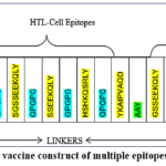 |
Figure 3A: Depicts the vaccine construct of multiple epitopes arranged with linkers. Click here to View Figure |
Final Multi-Epitope Construct
Total epitopes: 11
Final length: 147 amino acids
Molecular weight: 15,615.75 Da
Antigenicity: Probable antigen (VaxiJen: > 0.4)
Non-allergen
Non-toxic
Table 5: Potential immunotherapeutic peptides
|
S.No |
Type of Epitope | Peptide Sequence | Antigenicity score | Hydropathicity
|
Theoretical PI |
|
1. |
B-cell | PDAVATW | 0.9912 | 0.16 |
3.8 |
|
2. |
B-cell | HKQSRL | 2.1243 | -2.02 |
11.01 |
|
3. |
MHC-I of T-cell | GSSEEKQLY | 1.2557 | -0.32 | 4.54 |
| 4. | MHC-I of T-cell | SGSSEEKQLY | 1.2584 | -0.31 |
4.54 |
|
5. |
MHC-I of T-cell | SSEEKQLY | 0.6806 | -0.38 | 4.54 |
| 6. | MHC-I of T-cell | HSHKQSRLY | 0.7785 | -0.48 |
10.01 |
|
7. |
MHC-II of T-cell | YKAIPVAQD | 0.7605 | -0.17 | 6.18 |
| 8. | Cytotoxic T-cell | GSSEEKQLY | 1.2557 | -1.54 |
4.54 |
|
9. |
Cytotoxic T-cell | HSDESDELV | 0.6528 | -1.2 | 3.92 |
| 10. | Cytotoxic T-cell | HSHKQSRLY | 0.7785 | -1.93 |
10.01 |
|
11. |
Cytotoxic T-cell | VSSEETNDF | 0.7932 | -1.03 |
3.58 |
Table.5- The List of epitopes were capable of serving immunotherapeutic peptides, their Antigenicity score, Hydropathicity, Theoretical PI were included were the output of Protparam tool.
Allergenicity, Antigenicity, and Toxicity screening of the constructed Vaccine
VaxiJen v3.0 analysis classified the vaccine construct as a “probable immunogen” with 100% probability, confirming its immunogenicity. The construct tested non-allergenic and non-toxic, exhibiting a hydropathicity of -1.101, theoretical pI of 6.37, molecular weight of 15,615.75 Da, and 147 amino acids. Its predicted half-life spanned 1 hour in mammalian reticulocytes, 30 minutes in yeast, and over 10 hours in E. coli.
3D Structure Design and Function Prediction
The 3D structure of immunotherapeutic construct model was created (shown in Figure 3B), with I-Tasser and evaluated using C-score, T-M score, and cluster density. From the five generated models generated by I-TASSER, Model-1 was selected based on the highest C-score (-3.26), largest number of decoys (1182), and highest cluster density (0.0565). While the C-score and TM-score (0.35±0.12) indicate moderate confidence in the predicted structure (C-scores typically range from -5 to 2; TM-score >0.5 indicates high confidence), this model represents the best available prediction for this novel multi-epitope construct. Gene ontology report of I-Tasser reveals the strong influence of the model in defense response GO:0006952. The MolProbity consulted about the macromolecule structure, diagnosing local problems enabling corrections, and validating geometry and dihedral angle combinations. Structure refinement analysis of model results were structurally favorable with Root Mean square Deviation (RMSD) of 0.399, clash score of 5.0, and Molprobity score of 2.214 with the great coverage of 89.7% favored regions in Ramachandran plot without any poor rotamers (0.0). Thus, the predicted peptide structure was geometrically successful as a peptide. The structure was subsequently refined using GalaxyRefine to improve geometric quality, resulting in favorable Ramachandran statistics (89.7% in favored regions).
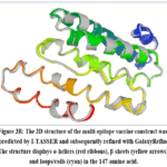 |
Figure 3B: The 3D structure of the multi-epitope vaccine construct was predicted by I-TASSER and subsequently refined with GalaxyRefine. The structure displays α-helices (red ribbons), β-sheets (yellow arrows), and loops/coils (cyan) in the 147-amino acid. |
Binding Affinity of the construct
Top 5 models of designed peptides were retrieved from the I-Tasser server and their weighted scores predicted using ClusPro server. Models were ranked based on the arbitrary composite docking towards the target protein TLR4 (2Z63). Scores represent composite energy terms of van der Waals, electrostatics and desolvation. The results of Model-1 (-875.4), Model-2 (-1009.), Model-3 (-1025.1), Model-4 (-906.5) and Model-5 (-1168.7) were shown in Figure 3C-3G. Negative binding energy represents greater arbitrary energy for docking orientation. The weighted score of Model-5 contributed by Van der Waals force of -251.8 and a balanced score of -875.4 an Electrostatic favor of -1009.0 and a Hydrophobic favor of -1007.9. More negative scores indicate stronger predicted binding interactions. The Model-5 shows dominant weighted scores proceeded for further analysis. Interaction of the peptide construct with adjuvant and without adjuvant towards the receptor (TLR4) shows similar results. Thus, the usage of adjuvant for the peptide construct may not influence its activity.
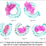 |
Figure 3C-G: Represents the molecular docking of 2Z63 with model 1-5. Cyan represents the receptor, and magenta represents the peptide. |
Peptide Behavioral Studies
The simulation studies of multi-epitope vaccine candidates were carried out in In-silico mode against each immune mechanism. Overall immune response elicited by the vaccine candidate was documented for 35 days. From the immune simulation results of the Immunotherapeutic peptide vaccine candidate, B-cell activity can be achieved from day and the memory cell population can bring by day 5, between 5-35 days of immunization. B isotype IgM-induced immunity was achieved with 300/mm3 of memory cells, as shown in Figure 4A. In the peripheral blood, the Ig population increased as IGM (15/mm3) and IgG1 (5/mm3) within 5-7 days shown in Figure 4B. As a part of the immune response, B-cells begin to proliferate from day 1, and within 5 days, the count reaches 500/mm3 shown in Figure 4C, and it was in a highly active state between the antigen presentation of 1-10 days. The T-helper cells population initially was 1500/mm3 (day-1), reaching a peak of 6500/mm3 with 600/mm3 shown in Figure 4D memory cells within 5-10 days and remained consistent up to 35 days post immunization. Anergic T-helper cells remained at negligible levels (<100/mm³) throughout the simulation period, indicating minimal tolerance induction. Activity against the multiplication of Th cells was ineffective, as shown in Figure 4E. Th1 cells actively proliferated, reaching 50 000 cells/mm3 within 10 days, achieving 90% immune activity, as in Figure 4F. The immune system was induced to generate NK cells (370/mm3), shown in Figure 4G, in response to peptide introduction. Dendritic cell counts also increased shown in Figure 4H, to recognize the peptide, and present antigen towards MHC-I, and MHC-II molecules. The antigenic ability of peptide induces the antigen to count up to 7 00 000/ml within 5 days, and gathers the immune responses of IgG, IgM, IgG1, and IgG2 shown in Figure 4I, immediately after antigen recognition. IFNg plays a crucial role by triggering macrophage activation, Th1 response, vaccine-induced immunity, immune regulation, Dendritic cell maturation, and cytokine production, and the acquired immunity by peptide efficiency to increase the count up to 4 25 000 ng/ml. Also said to increase IL-2 count (3 50 000 ng/ml), shown in Figure 4J, as it regulates the activation, differentiation, and function of T-cells, B-cells, and APCs.
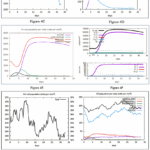 |
Figure 4: A Depicts B-cell population as a result of immunization with peptide; B. Depicts Human diploid myeloid leukemia cells (PLB) differentiation due to immune response with Immunoglobulins; C. Defines the TC memory cell population against antigenic particles; D. Depicts the T-cell population, activation, and generic reaction. |
In silico cloning and expression of peptide vaccine
Codon optimization and in silico cloning of the vaccine into the pET-28a(+) plasmid vector—between XhoI and BamHI restriction sites—in E. coli K12 strain achieved optimal expression potential, yielding a CAI of 1.0 (Figure 5). The optimized sequence exhibited a GC content of 54.65%, closely matching E. coli K12’s 50.80%.
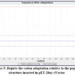 |
Figure 5: Depicts the codon adaptation relative to the peptide structure inserted in pET-28a(+)Vector Click here to View Figure |
Discussion
Immunotherapy is a predominant method for either preventing or treating cancer. Peptide-based cancer vaccines (namely, T and B cell epitopes) have garnered increased attention worldwide in recent years.35 Additionally, there are several benefits to this kind of vaccination, including ease of manufacturing, target specificity, safety, and preclinical model success.4 In this study, we used bioinformatics to create an effective subunit cancer vaccine.
Overexpressed MUC1 has been demonstrated as a vaccine against lung cancer, which prevents native cells from dying, and suppresses tumor growth, angiogenesis, proliferation, epithelial-mesenchymal transition (EMT), and metastasis.36 Likewise, Lung cancer invasion, migration, and cisplatin resistance are all influenced by the cancer-promoting gene SPP1. There is a correlation between poor patient survival, prognosis, and SPP1 overexpression. Hypomethylation of the promoter of SPP1 promotes its upregulation.34 Annadurai et. al., 2024 evidences the high expression of SPP1 in the worldwide Lung Cancer patients’ samples.37 Overexpression of SPP1 was more common in malignancies of the ovarian, breast, bladder, colon, head, neck, liver, lung, esophagus, found in Colorectal cancer, leukemia, liver cancer, and pancreatic cancer.34,38–40 Farias (2021) proved the negative correlation of SPP1 with GZMB+CD8+ T-cell infiltration, and increased regulatory T-cell.41 During Immunotheraphy, renal cancer patients exhibits worse Progress Free Survival of (HR = 2.73, p = .018), with only 14.8% response in high expression of SPP1, meanwhile its 66.7% in lower expression cases.42
Tumor-associated antigens (TAAs) are the targets of cancer peptide vaccines. Choosing the right antigens is essential for a successful immune response. Thus, the predicted peptides showed a good antigenic score of 2.1, without any antigenic or allergic violation. Sequences of the MUC1 gene effectively elicit T-cell responses to each of the MUC1 antigenic epitopes.36 In this study, epitopes derived from the SPP1 gene sequence were analyzed with great antigenicity (antigenic score 0.6 – 2.1). Korepanova., 2021 described that the intervention of transmembrane helices highly influences the protein activity during cloning, and expression.43 Thus, lower transmembrane helices ratio aided to achieve successful cloning, and expression in the host. The protein topology analysis did not find any violation of alpha-helical, and beta-barrel shown in Figure1.
OPN (Osteopontin) protein expressed by SPP1 gene that increases the generation, proliferation of B lymphocyte immunoglobulins, and is chemotactic different cell types, including dendritic cells, macrophages and T-cells. It promotes both pro- and anti-inflammatory processes under inflammatory conditions. Wang., 2008 demonstrated that OPN possesses Th1-cytokine activities, and affects cell-mediated immunity. Thus, the source sequence had a biological background to modulate immune response.44 “GPGPG” used as a preferable linker in peptide vaccine against cancer by Navid., 2014 respectively, stimulates HTL immunity.4 HTL cells are essential for developing humoral and cellular immune responses as they emit cytokines like IL-2 that help B cells differentiate into antibodies, and mature into cytotoxic T-cells.45 Herrera., 2020 designed a multi-epitope peptide vaccine (Fvax) for NSCLC with GPCR overexpression, showing 90% of favoured regions in Ramachandran Plot analysis, and hydrophilic.46 In this study, the constructed SPP1 gene-based peptide vaccine also shares the same Ramachandran plot range of 89.7% and a theoretical PI of 6.3, with a negative Gravy score (-1.101), indicating that the peptide is hydrophilic. A DNA-based peptide vaccine showed high interaction with TLR, and produced relative binding energy (− 839.0, − 957.8, − 908.0, − 1210.0, − 929.0, and − 1034.0), and the peptide activity against cancer was favorable in in-vivo studies.47 The constructed peptide produced a higher interaction with the TLR receptor (-1168.7 kcal/mol). TLR4 activation can trigger the production of cytokines (TGF-β, and IL-10), which reduce the immune response against the tumor. The release of pro-inflammatory cytokines (like IL-12, and IFN-γ), which signaling immune cells responsible for antitumor, including NK cells and T-cells, can be triggered by TLR4 agonists. Tumor-derived antigens are processed and presented to T-cells by TLR4-expressing antigen-presenting cells (APCs), thereby triggering antitumor immune responses.48 Targeting TLR4 as an immune receptor therefore serves as a strategic approach to elicit effective immunity. Immunotherapy resistance is an importance drawback reported in NSCLC patients due to weak tumor suppression, loss of tumor antigen, and lowest antigen presentation.
Mijanur., 2023 developed a multi-epitope vaccine against NSCLC via reverse vaccinology, incorporating MHC-I, MHC-II, CTL, and B-cell epitopes from oncoproteins MAGE-A3, EGF, and MUC-1. In silico cloning and expression yielded a CAI of 0.9607, indicating effective vaccine potential.49 In contrast, our SPP1-based peptide vaccine, featuring MHC-I, MHC-II, CTL, and B-cell epitopes, demonstrated superior expression with a CAI of 1.0. The designed peptide vaccine underwent comprehensive evaluation against key immunotherapeutic criteria. Satisfying physicochemical, structural, and hydropathy requirements positions it as a promising candidate for immunotherapy.
Conclusion
This study presents the in-silico design of a peptide vaccine with 11-epitopes, incorporating CTL (4 epitopes), HTL (1 epitope), and B-cell (2 epitopes) complemented by T-cell epitopes (4 epitopes), targeting the SPP1 antigen for NSCLC immunotherapy. The vaccine construct demonstrated favorable antigenicity (probable antigen, VaxiJen score >0.4) with non-allergenic, and non-toxic properties. Stable 3D structure (89.7% residues in Ramachandran favored regions, RMSD 0.399 Å), strong predicted binding to TLR4 receptor (ClusPro weighted score: -1168.7), Robust in silico immune responses including B-cell proliferation (500/mm3), memory T-cell generation (600/mm3), NK cell activation (370/mm3), and immunoglobulin production (IgM: 15/mm3, IgG1: 5/mm3), High expression potential in E. coli K12 (CAI:1.0, GC content: 54.65%). However, experimental validation remains imperative. Future research should prioritize immunogenicity assays, and structure validation by peptide synthesis.
Acknowledgement
The author would like to thank Central Research Laboratory for Biomedical Research, Vinayaka Mission’s Medical College and Hospital, Karaikal, Puducherry, India, for computational resources.
Funding Sources
The author(s) received no financial support for the research, authorship, and/or publication of this article.
Conflict of Interest
The authors do not have any conflict of interest.
Data Availability Statement
All data utilized in this study are publicly accessible, and cited within the manuscript; additionally, data generated from this research are included both in the manuscript, and in the supplementary materials.
Ethics Statement
This research did not involve human participants, animal subjects, or any material that requires ethical approval.
Informed Consent Statement
This study did not involve human participants, and therefore, informed consent was not required.
Permission to reproduce material from other sources
Not Applicable
Clinical Trial Registration
This research does not involve any clinical trials.
Authors Contribution
- Yamuna Annadurai: Study Conceptualisation, Methodology, drafting of Manuscript, review and editing.
- Murugesh Easwaran: Data curation, Formal analysis, Investigation, and software.
- Prabhu Dhamodharan: Study validation, and visualization.
- Shanmughavel Piramanayagam: Project administration, Resources, and Supervision.
- Luke Elizabeth Hanna: Study validation, review and editing.
References
- Guan Y, Ren M, Guo D, He Y. Research progress on lung cancer screening. Chinese Journal of Lung Cancer. 2020;23(11):954-960. doi:10.3779/j.issn.1009-3419.2020.101.37
- Bray F, Laversanne M, Sung H, et al. Global cancer statistics 2022: GLOBOCAN estimates of incidence and mortality worldwide for 36 cancers in 185 countries. CA Cancer J Clin. 2024;74(3):229-263. doi:10.3322/CAAC.21834
CrossRef - Lung and Bronchus Cancer — Cancer Stat Facts. Accessed October 15, 2025. https://seer.cancer.gov/statfacts/html/lungb.html
- Nezafat N, Ghasemi Y, Javadi G, Javad M. A novel multi-epitope peptide vaccine against cancer : An in silico approach. J Theor Biol. 2014;349:121-134. doi:10.1016/j.jtbi.2014.01.018
CrossRef - Global Cancer Observatory. Accessed October 15, 2025. https://gco.iarc.who.int/en
- Noronha V, Budukh A, Chaturvedi P, et al. Uniqueness of lung cancer in Southeast Asia. The Lancet Regional Health – Southeast Asia. 2024;27:100430. doi:10.1016/j.lansea.2024.100430
CrossRef - Walser T, Cui X, Yanagawa J, et al. Smoking and lung cancer: The role of inflammation. Proc Am Thorac Soc. 2008;5(8):811-815. doi:10.1513/pats.200809-100TH
CrossRef - Lung Cancer Statistics | How Common Is Lung Cancer? | American Cancer Society. Accessed October 15, 2025. https://www.cancer.org/ cancer/types/lung-cancer/about/key-statistics.html
- F AMRA, F ADA. Multiple primary lung cancer : A literature review. Published online 2018. doi:10.17219/acem/68631
CrossRef - Manuel V, Sousa L De, Carvalho L. Heterogeneity in Lung Cancer. Published online 2018:96-107. doi:10.1159/000487440
CrossRef - Iversen TZ. Immune modulations during chemoimmunotherapy & novel vaccine strategies – ‐ In metastatic melanoma and non small- ‐ cell lung cancer. Published online 2013:1-21.
- Salem PP, Chami P, Daou R, et al. Proton Radiation Therapy: A Systematic Review of Treatment-Related Side Effects and Toxicities. Int J Mol Sci. 2024;25(20):10969. doi:10.3390/ijms252010969
CrossRef - Chauvin JM, Zarour HM. TIGIT in cancer immunotherapy. Published online 2020. doi:10.1136/jitc-2020-000957
CrossRef - Laidlaw BJ, Craft J, Kaech SM, Haven N. HHS Public Access. 2017;16(2):102-111. doi:10.1038/nri.2015.10.
CrossRef - Bardhan K, Anagnostou T, Boussiotis VA. The PD1: PD-L1/2 pathway from discovery to clinical implementation. Front Immunol. Frontiers Media S.A. 2016;7(DEC). doi:10.3389/fimmu.2016.00550
CrossRef - O’Donnell JS, Teng MWL, Smyth MJ. Cancer immunoediting and resistance to T cell-based immunotherapy. Nat Rev Clin Oncol. Nature Publishing Group. 2019;16(3):151-167. doi:10.1038/s41571-018-0142-8
CrossRef - Kumar A, Kanak KR, Lakshmi PT V. Comparative transcriptome pro fi ling and weighted gene co-expression network analysis to identify core genes in maize ( Zea mays L .) silks infected by multiple fungi. 2022;(October):1-19. doi:10.3389/fpls.2022.985396
CrossRef - North S, Butts C. Vaccination with BLP25 liposome vaccine to treat non-small cell lung and prostate cancers. Expert Rev Vaccines. 2005;4(3):249-257. doi:10.1586/14760584.4.3.249
CrossRef - Weerarathna IN, Doelakeh ES, Kiwanuka L, Kumar P, Arora S. Prophylactic and therapeutic vaccine development: advancements and challenges. Molecular Biomedicine. 2024;5(1):57. doi:10.1186/s43556-024-00222-x
CrossRef - Hallgren J, Tsirigos KD, Pedersen MD, et al. DeepTMHMM predicts alpha and beta transmembrane proteins using deep neural networks. Preprint posted online April 10, 2022. doi:10.1101/2022.04.08.487609
CrossRef - Jespersen MC, Peters B, Nielsen M, Marcatili P. BepiPred-2.0: improving sequence-based B-cell epitope prediction using conformational epitopes. Nucleic Acids Res. 2017;45(W1):W24-W29. doi:10.1093/nar/gkx346
CrossRef - Atkins TK, Solanki A, Vasmatzis G, Cornette J, Riedel M. Evaluating NetMHCpan performance on non-European HLA alleles not present in training data. Front Immunol. 2024;14. doi:10.3389/fimmu.2023.1288105
CrossRef - Doytchinova IA, Flower DR. VaxiJen : a server for prediction of protective antigens , tumour antigens and subunit vaccines. 2007;7:1-7. doi:10.1186/1471-2105-8-4
CrossRef - Dimitrov I, Flower DR, Doytchinova I. AllerTOP – a server for in silico prediction of allergens. 2013;14(Suppl 6).
CrossRef - Wilde EJ, Hughes A, Blagova E V., et al. Interactions of the periplasmic binding protein CeuE with Fe(III) n-LICAM4− siderophore analogues of varied linker length. Sci Rep. 2017;7(1):45941. doi:10.1038/srep45941
CrossRef - Khamjan NA, Lohani M, Khan MF, Khan S, Algaissi A. Immunoinformatics Strategy to Develop a Novel Universal Multiple Epitope-Based COVID-19 Vaccine. Vaccines (Basel). 2023;11(6):1090. doi:10.3390/vaccines11061090
CrossRef - Contributions A. performed research and created and tested the I-TASSER Suite package. Z Nat Methods. 2015;12(1):7-8. doi:10.1038/nmeth
CrossRef - Hon J, Marusiak M, Martinek T, et al. SoluProt: prediction of soluble protein expression in Escherichia coli. Bioinformatics. 2021;37(1):23-28. doi:10.1093/bioinformatics/btaa1102
CrossRef - Heo L, Park H, Seok C. GalaxyRefine: Protein structure refinement driven by side-chain repacking. Nucleic Acids Res. 2013;41(Web Server issue). doi:10.1093/nar/gkt458
CrossRef - Jones G, Kotelnikov S, Egbert M, et al. Elucidation of protein function using computational docking and hotspot analysis by ClusPro and FTMap research papers. Published online 2022:690-697. doi:10.1107/S2059798322002741
CrossRef - Rapin N, Lund O, Bernaschi M, Castiglione F. Computational Immunology Meets Bioinformatics : The Use of Prediction Tools for Molecular Binding in the Simulation of the Immune System. 2010;5(4). doi:10.1371/journal.pone.0009862
CrossRef - No B, Grote A, Hiller K, et al. JCat : a novel tool to adapt codon usage of a target gene to its potential expression host. 2005;33:526-531. doi:10.1093/nar/gki376
CrossRef - Matsubara E, Yano H, Pan C, et al. The Significance of SPP1 in Lung Cancers and Its Impact as a Marker for Protumor Tumor-Associated Macrophages. Cancers (Basel). 2023;15(8):2250. doi:10.3390/cancers15082250
CrossRef - Tang H, Chen J, Han X, Feng Y, Wang F. Upregulation of SPP1 Is a Marker for Poor Lung Cancer Prognosis and Contributes to Cancer Progression and Cisplatin Resistance. 2021;9(April):1-9. doi:10.3389/fcell.2021.646390
CrossRef - Chiarella P, Massi E, Robertis M De, Fazio VM, Signori E. Recent Advances in Epitope Design for Immunotherapy of Cancer Pieranna Chiarella. Published online 2009:227-240.
CrossRef - Sharma S, Srivastava MK, Harris-White M, Lee JM, Dubinett S. MUC1 peptide vaccine mediated antitumor activity in non-small cell lung cancer. Expert Opin Biol Ther. 2011;11(8):987-990. doi:10.1517/14712598.2011.598146
CrossRef - Annadurai Y, Easwaran M, Sundar S, et al. SPP1, a potential therapeutic target and biomarker for lung cancer: functional insights through computational studies. J Biomol Struct Dyn. 2024;42(3):1336-1351. doi:10.1080/07391102.2023.2199871
CrossRef - Gao W, Liu D, Sun H, et al. SPP1 is a prognostic related biomarker and correlated with tumor-infiltrating immune cells in ovarian cancer. BMC Cancer. 2022;22(1):1367. doi:10.1186/s12885-022-10485-8
CrossRef - Eremo AG, Kajsa L, Othman L, Montgomery S, Andersson G, Tina E. Evaluation of SPP1 / osteopontin expression as predictor of recurrence in tamoxifen treated breast cancer. Published online 2020:1-9. doi:10.1038/s41598-020-58323-w
CrossRef - Tu Y, Chen C, Fan G. Association between the expression of secreted phosphoprotein – related genes and prognosis of human cancer. BMC Cancer. 2019;19(1):1230. doi:10.1186/s12885-019-6441-3
CrossRef - Farias A, Soto A, Puttur F, et al. A TLR4 agonist improves immune checkpoint blockade treatment by increasing the ratio of effector to regulatory cells within the tumor microenvironment. Sci Rep. 2021;11(1):15406. doi:10.1038/s41598-021-94837-7
CrossRef - Xu X, Lin J, Wang J, et al. SPP1 expression indicates outcome of immunotherapy plus tyrosine kinase inhibition in advanced renal cell carcinoma. Hum Vaccin Immunother. 2024;20(1). doi:10.1080/21645515.2024.2350101
CrossRef - Korepanova A, Gao FEIP, Hua Y, Qin H, Nakamoto RK, Cross TA. Cloning and expression of multiple integral membrane proteins from Mycobacterium tuberculosis in Escherichia coli. 2005;3:148-158. doi:10.1110/ps.041022305.
CrossRef - Wang KX, Denhardt DT. Osteopontin : Role in immune regulation and stress responses. 2008;19:333-345. doi:10.1016/j.cytogfr.2008.08.001
CrossRef - Yata J, Tsukimoto I, Arimoto T, Goyat N, Tachibanat T. HUMAN THYMUS LYMPHOID TISSUE (HTL) ANTIGEN, COMPLEMENT RECEPTOR AND ROSETTE FORMATION WITH SHEEP ERYTHROCYTES OF THE LYMPHOCYTES FROM PRIMARY IMMUNODEFICIENCY DISEASES. Vol 14. 1973.
CrossRef - Herrera LRM. In Silico Approach in Designing a Novel Multi-Epitope Vaccine Candidate against Non-Small Cell Lung Cancer with Overexpressed G Protein-Coupled Receptor 56. Asian Pacific Journal of Cancer Prevention. 2020;21(8):2297-2306. doi:10.31557/APJCP.2020.21.8.2297
CrossRef - Kayyal M, Bolhassani A, Noormohammadi Z, Sadeghizadeh M. In Silico Design and Immunological Studies of Two Novel Multiepitope DNA ‑ Based Vaccine Candidates Against High ‑ Risk Human Papillomaviruses. Springer US; 2021. doi:10.1007/s12033-021-00374-z
CrossRef - Urban-Wojciuk Z, Khan MM, Oyler BL, et al. The Role of TLRs in Anti-cancer Immunity and Tumor Rejection. Front Immunol. 2019;10. doi:10.3389/fimmu.2019.02388
CrossRef - Rahman MM, Masum MHU, Talukder A, Akter R. An in silico reverse vaccinology approach to design a novel multiepitope peptide vaccine for non-small cell lung cancers. Inform Med Unlocked. 2023;37:101169. doi:10.1016/j.imu.2023.101169
CrossRef
Abbreviations
NSCLC – Non-small cell lung cancer
SPP1 – Secreted phosphoprotein 1 (osteopontin)
OPN – Osteopontin
MUC1 – Mucin 1
TLR – Toll-like receptor
TLR3 – Toll-like receptor 3
TLR4 – Toll-like receptor 4
CTL – Cytotoxic T lymphocyte
HTL – Helper T lymphocyte
Trm – Tissue-resident memory T lymphocyte
Th – T helper cell
NK cell – Natural killer cell
APC – Antigen-presenting cell
MHC – Major histocompatibility complex
MHC-I – Major histocompatibility complex class I
MHC-II – Major histocompatibility complex class II
HLA – Human leukocyte antigen
HLA-A – Human leukocyte antigen A
HLA-DP – Human leukocyte antigen DP
IL – Interleukin
IFNγ – Interferon-gamma
TMHMM / DeepTMHMM – Transmembrane Hidden Markov Model
TMP – Transmembrane protein
IEDB – Immune Epitope Database
BepiPred – B-cell epitope prediction method
NetMHCIIpan – Neural-network based MHC class II epitope prediction tool
netCTL – Neural-network based CTL epitope prediction tool
VaxiJen – Antigenicity prediction server
AllerTOP – Allergenicity prediction server
ToxinPred / ToxinPred2 – Peptide toxicity prediction server
I-TASSER – Iterative Threading ASSEmbly Refinement (3D structure prediction server)
RMSD – Root mean square deviation
CAI – Codon adaptation index
GC content – Guanine–cytosine content
EMT – Epithelial–mesenchymal transition
RNA (if mentioned in references context) – Ribonucleic acid
AI (ProtParam) – Aliphatic index
pI – Isoelectric point
GRAVY – Grand average of hydropathicity

Accepted on: 26-08-2026
Second Review by: Dr. Sarraa Dhiaa Kasim
Final Approval by: Dr. Hanefi ÖZBEK