Immune-Mediated Thyroid Dysfunction: Contemporary Mechanisms Linking Hashimoto thyroiditis to Hypothyroidism
 , Deepthi Mandya Nagesh, Ravi Kumar Arikeriand Yenduri Suvarna
Department of Pharmaceutical Chemistry and Analysis, Sri Adichunchanagiri College of Pharmacy, Adichunchangiri University, B.G.Nagara, India.
Corresponding author Email: knp.pharma@gmail.com
Download this article as:
ABSTRACT:Autoimmunity in the thyroid refers to the breakdown of immune tolerance toward thyroid-specific antigens, particularly thyroid peroxidase (TPO), thyroglobulin (Tg) and the thyroid-stimulating hormone receptor (TSHR). This process involves activation of autoreactive T lymphocytes, B-cell mediated autoantibody production and cytokine-driven inflammatory responses that progressively damage thyroid follicular cells and impair hormone synthesis. The thyroid gland is especially susceptible to autoimmune reactions due to its unique antigenic proteins, high vascularity and continuous exposure to environmental and hormonal influences, which together facilitate immune-mediated injury. Autoimmunity and thyroid dysfunction are interconnected through genetic predisposition, immune dysregulation and environmental triggers that impair tolerance. The global rise in Autoimmune thyroid diseases, influenced by lifestyle, iodine status and epigenetics highlights the need for early recognition. Hashimoto Thyroiditis advances from subclinical stages to overt hypothyroidism through follicular loss, fibrosis and reduced hormone synthesis. Preventive strategies include optimal iodine intake, limiting radiation exposure and monitoring high risk individuals. Management primarily relies on levothyroxine replacement therapy to restore euthyroid status and minimize complications. Clinical implications extend to cardiovascular, metabolic and reproductive health, necessitating regular follow-up. Future directions focus on predictive biomarkers, immune based therapies and precision medicine. This review outlines contemporary mechanism linking Hashimoto thyroiditis to hypothyroidism and provides updated insights into prevention and management.
KEYWORDS:Autoimmune thyroid disease; Hashimoto thyroiditis; Thyroid peroxidase antibodies; Thyroglobulin antibodies; Ultrasonography
Introduction
Autoimmune diseases (ATD) are a wide class of disorders characterized by a decline in immunological self-tolerance, which results in abnormal attacks on the tissues and organs of the host.1 Women are more likely to develop autoimmune diseases than men, and in Hashimoto thyroiditis that effect is maximized, occurring 10-15 times more frequently in women than in men. Thyroid dysfunction can be symptomatic (0.1-2% of the population) or subclinical (10-15%).2 Even though the majority of Autoimmune diseases are not monogenic, they may occur as a result of various genetic factors such as particular genetic mutations, HLA(Human Leukocyte Antigen) variation, mutations at non-HLA loci and epigenetic process.3 Among the most prevalent organ specific autoimmune diseases, AITD is characterized by lymphocyte malfunction carried by an infection, trauma and other stressors that are inherited and most common in children and adolescents. In AITDs, the major two types which is of organ specific are Hashimoto thyroiditis and Graves’ disease.4 A recent large prospective case-control study has demonstrated a significantly higher occurence of various systemic autoimmune disorders among patients with autoimmune thyroiditis(AT) including systemic lupus erythematosus(SLE) chronic autoimmune gastritis(CAG), Sjogren’s disease(SS)z, alopecia ,vitiligo (Vit), psoriatic arthritis(PsA) multiple sclerosis, Celiac disease, rheumatoid arthritis(RA) diabetes, hepatitis C VIRUS(HCV)-related mixed cryoglobulinemia(MC), sarcoidosis(S), systemic sclerosis(SSc) and polymyalgia rheumatica (Polym). Additionally, a nearly significant association was observed with Addison’s disease and ulcerative colitis.5
Hashimoto thyroiditis (HT), known also as autoimmune thyroiditis (AIT) is an autoimmune disorder of the thyroid gland which is usually characterized by thyroid enlargement, prominent infiltration of lymphocytes and increased serum concentrations of thyroid specific autoantibodies.6 Hashimoto thyroiditis commonly occurs between the ages of 30-60, with a female prevalence between 4.8-25.8% and male prevalence between 0.9-7.9%. About 14.3% of patients present with coexisting autoimmune disorders. In children, HT occurs in 0.3-9.6% rarely before age 3 and peaks during early to adolescence. A strong female predominance is observed with female to male ratios ranging from 2:1 to 9.7:1, and not as strong in younger. Prepubertal children (female-to- male ratio 1.6:1) compared with adolescents. This indicates that sex hormones, which become more active after puberty, may contribute to the higher risk of HT observed in females.4,7
The pathogenesis of Hashimoto thyroiditis is primarily driven by autoimmune responses against thyroid-specific proteins, particularly thyroid peroxidase (TPO), thyroglobulin (Tg) and, in some cases, the thyroid-stimulating hormone receptor (TSHR), which act as key autoantigens. Loss of immune tolerance enables antigen-presenting cells to present thyroid antigens to CD4⁺ T helper cells, leading to activation of B lymphocytes and production of autoantibodies such as anti-TPO and anti-Tg. These immune responses promote infiltration of the thyroid gland by T lymphocytes, B lymphocytes, macrophages and plasma cells, resulting in chronic inflammation and progressive destruction of thyroid follicular cells. Cytotoxic CD8⁺ T cells induce thyrocyte apoptosis through Fas–Fas ligand interactions and perforin-mediated pathways, while pro-inflammatory cytokines including interferon-γ (IFN-γ) and tumor necrosis factor-α (TNF-α) further enhance tissue damage and impair thyroid hormone synthesis. Over time, this sustained immune-mediated injury leads to follicular atrophy, fibrosis and reduced thyroid hormone production, ultimately resulting in hypothyroidism.8,9
According to a comprehensive Global Burden of Disease analysis, thyroid disorders account for a remarkable portion of the spectrum of endocrine diseases. These conditions are major contributors to disability- adjusted life years and mortality worldwide across endocrine, metabolic, blood and immune disorders.10 Even Benign thyroid disorders have large personal and economic consequences, as evidenced by a review of thyroid nodules that described their high prevalence, high healthcare utilization and personal worry.11 Hypothyroidism disrupts endothelial function, alters lipid metabolism and increases cardiovascular risk, highlighting how thyroid disorders lie at the junction of endocrine regulation and cardiometabolic disease.12 Robust reviews connect bisphenols, phthalates and perchlorates to altered TH synthesis, transport and receptor signaling with vulnerable populations(pregnant people, infants) at special risk. Lifestyle factors (Smoking, alcohol, diet, physical activity) and microbiome shifts also modulate TSH/TH distributions.13
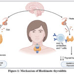 |
Figure 1: Mechanism of Hashimoto thyroiditis Click here to View Figure |
Global synthesis indicates substantial and regionally variable HT prevalence in adults with higher estimates in several Asian and Middle-eastern cohorts; broader ultrasound use and antibody testing likely contribute to rising detection.6 Hashimoto’s thyroiditis is not just a thyroid problem, it can affect many body systems. It raises the risk of heart and vascular disease, especially in younger or poorly treated patients, so lifestyle care is important along with thyroid medication.14 In women, it is linked to pregnancy problems like miscarriage and preterm birth, so close thyroid monitoring during pregnancy is needed. There is still debate going on about whether treating euthyroid women with antibodies improves IVF outcomes or not.15 Research also shows that gut bacteria imbalances may play a role in Hashimoto’s, making the gut a possible future treatment target.16 New imaging tools like shear-wave and strain elastography measures tissue stiffness and add value to traditional ultrasound in Hashimoto thyroiditis, even when the thyroid is fibrotic.17 These techniques are being studied in both children and adults, with growing datasets. Artificial intelligence and radiomics are now being applied to ultrasound images, helping to detect nodules and even identify early or antibody-negative cases. However, large clinical studies show that elastography alone is not always better than standard ultrasound with fine-needle aspiration.18
This review is about the frequent coexistence of thyroid disease with other autoimmune conditions underscores the need for vigilant screening, early recognition of co-morbidities and multidisciplinary management strategies to enhance durability. Overall, the study of Hashimoto’s thyroiditis highlights its dual role as both a thyroid specific disease and a marker of systemic autoimmunity, offering important intuition into the common pathways that drive autoimmune clustering.
Immunogenetics and systems biology of AITD
The etiology of Hashimoto thyroiditis is complex, including a complex interaction between environmental factors and genetic predisposition that together impair immunological tolerance and cause the thyroid gland to be destroyed by an autoimmune reaction.19
Genetic Susceptibility
Inherited variations in key immune regulatory gens can make some individuals more prone to losing immune tolerance, setting the stage for autoimmune diseases.20 Autoimmunity can be set off by a mix of environmental triggers such as smoking, stress and infections, along with genetic factors like specific HLA and CTLA4 gene variants.21 In addition to those included within the HLA (Human Leukocyte Antigen) complex, there are multiple other immunoregulatory genes that modulate immune responses. Polymorphism of class II human leukocyte antigens (HLA-DR) genes, as well as cytotoxic T-lymphocyte associated protein-4 (CTLA-4), cluster of differentiation-40 (CD40), protein tyrosine phosphatase non-receptor type 22(PTPN22), forkhead transcription box protein P3(FOXP3) and C25, disrupt the mechanisms of immune tolerance.22 Genetic variations in CTLA4,CD40 and PTPN22 can modify pathways involved in co-stimulation and the way immune cells display antigens at the molecular level, hence fostering an elevated autoimmune state.21 FOXP3 located on the X chromosome, regulates T cells(Tregs) and its polymorphisms are linked to Hashimoto’s thyroiditis (HT). CD40, a TNF receptor is essential for B-cell expansion, (antigen presenting cells) APC-T cell interaction and antibody class switching. On the other hand, CTLA4 and PTPN22 help to keep the immune response in check by limiting T-cell activation.23
Two of the three genes encoding the major thyroid autoantigens- Thyroglobulin (Tg) and thyroid stimulating hormone receptor (TSHR) have been associated with AITD. The other autoantigen, thyroid peroxidase (TPO), has been implicated but not in the mutations of the gene.24 Genome-wide and targeted studies identify TG-related loci and several non-thyroid immune loci that modulate thyroid- antibody levels, implying that both thyroid-specific structural changes and broader immune-regulatory genes determine who develops high-titer, epitope-restricted Tg/TPO antibodies and progressive HT.25
Environment factors
Environmental influences such as bacterial or viral infections, smoking, maternal-fetal micromerism and exposure to chemicals like flame retardants and phthalates can contribute to immune disturbances.8 Populations with mild iodine deficiency show a lower risk, while selenium deficiency is strongly linked to higher thyroid autoimmunity. Hashimoto thyroiditis often coexists with iron deficiency, especially in women of fertile period. And it is caused due to comorbid conditions like autoimmune gastritis or celiac disease, along with menstrual blood loss. Such factors make women particularly vulnerable to combined thyroid and iron related complications. Selenium and iron deficiencies further worsen immune dysregulation in thyroid disease. Alongside these influences, Gut microbiota community is crucial for guiding and maintaining health y systemic immune responses. It influences thyroid hormone metabolism and immune tolerance. Dysbiosis of hut flora may therefore predispose individuals to thyroid autoimmunity.19
Epigenetic Programming
Epigenetics studies how gene expression and phenotypic changes regulate non-genetic factors without modifying the encoded genome. The main actions are methylated DNA, chemical modification of histones and expression of short and long non-coding RNAs.26
DNA Methylation
DNA Methylation is a biochemical process involving the addition of a methyl group to cytosine nucleotides, mainly within CpG regions of the genome. This modification typically downregulates gene expression by obstructing the interaction between transcription factors and the DNA template.27 Thyroid hormones (THs) influence immune function, though the mechanisms remain unclear. In-vitro supraphysiological T3 exposure in human monocytes altered DNAm patterns, modulated immune-related gene expression and changed responses to lipopolysaccharide stimulation.28 Several studies report global DNA hypomethylation in autoimmune thyroid disease (AITD), leading to upregulation of immune-related genes and activation of auto-reactive cells that damage thyroid tissue. Polymorphisms in DNA methylation-regulating genes may further disrupt methylation processes, thereby increasing susceptibility to AITD.26 In Hashimoto’s thyroiditis, the highest number of DMPs (Differentially methylated positions-30) are on chromosome 6 and most DMGs (Differentially methylated genes-15) on chromosome 12. Chromosome 6 nurture HLA-DPβ1, a key gene for T-cell activation linked to systemic autoimmunity. This non-random distribution suggests that chromosomes enriched with DMPs/DMGs play critical roles in HT pathogenesis and merit focused investigation. There are an expanded table-1 highlights key gene candidates and the type of epigenetic alterations observed in HT/AITD contexts, offering a clearer perspective.27 DNA methylation is also an emerging therapeutic target for autoimmune disease.29
Table 1: Key gene responsible for the cause of Hashimoto thyroiditis
|
Chromosome |
Key gene | Function |
Relevance to Hashimoto Thyroiditis |
|
1 |
PTPN22 | Lymphoid tyrosine phosphatase-negative regulator of T-cell receptor signaling. |
Well established susceptibility locus for AITD; rare variants and epigenetic changes (including promoter methylation differences) have been reported in families and patient series with HT.22 |
|
2 |
CTLA4 | Immune checkpoint receptor that down-regulates T-cell activation (Treg function) |
CTLA4 variants and epigenetic modulation associate with altered T-cell tolerance and increase AITD risk; recent OA studies report CTLA4 SNPs linked to HT and candidate biomarker variants.30 |
|
6 |
HLA-DPβ1 | MHC class II antigen presentation to CD4+ T cells. | Identified among differentially methylated genes/regions in HT; MHC loci on chr6 show concentrated DMPs in HT cohorts, consistent with antigen-presentation-driven autoimmunity.27 |
| 10 | IL2RA(CD25) | IL-2 receptor α-critical for regulatory T cell (Treg) survival/function. |
Polymorphisms and promoter methylation changes in IL2RA have been described in AITD cohorts; IL2RA dysregulation may impair Treg-mediated tolerance in HT.30 |
|
17 |
SLFN12 | Schlafen family member-implicated in immune cell function (monocytes/T cells) |
Identified as a strongly hypomethylated/overexpressed epigenetic marker in genome-wide methylation profiling of HT blood samples (potential HT epigenetic biomarker).27 |
Histone Modifications
Histone deacetylase inhibitors (HDACi) can boost the activity of many genes, including those play a role in autoimmune responses. Evidence indicates that post-translational modifications are crucial in regulating immune tolerance, autoimmune disorders and the development of diverse cell lineage.29 Changes in Histone such as adding of acetyl or methyl groups, phosphorylation and sumoylation are vital for gene regulation. HDAC inhibitors represent promising therapies for cancers and autoimmune diseases. Genetic polymorphisms in histone-modifying genes may impair their function. Such dysfunction can cause abnormal histone modifications contributing to AITD.26 Lysine acetylation is the most studied histone modification, where acetylation opens chromatin to enhance gene expression, while deacetylation compacts it. Acetylation occurs on lysine residues of histone N-terminal tails. Additionally, H3K4me3 marks active promoters, whereas H3K9me3 and H3K27me3 help switch genes off by tightening the chromatin structure. By making the DNA more compact, they prevent the transcription machinery from accessing those genes.29
Non-coding RNAs
Non-coding RNAs(ncRNAs) including lncRNAs, circRNAs and miRNAs are key regulators of gene transcription and protein translation despite lacking protein-coding ability. Non-coding RNAs shaping cytokine, apoptosis and fibrosis pathways in Hashimoto thyroiditis. Among them, microRNAs play a vital role in T-cell activation, multiplication and cytokine release. Dysregulated miRNAs are linked to autoimmune diseases, exerting either pro or anti-inflammatory effects.31 The role of miRNAs in immune regulation is supported by evidence showing their rapid induction in B and T cells upon exposure to antigens or inflammatory stimuli they also influence transcription factors that control the Helper T Lymphocytes 1(HTLs1)/Helper T Lymphocytes 2(HTLs2) balance and contribute to antiviral immune responses.32 In Hashimoto’s thyroiditis, levels of miR-146a in the blood are often higher and this increase is linked to raised helper T Lymphocytes 17(HTLs17) related cytokines and lower IL-10, indicating a disturbance between HTLs17 and Treg cells.33 Mechanistically, miR-146a normally helps keep inflammation in check by regulating the TLR/NF-ĸB pathway through its effects on IRAK-1and TRAF6. By doing so, it reduces excessive NF-ĸB activity, affects inflammatory cytokine release and can influence the survival of immune cells like dendritic cells and macrophages. Recent experimental reviews show miR-146a can both limit excessive cytokine production and promote apoptosis of some immune cells depending on context.34 miR-155 is a master regulator of immune responses (affects B cells, T cells, Dendritic cells). It modulates HTLs1/HTLs17 differentiation and cytokine output (eg., IFN-γ, IL-17) acts via multiple targets like SOCS1(Suppressor of cytokine signalling-1, IKBKβ that feed into JAK/STAT and NF-ĸB/MAPK pathways. In autoimmune models, altered miR-155 changes disease severity and cytokine profiles. Functional outcomes include altered cytokine production (promotion of IL-17/ IFN-γ in some contexts) and effects on cell survival. miR-155 depletion or inhibition often reduces pathogenic inflammation in animal autoimmune models, indicating its pro-inflammatory role.35 The miR-29 family is well described as anti-fibrotic (targets collagens, fibrillin and other ECM genes) and participates in TGF-β signaling loops: reduced miR-29 allows TGF- β driven fibrotic gene expression and may change cytokine milieu and cell survival programs. Comprehensive reviews of miR-29 in innate immunity and fibrosis summarize these pathways.36 Collectively these ncRNAs link immune dysregulation of cytokine imbalance, apoptosis and fibrotic remodeling, may also explored as options for future therapy in HT.
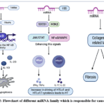 |
Figure 2: Flowchart of different miRNA family which is responsible for cause of HT Click here to View Figure |
From Autoimmunity to Hypothyroidism: Core Mechanisms
Autoantibodies (Anti-TPO, Anti-Tg, TSHR-blocking)
The Pathophysiology includes that immune system wrongly targets the thyroid by producing antibodies against TPO, thyroglobulin and the TSH receptor. The thyroid tissue also develops germinal centers, shows disrupted TSH regulation and becomes filled with various immune cells such as lymphocytes, monocytes, NK cells and macrophages.37
The primary mechanism of HT is a cellular response that targets thyroid autoantigens, resulting in fibrosis, inflammation and impaired thyroid gland function.38 The primary autoantigen in AITD is TPO, where autoantibodies and T cells that are specific for the enzyme are crucial in the destruction of the thyroid gland.39 Thyroid peroxidase antibodies (TPOAbs) are key markers of autoimmune thyroid disease and, even in euthyroid individuals, predict higher risk of future thyroid dysfunction. They cross the placenta, and their detection during pregnancy helps forecast postpartum thyroiditis. Maternal TPOAb determined to be related with sensorineural hearing loss in children, miscarriage and preterm delivery. Thyroid autoantibodies act as important predictors of adverse reproductive outcomes. In hypothyroid women, irregular menstrual cycles occur in 23-68% of cases, reflecting a high prevalence of ovulatory dysfunction.40 These antibodies faultyly target normal tissue, like thyrocytes, within the thyroid gland. Following the assault of anti-TPO antibodies, there is inflammation and reduced function of the thyroid gland.41 Apoptosis is a noteworthy aspect of the pathology of HT, which has characteristics of both type II and type IV hypersensitivity reactions is ADCC in patients with HT and a positive blood test for anti-TPO antibodies.42 Thyroid hormone synthesis is stimulated by TSH with thyroglobulin (Tg) as the essential precursor. Normally, Tg promotes T-cell tolerance and prevents anti-Tg antibody formation, but in autoimmune thyroid disease (AITD) oligoclonal anti-Tg antibodies develop. Elevated Tg from tissue damage, altered conformation due to high iodide or excessive TSH can expose new epitopes and break immune tolerance. Excess iodine intake further amplifies thyroid autoimmunity, enhancing anti-Tg and anti-TPO antibody production. Rising antibody titers drive thyroid inflammation and progressive loss of function, leading to hypothyroidism.43 Anti-TPO autoantibodies in HT target specific thyroid gland regions, resulting in gradual thyroid destruction, as well as diminished resistance to cold, dry skin, depression, muscle aches and decreased tolerance to exercise.44
Fibrous thyroiditis involves progressive fibrous replacement of thyroid tissue, leading to gland enlargement, epithelial loss and primary hypothyroidism. It is classified within systemic IgG4-related disease and must be distinguished from other hard goiters, often coexisting with fibro inflammatory disorders such as retroperitoneal fibrosis, mediastinitis or sclerosing cholangitis.45
TRAb is now understood to include several types of antibodies, some that stimulate the TSH receptor (TSI), some that block it (TBI) and others that have no noticeable effect.46 TSAb bind and stimulate TSHR, increasing cAMP and enhancing T4 and T3 synthesis. TBAb inhibit TSHR function, decreasing hormone production and suppressing thyroid cell growth. Neutral antibodies activate alternative signaling pathways, including PKC/MAPK, mTOR, NF-ĸB, cytokines and ROS. These distinct mechanisms explain the variable clinical outcomes observed in autoimmune thyroid disease.47 Treatment with antithyroid drugs or radioiodine therapy can alter the dynamics of TSHR-Abs, potentially leading to a switch from hyperthyroidism to hypothyroidism. Pregnancy and postpartum period have hormonal changes during and after pregnancy can impact the immune system and antibody profiles, sometimes resulting in a shift in thyroid function. Immune modulation is a factor which includes infections or immunosuppressive therapies can influence the production and activity of TSHR-Abs, leading to changes in thyroid function.48
T-cell mediated Thyroid destruction
The contribution of T cells to the pathophysiology of chronic lymphocytic thyroiditis is now appreciated. T cells can be divided into two types, CD8+ and CD4+ T cells. The T cell subset dysfunction is complex and arises from perturbed expression of important molecules or specific cytokines that process or regulate T cell actions.49 During the initial stage of Hashimoto’s thyroiditis, dendritic cells and macrophages that express MHC (Major Histocompatibility Complex) class II migrate to the thyroid and present autoantigens to naive CD4+ T cells, stimulating their activation and expression. Thyroid damage results from three main mechanism: cytotoxic T cells, death receptor signaling and autoantibodies. HTLs1 and HTLs17 subsets are mediating HTLs1 cells induced by IL-12, IL-2 and IFN-γ, which activate macrophages, B cells and cytotoxic T lymphocytes. It is a cytokine further released by these cells under IFN-γ, TNF-α and IL-2 which intensify the immune response. More importantly, thyrocytes can express MHC class II, acting as antigen-presenting cells to amplify T cell activation. Overall, excessive HTLs1 driven immunity contributes significantly to thyroid inflammation and tissue destruction.22 Recent research has also revealed metabolic reprogramming in CD4+ T cells during HT, marked by elevated glycolysis and activation of the mTOR/HIF-1α pathway, which are essential for HTLs1 and HTLs17 differentiation and activity. Targeting these metabolic and signaling pathways has demonstrated potential in reducing thyroid inflammation in experimental models. Elucidating these processes provides promising avenues for therapeutic intervention in autoimmune thyroid disease.50
Tregs are responsible for maintaining immune tolerance and preventing autoimmunity. In autoimmune thyroid diseases, Tregs can be dysfunctional or reduced in number, resulting in uncontrolled activation of autoreactive T cells. This dysfunction seems to be correlated to impaired expression of immune checkpoint molecules such as CTLA-4 and PD-1. Studies have shown that low expression of CTLA-4 on Tregs correlates with increase disease activity on systemic lupus erythematosus suggesting a similar mechanism in autoimmune thyroiditis. Additionally, PD-1 blockade has been linked to thyroiditis in cancer immunotherapy, highlighting the role of checkpoint pathways in thyroid autoimmunity. Restoring Tregs function or enhancing checkpoint signaling may offer therapeutic strategies to mitigate autoimmune thyroid disease progression.51 The PD-1/PD-L1 pathway, though active in Hashimoto’s thyroiditis (HT) is insufficient to prevent disease progression. Deficient Tregs and aberrant Helios/ PD-1 expression impair immune regulation. These alterations drive thyroid autoimmunity and suggest PD-1/ PD-L1 as a therapeutic target.52
Clinical Phenotypes and Trajectories to Hypothyroidism
Subclinical hypothyroidism (sHypo) happens when your thyroid stimulating hormone (TSH) level is higher than normal, but your free T4 hormone is still within the normal range. This is different from overt hypothyroidism, where the TSH level is high and the free T4 level is low, clearly showing reduced thyroid function.53 The prevalence of overt hypothyroidism is relatively low, ranging from 0.2-1% of adults, while subclinical hypothyroidism is much more common, affecting 4-20% of the adult population, particularly in iodine-sufficient regions. About one-fifth of people with subclinical hypothyroidism have TSH values in the moderately elevated range(7-9.9 mIU/L) and a smaller group of around 5% show markedly high levels(10 mIU/L or more).54 In regions with adequate iodine such as the U.S, autoimmune thyroid diseases, especially Hashimoto’s thyroiditis, are the leading cause of both subclinical and overt hypothyroidism.55 Individuals who are female, older or positive for thyroid autoantibodies (TPOAb or TgAb) have a higher likelihood of progressing from subclinical to overt hypothyroidism. TPOAb positivity approximately doubles the risk, with an annual incidence of 4% in positive women versus 2.6% in antibody negative individuals and TSH levels > 12-15 mIU/L further increase this risk.56 Clinically, subclinical hypothyroidism is often asymptomatic but may progress, while overt hypothyroidism manifests with significant symptoms and metabolic changes such as elevated triglycerides.57 Levothyroxine treatment in sHypo may help prevent progression and the onset of more severe disease.54 The overall aspect of subclinical and overt hypothyroidism will be mentioned in Table 2.
Table 2: Comparison of subclinical Hypothyroidism with Overt hypothyroidism
|
Aspect |
Subclinical Hypothyroidism |
Overt Hypothyroidism |
|
Definition |
Elevated TSH, normal free T4.58 | Elevated TSH, low free T4.53 |
| Prevalence | ~4-20 % adults; up to ~10% in general population58 |
~0.2- 1% of adults.54 |
|
Severity |
20% have TSH 7-9.9 mIU/L(moderate)
~5% have TSH ≥10 mIU/L (severe).54 |
N/A |
| Causes | More common in iodine-sufficient regions; often autoimmune (Hashimoto thyroiditis).55 |
Frequently autoimmune (chronic lymphocytic thyroiditis/Hashimoto’s thyroiditis). 55 |
|
Risk factors |
Female sex
Older age Iodine sufficiency Positive TPOAb/ TgAb.56 |
Similar risk factors, usually more advanced disease.56 |
|
Progression |
TPOAb positivity doubles risk
Annual progression: 4% (TPOAb+) vs 2.6% (TPOAb-) TSH > 12-15 mIU/L – higher risk.56 |
Already established dysfunction.56 |
|
Clinical note |
Early stage, may be asymptomatic but can progress.57 | Symptomatic with significant thyroid hormone deficiency.57 |
| Management | Levothyroxine may prevent progression and symptoms.54 |
Requires thyroid hormone replacement.54 |
|
Metabolic effects |
Lipids generally unchanged.57 |
Often increased triglycerides due to reduced lipogenesis and lipase activity.57 |
Postpartum thyroiditis (PPT) is a painless thyroid dysfunction occurring within the first year after delivery in women devoid of any toxic nodules or TRAb. It is strongly associated with anti-TPO antibodies, which also increase the risk of postpartum depression, though no causal link is proven.59 About 30-50% women with TPOAb positivity with elevated titers in early pregnancy are at enhanced risk for postpartum thyroiditis.60 After PPT, we define the progression of HT by the need for LT4(levothyroxine) therapy for 12 months postpartum in previously euthyroid women(TSH within normal limits) or by an increase in LT4 dose of greater than or equal to 20% to maintain preconception TSH levels in women who were already hypothyroid.61
In kids and youths, HT is the leading cause of acquired hypothyroidism; presentations range from euthyroid with thyroid antibodies through subclinical hypothyroidism to overt hypothyroidism. Older children and adolescents are more commonly show antibody- positive (TPO/Tg) disease, goiter and disease course can be variable like some remain stable, other progress to permanent hypothyroidism requiring lifelong levothyroxine. Pediatric HT often coexists with other autoimmune diseases and may affect growth, puberty and lipid profiles, so regular monitoring of growth, development, TSH/free T4 and antibodies is recommended.62 Identifying hypothyroidism in older adults is difficult because age-related TSH variations may resemble normal aging. Failure to follow guidelines often results in poor outcomes, with many patients not reaching target TSH levels. Individuals with multiple health issues or limited access to care are more prone to undertreatment or misdiagnosis. Regular follow-up testing is essential for dose adjustments and maintaining appropriate TSH control. Improved outcomes require adherence to guidelines, personalized management and better healthcare systems.63
Antithyroid drugs (ATDs) may induce remission in a subset, but reversion is common; conversion to permanent hypothyroidism after ATD is less frequent than after abrasion therapy, though eventual hypothyroidism can still occur (especially after repeated treatments or if destructive changes develop).64
Reported rates of eventual permanent hypothyroidism vary (commonly cited 5-15% with some series reporting a broader range) and risk is higher, when anti-TPO antibodies are present with greater lymphocytic destruction or with older age/other autoimmune features. Long term(annual) TFT monitoring is recommended because late- onset hypothyroidism can occur.65
Diagnostics for Today’s clinic
Thyroid ultrasound patterns
Thyroid ultrasound (US) is a valuable tool for assessing gland size, anatomy and parenchymal changes. US patterns are graded by the degree of hypoechogenecity (compared with strap muscles) and heterogeneity (areas of unclear boundaries with variable echogenicity). In Hashimoto thyroiditis (HT), typical findings include patchy hypoechoic areas and micronodular patterns of 2-6 mm nodules. Graves’s disease generally shows increased vascularity, while vascularity in HT is variable. Hypoechogenecity reflects infiltration and destruction by inflammatory cells, commonly observed in both HT and GD. The degree of hypoechogenecity and heterogeneity correlates with thyroid dysfunction in juveniles and teenagers with AITD.66 Shear wave elastography is a new imaging method that alone with B-mode and Doppler ultrasound, helps evaluate thyroid size, structure and blood flow. It also measures how cystine buildup and disease progression make the thyroid tissue stiffer in cystinosis patients.67 Strain elastography (SE) evaluates tissue elasticity using a color map, where green indicates soft tissue and blue/red denote stiffness. It is useful in thyroid nodule risk stratification and reduces unnecessary fine-needle aspiration biopsies. Emerging evidence shows elastography, particularly shear wave elastography (SWE), also aids in diagnosing diffuse thyroid diseases. Few studies have assessed SE in this context compared to SWE. Tissue stiffness increases progressively from normal thyroid < Hashimoto’s thyroiditis < subacute thyroiditis.68
Based on current evidence, thyroid ultrasonography remains the cornerstone for evaluating autoimmune thyroid diseases, with hypoechogenicity and heterogeneity serving as reliable markers of thyroid dysfunction. Doppler studies add functional insight, while elastography particularly SWE, is emerging as a promising adjunct for assessing tissue stiffness, fibrosis and disease progression. SE further enhances nodule risk stratification, reducing unnecessary biopsies. Together, these imaging modalities, when integrated with serological markers, offer a comprehensive and increasingly precise diagnostic approach for thyroid disorders in today’s clinical practice.
Emerging Inflammatory and fibrosis biomarkers
The HTLs1 cytokine/chemokine pathway is a major driver in AITDs. Cytokines like TNF-α and IFN-γ activate thyroid cells and the orbital tissues, leading them to release chemokines like Monokine induced by gamma interferon (MIG), Interferon gamma-induced protein 10(IP-10) and Interferon gamma-induced protein 9(IP-9). Among these, IP-10 is particularly important with elevated serum levels consistently observed in patients with HT.69 IP-10 binds to its receptor (C-X-C chemokine receptor type-3) CXCR3, promoting HTLs1 cell recruitment and amplifying inflammatory signaling within thyroid tissue. This self-perpetuating cycle of immune activation leads to progressive thyroid destruction and hypothyroidism. Mendelian Randomization analysis further showed that hypothyroidism alters plasma proteins with IP-10 acting as a mediator linking thyroid dysfunction to idiopathic pulmonary fibrosis (IPF). Interestingly, thyroid activity and autoantibody levels did not affect these protein changes, underscoring IP-10 as a key driver. Thus, chemokines like IP-10 may serve as a link between thyroid autoimmunity and systemic tissue remodeling, extending the impact of AITD beyond the thyroid.70 TGF-β is a major regulator of the human immune system, maintaining balance between tolerance and autoimmunity. It has been strongly implicated in the pathophysiology of AITDs. The onset of thyroid autoimmunity occurs as the result of genetic susceptibility and dysregulated immune system activity. Scientific studies highlight that TGF- β has a disease specific influence on AITDs. It can determine the rigidity, severity and overall course of different thyroid autoimmune disorders. With decreased serum TGF-β concentrations, immune tolerance is compromised. This increases susceptibility to Hashimoto’s thyroiditis. Such deficiency is believed to trigger the early immune phenomena that initiate the pathophysiological cascade of the disease.71
Management Implications – Hormone Replacement
LT4 dosing in hypothyroidism depends on residual thyroid function and patient factors such as weight, BMI or lean body mass (LBM). Age, sex and estrogen therapy further influence requirements, making personalized dosing essential. Using 2.3 µg/kg of LBM offers a more precise approach, especially in obese patients, to maintain stable thyroid levels and avoid overdosing.72
Typical full-replacement dosing for otherwise healthy adults is around 1.6 µg/kg/day (range 1.6-1.8 µg/kg for euthyroid patients) based on weight. Start with the estimated full dose in young, healthy adults.73 Begin lower (e.g., 12.5-50 µg/day) and up-titrate cautiously in older patients or those with cardiac disease to avoid ischemia or arrythmia; titrate in small increments with close monitoring. Rapid full replacement may be considered for severe, acute hypothyroidism but is usually avoided in elderly patients. Requirements typically increase by 25-50% during pregnancy; many guidelines advise increasing the dose as soon as pregnancy is confirmed and rechecking TSH frequently to keep levels within pregnancy-specific targets. Preconception optimization and early adjustment are recommended for maternal and fetal health.74
For patients with subclinical hypothyroidism linked to significant autoimmune thyroid damage, evaluating TSH and TPO antibody levels is essential. When TSH levels exceed 10mIU/L, treatment with levothyroxine is generally recommended. LT4 is the suggested treatment with moderate titration starting at about 1.5 µg/kg/day (lower in the elderly people); T3 or LT4/ T3 combinations are not advised.53
Levothyroxine (LT4) monotherapy is recommended approach to treating hypothyroidism, ensuring consistent potency and preventing long-term complication. While effective for most, 5-10% of patients still experience symptoms like fatigue and mood changes despite normalized TSH levels. This suggests that TSH may not fully reflect tissue thyroid hormone status, pointing to limitations of LT4 alone. Genetic and metabolic variations affecting T4 to T3 conversion further complicate treatment. As a result, combined LT4/LT3 therapy or desiccated thyroid extract (DTE) has been proposed to better mimic natural hormone secretion and address residual symptoms.75 Levothyroxine should be taken daily on an empty stomach with dose adjustments of 12.5-25 mcg based on TSH levels. Proper management is crucial as untreated hypothyroidism increases risks of miscarriage, preeclampsia, preterm birth, placental abruption and fetal death.76 In combination therapy, LT4 and LT3 doses vary with regimens ranging from fixed daily concentrations (µg/day) to weight-based dosing (about 0.16 µg/kg/day for LT3 and 1.44 µg/kg/day for LT4), further adjusted according to TSH levels.77 Ovarian stimulation and assisted reproductive technology (ART) elevated estradiol, which can induce hypothyroidism in women with thyroid autoimmunity, making LT4 supplementation necessary. Optimal outcomes are seen when TSH is maintained <2.5 mIU/L, while TSH >4.0 mIU/L reduces fertilization and embryo quality improves with LT4 therapy. Women with overt hypothyroidism and TSH level above 4.0 mIU/L should receive with LT4 therapy, even if thyroid autoimmunity is not present. High TPOAb levels indicate increased risk of gestational hypothyroidism and postpartum thyroiditis.78
Therefore, LT4 remains the gold standard for managing autoimmune thyroid disease with dosing tailored to residual thyroid function, body composition and patient-specific factors. Regular monitoring and dose adjustments are essential, particularly during pregnancy and in older adults, to maintain optimal thyroid function. While most patients respond well, persistent symptoms in some suggest a possible role of LT4/LT3 combination therapy. Overall, individualized LT4 management is vital for ensuring favorable clinical and reproductive outcomes.
Conclusion
In conclusion, Hashimoto thyroiditis is a multifactorial autoimmune disorder driven by the interaction of genetic susceptibility, immune dysregulation and environmental triggers, ultimately leading to progressive thyroid destruction and hypothyroidism. This review highlights contemporary mechanisms linking thyroid autoimmunity to hypothyroidism, including breakdown of immune tolerance, activation of autoreactive lymphocytes and production of thyroid-specific autoantibodies that promote chronic inflammation and follicular damage. Advances in immunogenetic and molecular studies have improved understanding of the pathways involved in disease initiation and progression, explaining the transition from early immune activation and subclinical thyroid dysfunction to overt hypothyroidism. Clinically, the disease presents with a wide spectrum ranging from mild subclinical changes to significant hormone deficiency with systemic effects.
Although levothyroxine replacement therapy remains the cornerstone of treatment, it primarily corrects hormone deficiency rather than targeting the underlying autoimmune process. Future research focusing on predictive biomarkers, immune-modulating therapies and integration of genomic data may support more personalized and preventive approaches in the management of autoimmune thyroid disease.
Acknowledgement
The authors sincerely acknowledge Sri Adichunchanagiri College of Pharmacy, Adichunchanagiri University (ACU) for providing the necessary academic environment, infrastructure, and support to carry out this review work. We extend our gratitude to the faculty members and the Department of Pharmaceutical Chemistry and Analysis for their valuable guidance, constructive suggestions, and continuous encouragement throughout the preparation of this manuscript. The support and resources provided by the institution played a vital role in the successful completion of this review article.
Funding Sources
The author(s) received no financial support for the research, authorship, and/or publication of this article.
Conflict of Interest
The authors do not have any conflict of interest.
Data Availability Statement
This statement does not apply to this article.
Ethics Statement
This research did not involve human participants, animal subjects, or any material that requires ethical approval.
Informed Consent Statement
This study did not involve human participants, and therefore, informed consent was not required.
Clinical Trial Registration
This research does not involve any clinical trials.
Permission to reproduce material from other sources
Not Applicable
Author contributions:
- Naga Prashant Koppuravuri -Provided academic guidance throughout the manuscript preparation.
- Deepthi Mandya Nagesh – Conceptualized and writing of manuscript with scientifically relevant information.
- Ravi Kumar Arikeri– Structured and formatted the manuscript in accordance with journal requirements.
- Yenduri Suvarna– Verified the completeness and quality of the manuscript.
References
- Song X, Liang H, Nan F, et al. Autoimmune Diseases: Molecular Pathogenesis and Therapeutic Targets. MedComm (Beijing). John Wiley and Sons Inc. 2025;6(7). doi:10.1002/mco2.70262
CrossRef - Święchowicz B, Kasielska-Trojan A, Manning JT, Antoszewski B. Can Digit Ratio (2D:4D) Be Indicative of Predispositions to Autoimmune Thyroid Diseases in Women – Hashimoto Thyroiditis and Graves’ Disease? Front Endocrinol (Lausanne). 2022;13. doi:10.3389/fendo.2022.914471
CrossRef - Li SJ, Wu YL, Chen JH, Shen SY, Duan J, Xu HE. Autoimmune diseases: targets, biology, and drug discovery. Acta Pharmacol Sin. Springer Nature. 2024;45(4):674-685. doi:10.1038/s41401-023-01207-2
CrossRef - Kyritsi EM, Kanaka-Gantenbein C. Autoimmune Thyroid Disease in Specific Genetic Syndromes in Childhood and Adolescence. Front Endocrinol (Lausanne). Frontiers Media S.A. 2020;11. doi:10.3389/fendo.2020.00543
CrossRef - Antonelli A, Benvenga S. Editorial: The association of other autoimmune diseases in patients with thyroid autoimmunity. Front Endocrinol (Lausanne). Frontiers Media S.A. 2018;9(SEP). doi:10.3389/fendo.2018.00540
CrossRef - Dadashi M, Guo M, First People G, Ke Ding C. Global Prevalence and Epidemiological Trends of Hashimoto’s Thyroiditis in Adults: A Systematic Review and Meta-Analysis. https://www.crd.york.ac.uk/prospero/,
- Boutzios G, Koukoulioti E, Goules A V., et al. Hashimoto Thyroiditis, Anti-Parietal Cell Antibodies: Associations With Autoimmune Diseases and Malignancies. Front Endocrinol (Lausanne). 2022;13. doi:10.3389/fendo.2022.860880
CrossRef - Klubo-Gwiezdzinska J, Wartofsky L. Hashimoto thyroiditis: an evidence-based guide to etiology, diagnosis and treatment. Pol Arch Intern Med. Medycyna Praktyczna Cholerzyn. 2022;132(3). doi:10.20452/pamw.16222
CrossRef - Brčić L, Barić A, Gračan S, et al. Genome-wide association analysis suggests novel loci underlying thyroid antibodies in Hashimoto’s thyroiditis. Sci Rep. 2019;9(1). doi:10.1038/s41598-019-41850-6
CrossRef - Wu J, Lin X, Huang X, Shen Y, Shan PF. Global, regional and national burden of endocrine, metabolic, blood and immune disorders 1990-2019: a systematic analysis of the Global Burden of Disease study 2019. Front Endocrinol (Lausanne). 2023;14. doi:10.3389/fendo.2023.1101627
CrossRef - Uppal N, Collins R, James B. Thyroid nodules: Global, economic, and personal burdens. Front Endocrinol (Lausanne). Frontiers Media S.A. 2023;14. doi:10.3389/fendo.2023.1113977
CrossRef - Patrizio A, Ferrari SM, Elia G, et al. Hypothyroidism and metabolic cardiovascular disease. Front Endocrinol (Lausanne). Frontiers Media SA. 2024;15. doi:10.3389/fendo.2024.1408684
CrossRef - Street ME, Shulhai AM, Petraroli M, et al. The impact of environmental factors and contaminants on thyroid function and disease from fetal to adult life: current evidence and future directions. Front Endocrinol (Lausanne). Frontiers Media SA. 2024;15. doi:10.3389/fendo.2024.1429884
CrossRef - Soetedjo NNM, Agustini D, Permana H. The impact of thyroid disorder on cardiovascular disease: Unraveling the connection and implications for patient care. IJC Heart and Vasculature. Elsevier Ireland Ltd. 2024;55. doi:10.1016/j.ijcha.2024.101536
CrossRef - Bogović Crnčić T, Ćurko-Cofek B, Batičić L, et al. Autoimmune Thyroid Disease and Pregnancy: The Interaction Between Genetics, Epigenetics and Environmental Factors. J Clin Med. Multidisciplinary Digital Publishing Institute (MDPI). 2025;14(1). doi:10.3390/jcm14010190
CrossRef - Fang L, Ning J. Recent advances in gut microbiota and thyroid disease: pathogenesis and therapeutics in autoimmune, neoplastic, and nodular conditions. Front Cell Infect Microbiol. Frontiers Media SA. 2024;14. doi:10.3389/fcimb.2024.1465928
CrossRef - Zhu X, Zhang C, Feng S, He R, Zhang S. Intestinal microbiota regulates the gut-thyroid axis: the new dawn of improving Hashimoto thyroiditis. Clin Exp Med. Springer Science and Business Media Deutschland GmbH. 2024;24(1). doi:10.1007/s10238-024-01304-4
CrossRef - Borysewicz-Sańczyk H, Bossowski F, Anikiej K, et al. Application of shear wave elastography in the management of thyroid nodules in children and adolescents: our experience and a review of the literature. Front Endocrinol (Lausanne). 2024;15. doi:10.3389/fendo.2024.1486285
CrossRef - Karunarathna I, Pn R, Ranasinghe S, et al. Hashimoto Thyroiditis: A Comprehensive Review of Pathogenesis, Diagnosis, and Management Strategies in Autoimmune Thyroid Disease. 2025.
- Song JL, Hu JW, Li LR, et al. Association of thyroid autoimmunity with extra-thyroid diseases and the risk of mortality among adults: evidence from the NHANES. Front Endocrinol (Lausanne). 2024;15. doi:10.3389/fendo.2024.1323994
CrossRef - Popa EC, Maghiar L, Maghiar TA, et al. Hashimoto’s Thyroiditis and Female Fertility: Endocrine, Immune, and Microbiota Perspectives in Assisted Reproduction—A Narrative Review. Biomedicines. Multidisciplinary Digital Publishing Institute (MDPI). 2025;13(6). doi:10.3390/biomedicines13061495
CrossRef - Wrońska K, Hałasa M, Szczuko M. The Role of the Immune System in the Course of Hashimoto’s Thyroiditis: The Current State of Knowledge. Int J Mol Sci. Multidisciplinary Digital Publishing Institute (MDPI). 2024;25(13). doi:10.3390/ijms25136883
CrossRef - Casto C, Pepe G, Pomi AL, Corica D, Aversa T, Wasniewska M. Hashimoto’s thyroiditis and graves’ disease in genetic syndromes in pediatric age. Genes (Basel). Multidisciplinary Digital Publishing Institute (MDPI). 2021;12(2):1-22. doi:10.3390/genes12020222
CrossRef - Lee HJ, Stefan–Lifshitz M, Li CW, Tomer Y. Genetics and epigenetics of autoimmune thyroid diseases: Translational implications. Best Pract Res Clin Endocrinol Metab. Bailliere Tindall Ltd. 2023;37(2). doi:10.1016/j.beem.2022.101661
CrossRef - Brčić L, Barić A, Gračan S, et al. Genome-wide association analysis suggests novel loci underlying thyroid antibodies in Hashimoto’s thyroiditis. Sci Rep. 2019;9(1). doi:10.1038/s41598-019-41850-6
CrossRef - Wang B, Shao X, Song R, Xu D, Zhang JA. The emerging role of epigenetics in autoimmune thyroid diseases. Front Immunol. Frontiers Media S.A. 2017;8(APR). doi:10.3389/fimmu.2017.00396
CrossRef - Zhou Z, Liu J, Chen Y, et al. Genome-wide DNA methylation pattern in whole blood of patients with Hashimoto thyroiditis. Front Endocrinol (Lausanne). 2023;14. doi:10.3389/fendo.2023.1259903
CrossRef - Lafontaine N, Wilson SG, Walsh JP. DNA Methylation in Autoimmune Thyroid Disease. Journal of Clinical Endocrinology and Metabolism. Endocrine Society. 2023;108(3):604-613. doi:10.1210/clinem/dgac664
CrossRef - Bagni G, Biancalana E, Chiara E, et al. Epigenetics in autoimmune diseases: Unraveling the hidden regulators of immune dysregulation. Autoimmun Rev. Elsevier B.V. 2025;24(6). doi:10.1016/j.autrev.2025.103784
CrossRef - Borysewicz-Sańczyk H, Sawicka B, Wawrusiewicz-Kurylonek N, et al. Genetic Association Study of IL2RA, IFIH1, and CTLA-4 Polymorphisms With Autoimmune Thyroid Diseases and Type 1 Diabetes. Front Pediatr. 2020;8. doi:10.3389/fped.2020.00481
CrossRef - Liang Y, Zeng C, Yao J, Shen J. Emerging roles for non-coding RNAs in Autoimmune thyroid disease. Published online 2020. doi:10.1210/endocr/bqaa053/5818080
- Taheri M, Eghtedarian R, Dinger ME, Ghafouri-Fard S. Dysregulation of non-coding RNAs in autoimmune thyroid disease. Exp Mol Pathol. Academic Press Inc. 2020;117. doi:10.1016/j.yexmp.2020.104527
CrossRef - Li YN, Shen J, Feng Y, Zhang Y, Wang Y, Ren X. The relationship of peripheral blood lncRNA-PVT1 and miR-146a levels with Th17/Treg cytokines in patients with Hashimoto’s thyroiditis and their clinical significance. Biomolecules and Biomedicine. 2024;24(5):1170-1177. doi:10.17305/bb.2024.10237
CrossRef - Gronau L, Duecker RP, Jerkic SP, et al. Dual Role of microRNA-146a in Experimental Inflammation in Human Pulmonary Epithelial and Immune Cells and Expression in Inflammatory Lung Diseases. Int J Mol Sci. 2024;25(14). doi:10.3390/ijms25147686
CrossRef - Cichalewska-Studzinska M, Szymanski J, Stec-Martyna E, et al. The Role of miR-155 in Modulating Gene Expression in CD4+ T Cells: Insights into Alternative Immune Pathways in Autoimmune Encephalomyelitis. Int J Mol Sci. 2024;25(21). doi:10.3390/ijms252111355
CrossRef - Yao XC, Wu JJ, Yuan ST, Yuan FL. Recent insights and perspectives into the role of the miRNA‑29 family in innate immunity (Review). Int J Mol Med. Spandidos Publications. 2025;55(3). doi:10.3892/ijmm.2025.5494
CrossRef - Martin TC, Ilieva KM, Visconti A, et al. Dysregulated Antibody, Natural Killer Cell and Immune Mediator Profiles in Autoimmune Thyroid Diseases. Cells. 2020;9(3). doi:10.3390/cells9030665
CrossRef - Vukovic R, Zeljkovic A, Bufan B, Spasojevic-Kalimanovska V, Milenkovic T, Vekic J. Hashimoto Thyroiditis and Dyslipidemia in Childhood: A Review. Front Endocrinol (Lausanne). Frontiers Media S.A. 2019;10. doi:10.3389/fendo.2019.00868
CrossRef - Godlewska M, Banga PJ. Thyroid peroxidase as a dual active site enzyme: Focus on biosynthesis, hormonogenesis and thyroid disorders of autoimmunity and cancer. Biochimie. Elsevier B.V. 2019;160:34-45. doi:10.1016/j.biochi.2019.02.003
CrossRef - de Souza RSX, Quintino-Moro A, Engelbrecht Zantut-Wittmann D, Fernandes A. Antithyroid Antibodies and Reproductive Parameters of Women with Hashimoto’s Thyroiditis. Endocr Res. 2025;50(1):57-64. doi:10.1080/07435800.2024.2405147
CrossRef - Neupane N. Perspective Chapter: Role of Anti-TPO Association with the Autoimmune Thyroid Diseases. In: Probing Selected Autoimmune Diseases for Focused Perspectives. IntechOpen; 2024. doi:10.5772/intechopen.1004070
CrossRef - Phagoora J, Saini S, Raghunathan A, et al. Hashimoto Thyroiditis – A Comprehensive Review. Physician’s Journal of Medicine. 2023;2(1). doi:10.55070/pjom.v2i1.35
CrossRef - Coscia F, Taler-Verčič A, Chang VT, et al. The structure of human thyroglobulin. Nature. 2020;578(7796):627-630. doi:10.1038/s41586-020-1995-4
CrossRef - Kalarani IB, Veerabathiran R. Impact of iodine intake on the pathogenesis of autoimmune thyroid disease in children and adults. Ann Pediatr Endocrinol Metab. Korean society of pediatric endocrinology. 2022;27(4):256-264. doi:10.6065/apem.2244186.093
CrossRef - Navarro-Sánchez V, Marín-Castañeda LA, Gallegos CA, Quiroz O, Ahumada-Ayala M. Igg4-related fibrous thyroiditis (Riedel’s thyroiditis): A case report. American Journal of Case Reports. 2020;21:1-5. doi:10.12659/AJCR.928046
CrossRef - Dwivedi SN, Kalaria T, Buch H. Thyroid autoantibodies Endocrinology and Diabetes, Royal Wolverhampton Hospitals NHS Trust. J Clin Pathol. 2023;76(1):19-28. doi:10.1136/jclinpath-2022-208290
CrossRef - George A, Diana T, Längericht J, Kahaly GJ. Stimulatory Thyrotropin Receptor Antibodies Are a Biomarker for Graves’ Orbitopathy. Front Endocrinol (Lausanne). 2021;11. doi:10.3389/fendo.2020.629925
CrossRef - McLachlan SM, Rapoport B. Thyrotropin-blocking autoantibodies and thyroid-stimulating autoantibodies: Potential mechanisms involved in the pendulum swinging from hypothyroidism to hyperthyroidism or vice versa. Thyroid. 2013;23(1):14-24. doi:10.1089/thy.2012.0374
CrossRef - Li Q, Wang B, Mu K, Zhang JA. The pathogenesis of thyroid autoimmune diseases: New T lymphocytes – Cytokines circuits beyond the Th1−Th2 paradigm. J Cell Physiol. Wiley-Liss Inc. 2019;234(3):2204-2216. doi:10.1002/jcp.27180
CrossRef - Wang Y, Fang S, Zhou H. Pathogenic role of Th17 cells in autoimmune thyroid disease and their underlying mechanisms. Best Pract Res Clin Endocrinol Metab. Bailliere Tindall Ltd. 2023;37(2). doi:10.1016/j.beem.2023.101743
CrossRef - Honing DY, Luiten RM, Matos TR. Regulatory T Cell Dysfunction in Autoimmune Diseases. Int J Mol Sci. Multidisciplinary Digital Publishing Institute (MDPI). 2024;25(13). doi:10.3390/ijms25137171
CrossRef - Ragusa F, Fallahi P, Elia G, et al. Hashimotos’ thyroiditis: Epidemiology, pathogenesis, clinic and therapy. Best Pract Res Clin Endocrinol Metab. Bailliere Tindall Ltd. 2019;33(6). doi:10.1016/j.beem.2019.101367
CrossRef - Yoo WS, Chung HK. Subclinical hypothyroidism: Prevalence, health impact, and treatment landscape. Endocrinology and Metabolism. 2021;36(3):500-513. doi:10.3803/ENM.2021.1066
CrossRef - Biondi B. Subclinical Hypothyroidism in Patients with Obesity and Metabolic Syndrome: A Narrative Review. Nutrients. Multidisciplinary Digital Publishing Institute (MDPI). 2024;16(1). doi:10.3390/nu16010087
CrossRef - Drake MT. Hypothyroidism in Clinical Practice. Mayo Clin Proc. Elsevier Ltd. 2018;93(9):1169-1172. doi:10.1016/j.mayocp.2018.07.015
CrossRef - Lamine F, De Giorgi S, Marino L, Michalaki M, Sykiotis GP. Subclinical hypothyroidism: new trials, old caveats. Hormones. Springer. 2018;17(2):231-236. doi:10.1007/s42000-018-0004-x
CrossRef - Calsolaro V, Niccolai F, Pasqualetti G, et al. Overt and subclinical hypothyroidism in the elderly: When to treat? Front Endocrinol (Lausanne). Frontiers Media S.A. 2019;10(MAR). doi:10.3389/fendo.2019.00177
CrossRef - Biondi B, Cappola AR, Cooper DS. Subclinical Hypothyroidism: A Review. JAMA – Journal of the American Medical Association. American Medical Association. 2019;322(2):153-160. doi:10.1001/jama.2019.9052
CrossRef - Epp R, Malcolm J, Jolin-Dahel K, Clermont M, Keely E. Postpartum thyroiditis. The BMJ. 2021;372. doi:10.1136/bmj.n495
CrossRef - Nguyen CT, Mestman JH. Postpartum Thyroiditis. 2019. www.clinicalobgyn.com
CrossRef - Moleti M, Di Mauro M, Alibrandi A, Vita R, Benvenga S, Vermiglio F. Postpartum thyroiditis in women with euthyroid and hypothyroid hashimoto’s thyroiditis antedating pregnancy. Journal of Clinical Endocrinology and Metabolism. 2020;105(7). doi:10.1210/clinem/dgaa197
CrossRef - Metwalley KA, Farghaly HS. Subclinical hypothyroidism in children: Updates for pediatricians. Ann Pediatr Endocrinol Metab. Korean society of pediatric endocrinology. 2021;26(2):80-85. doi:10.6065/apem.2040242.121
CrossRef - Lage Ramon Espaillat Jamie Vora Zsolt Hepp MJ. Hypothyroidism Treatment Among Older Adults: Evidence from a Claims Database. doi:10.6084/m9.figshare.11961417
- Kim HJ. Long-term management of Graves disease: a narrative review. Journal of Yeungnam Medical Science. Yeungnam University School of Medicine and College of Medicine. 2023;40(1):12-22. doi:10.12701/jyms.2022.00444
CrossRef - Ando Y, Ono Y, Sano A, Fujita N, Ono S. Subacute Thyroiditis after COVID-19: A Literature Review. American Journal of Tropical Medicine and Hygiene. American Society of Tropical Medicine and Hygiene. 2022;107(5):1074-1082. doi:10.4269/ajtmh.21-1223
CrossRef - Park JE, Hwang SM, Hwang JY, et al. The relationship between ultrasound findings and thyroid function in children and adolescent autoimmune diffuse thyroid diseases. Sci Rep. 2021;11(1). doi:10.1038/s41598-021-99016-2
CrossRef - Bako D, Kılavuz S, Yasin Köksoy A, Uzan Tatli Z, Beydogan E. A different approach to cystinosis: ultrasound, doppler, and shear wave elastography findings of thyroid gland. Orphanet J Rare Dis. 2023;18(1). doi:10.1186/s13023-023-02783-6
CrossRef - Cepeha CM, Paul C, Borlea A, et al. The value of strain elastography in predicting autoimmune thyroiditis. Diagnostics. 2020;10(11). doi:10.3390/diagnostics10110874
CrossRef - Bogusławska J, Godlewska M, Gajda E, Piekiełko-Witkowska A. Cellular and molecular basis of thyroid autoimmunity. Eur Thyroid J. BioScientifica Ltd. 2022;11(1). doi:10.1530/ETJ-21-0024
CrossRef - Xing X, Zhao C, Cai S, et al. Deciphering the mediating role of CXCL10 in hypothyroidism-induced idiopathic pulmonary fibrosis in European ancestry: a Mendelian randomization study. Front Immunol. 2024;15. doi:10.3389/fimmu.2024.1379480
CrossRef - Kardalas E, Maraka S, Papagianni M, Paltoglou G, Siristatidis C, Mastorakos G. TGF-β physiology as a novel therapeutic target regarding autoimmune thyroid diseases: Where do we stand and what to expect. Medicina (Lithuania). MDPI AG. 2021;57(6). doi:10.3390/medicina57060621
CrossRef - Jonklaas J. Optimal Thyroid Hormone Replacement. Endocr Rev. Endocrine Society. 2022;43(2):366-404. doi:10.1210/endrev/bnab031
CrossRef - Ratanapornsompong G, Sriphrapradang C. Appropriate dose of levothyroxine replacement therapy for hypothyroid obese patients. J Clin Transl Endocrinol. 2021;25. doi:10.1016/j.jcte.2021.100264
CrossRef - Gavigan C, Abbey EJ, McGready J, Simonsick EM, Mammen JS. Levothyroxine Dosing in Older Adults: Recommendations Derived From The Baltimore Longitudinal Study of Aging. Endocrine Practice. 2023;29(8):612-617. doi:10.1016/j.eprac.2023.05.002
CrossRef - Nassar M, Hassan A, Ramadan S, Desouki MT, Hassan MA, Chaudhuri A. Evaluating the effectiveness of combined T4 and T3 therapy or desiccated thyroid versus T4 monotherapy in hypothyroidism: a systematic review and meta-analysis. BMC Endocr Disord. BioMed Central Ltd. 2024;24(1). doi:10.1186/s12902-024-01612-6
CrossRef - Wilson SA, Stem LA, Bruehlman RD. Hypothyroidism Diagnosis and Treatment. Am Fam Physician. Published online May 15, 2021:605-613.
- Vargas-Uricoechea H, Wartofsky L. LT4/LT3 Combination Therapy vs. Monotherapy with LT4 for Persistent Symptoms of Hypothyroidism: A Systematic Review. Int J Mol Sci. Multidisciplinary Digital Publishing Institute (MDPI). 2024;25(17). doi:10.3390/ijms25179218
CrossRef - Poppe K, Bisschop P, Fugazzola L, Minziori G, Unuane D, Weghofer A. 2021 European Thyroid Association Guideline on Thyroid Disorders prior to and during Assisted Reproduction. Eur Thyroid J. 2021;9(6):281-295. doi:10.1159/000512790
CrossRef
Abbreviation
Autoimmune diseases- ATD, Human Leukocyte Antigen-HLA, Autoimmune thyroid diseases- AITD, Hashimoto thyroiditis-HT, Cytotoxic T-lymphocyte-associated protein 4 –CTLA-4, Cluster of differentiation 40-CD40, Protein tyrosine phosphatase non-receptor type 22- PTPN22,Forkhead transcription box protein P3-FOXP3, Tumor Necrosis Factor- TNF, Antigen Presenting cells-APC, Thyroglobin- Tg, Thyroid peroxidase- TPO, Thyroid stimulating hormone receptor-TSHR, DNA methylation- DNAm, Differentially methylated positions- DMPs, Differentially methylated genes- DMGs, Human Leukocyte Antigen-DP Beta-1 – HLA-DPβ1 Histone deacetylase inhibitors- HDACi, Non-coding RNAs- ncRNAs, Interleukin-1 receptor associated kinase-1 – IRAK-1 and Tumor necrosis factor receptor associated factor-6 – TRAF6, Inhibitor of Nuclear factor kappa-B kinase subunit beta – IKBKβ, Thyroid peroxidase antibodies- TPOAbs, Thyroglobulin antibodies- TgAbs, Antibody-dependent cell-mediated cytotoxicity (ADCC), TSH receptor-stimulating immunoglobulins (TSI), Thyrotropin receptor antibody- TRAb, TSH receptor-stimulating immunoglobulins- TSI, TSH receptor-blocking immunoglobulins- TBI, HTLs cells– Helper T cells, Major Histocompatibility Complex- MHC, Regulatory T cells- Tregs, Programmed cell death protein 1- PD-1, Programmed death ligand 1- PD-L1, Subclinical Hypothyroidism- sHypo, Upper limit of normal- ULN, Postpartum thyroiditis- PPT, Antithyroid Drugs- ATD, Thyroid function test- TFT, Thyroid Ultrasound- TU, Grave’s disease- GD, Strain elastography- SE, Shear wave elastography- SWE, Transforming growth factor-β- TGF-β, Desiccated thyroid extract- DTE, Lean body mass- LBM

Accepted on: 15-06-2026
Second Review by: Dr. Nicolas Padilla
Final Approval by: Dr. Prabhishek Singh