Manuscript accepted on :
Published online on: 07-01-2016
Plagiarism Check: Yes
Arif Yezdani1
Professor in Dept. of Orthodontics and Dentofacial Orthopaedics, Bharath University, Sree Balaji Dental College and Hospital, Narayanapuram, Pallikaranai,Chennai-600100
DOI : https://dx.doi.org/10.13005/bpj/696
Abstract
This case report describes the possibility of treating an adult skeletal Class III malocclusion with the use of microimplants. A 29-year-old male patient presented with a skeletal Class III malocclusion with a mild prognathic mandible and a retrognathic maxilla, a high mandibular plane angle with Angle’s Class III malocclusion, severe anterior cross bite, deep bite, bilateral maxillary posterior cross-bite, rotated maxillary incisors and imbricated and rotated mandibular incisors. Pre-adjusted edgewise appliance, Roth’s prescription, (0.022 x 0.028 inch slot), was strapped. Microimplants of diameter 1.3mm and length 8mm drilled between the mandibular second premolar and the permanent first molar bilaterally was used for the enmasse retraction of the mandibular anteriors which effectively corrected the reverse overjet and normalised the Class III features of the patient.
Keywords
adult skeletal Class III malocclusion; microimplants; pre-adjusted appliance Roth’s prescription
Download this article as:| Copy the following to cite this article: Yezdani A. Correction of Adult Skeletal Class iii Malocclusion with Microimplants. Biomed Pharmacol J 2015;8(October Spl Edition) |
| Copy the following to cite this URL: Yezdani A. Correction of Adult Skeletal Class iii Malocclusion with Microimplants. Biomed Pharmacol J 2015;8(October Spl Edition). Available from: http://biomedpharmajournal.org/?p=3625> |
Introduction
Skeletal Class III malocclusions are very challenging to treat as the factors contributing to it are complex.1 They present with a combination of various skeletal types which vary among various racial and ethnic groups.2-6 Severe crowding in the maxillary arch is a feature of many skeletal Class III patients. Subjects with retrognathic maxilla appear to have a vertical facial pattern, showing a tendency towards vertical growth as a compensatory mechanism. 5 The vertical growth pattern thus becomes an important factor to be considered in the successful treatment of a skeletal Class III malocclusion.
Orthognathic surgery is the ideal treatment option for an adult with skeletal class III malocclusion.It consists of a 2-jaw or 1-jaw mandibular surgery. But, patient’s with no remaining growth left, and are unwilling for surgery can be camouflaged by orthodontic treatment with fixed appliances alongwith extractions, whereby the underlying jaw discrepancy is compensated by displacing the teeth in relation to their supporting bone. To camouflage a skeletal class III malocclusion, the maxillary incisors are proclined and the mandibular incisors are retroclined.7
In those patients who are apprehensive of orthognathic surgery and who refuse this treatment option, orthodontic camouflage of the adult skeletal Class III malocclusion with extractions and fixed appliance mechanotherapy can be effectively executed with the use of the temporary anchorage devices (TADs) like dental implants, miniplates, and titanium screws.8-14 The use of microimplants as absolute skeletal anchorage has revolutionised the treatment of those malocclusions that were once thought difficult to correct using conventional orthodontics. Of the sundry skeletal anchorage systems , the use of microimplants has gained widespread popularity, due to their small size, few anatomic limitations, low cost and ease of surgical placement and removal between the roots of the teeth.8,15,16,17 Microimplants can provide anchorage for en-masse retraction of whole arch,18 anterior tooth segments 16,17,19, intrusion of one tooth or more 20, protraction of molars 21 and correction of cant of maxillary occlusal plane. Microimplants can also bring about skeletal changes after surface bone resorption after bodily retraction of anterior teeth 22 and mandibular skeletal changes by auto-rotation of the mandible after intrusion of molars in open-bite cases 23. Microimplants can control tooth movement in all the 3-dimensions precisely and can provide stationary anchorage for tooth movement in all directions. An adult skeletal Class III malocclusion case is presented here and as the patient was unwilling for orthognathic surgery an orthodontic camouflage of the same was attempted with the use of microimplants.
Diagnosis and etiology
A 29-year-old male presented with forward placement of mandibular anteriors with difficulty in mastication and pronunciation. The patient had a leptoprosopic face, concave profile, anterior divergence, incompetent lips, with no signs of temporomandibular joint dysfunction. (Fig 1a-c).
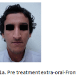 |
Figure 1a: Pre treatment extra-oral-Frontal. |
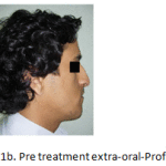 |
Figure 1b: Pre treatment extra-oral-Profile |
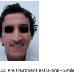 |
Figure 1c: Pre treatment extra-oral – Smiling |
In occlusion, bilateral maxillary posterior cross bite, severe anterior cross bite, and deep bite was observed. The maxillary and mandibular dental midlines coincided with each other and with the facial midline, and on both the sides the molar relation and the canine relation was Class III. The curve of spee was 2mm. (Fig 1d-f).
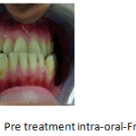 |
Figure 1d: Pre treatment intra-oral-Frontal |
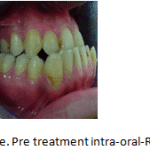 |
Figure 1e: Pre treatment intra-oral-Right. |
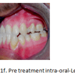 |
Figure 1f: Pre treatment intra-oral-Left |
Intraorally, oral hygiene was satisfactory. Maxillary arch was U-shaped, asymmetrical with crowding in maxillary left quadrant with distolabial rotations of 11,12, 21, 22, mesiopalatal rotation of 15, mesiobuccal rotation of 24, palatally locked 25 and distobuccal rotation of 26; and the mandibular arch was U-shaped, asymmetrical with imbricated mandibular incisors with distolingual rotation of 31, 32, mesiolingual rotation of 41, buccally placed 33 and 43, and lingually inclined 35,45.
Lateral cephalometric analysis revealed a severe Class III sagittal skeletal relationship ( ANB -3.50 , Witt’s appraisal BO ahead of AO by 11mm), with a mild prognathic mandible ( position of pogonion relative to nasion perpendicular was 5mm ( normal range, -2 to 4mm) ; Facial angle FHNPog 91.5 0 (normal range 87 0 ± 3) and a retrognathic maxilla ( Nasion perpendicular – point A -3.5mm ( normal value 2mm) ; Convexity of point A -6mm ( normal range 2mm ± 2); with the etiology assumed to be slight skeletal overgrowth of the mandible with undergrowth of maxilla with no facial asymmetry. The vertical facial proportions were increased ( N-Me 145mm). The SNB angle was 79.50, and the SNA angle was 760 and the distance from condylion to anatomic gnathion was 145 mm (normal value, (119.4 ± 5.3mm). The FMA was 27.50 (normal value 25 0), upper facial height was 62mm ( normal range 56 ± 3mm) and the lower facial height was 83mm ( normal range 70 ± 5mm). The maxillary incisors were proclined ( upper incisor to NA was 9mm, (normal value 4mm) and the mandibular incisors were proclined ( lower incisor to NB 6mm, (normal value 4mm) ; Lower incisor to A-Pog 7mm, (normal range 1 ± 2mm), showing dental compensation moreso for the severe sagittal skeletal discrepancy of the maxilla. (Table 1).
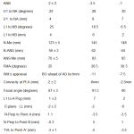 |
Table 1: cephalometric analysis |
The panoramic radiograph and lateral cephalometric radiograph confirmed the presence of all permanent teeth, and normal alveolar bone levels with no obvious signs of any pathology (Fig 2, 3).
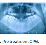 |
Figure 2: Pre treatment OPG |
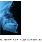 |
Figure 3: Pre treatment lateral cephalometric radiograph |
Treatment Objectives
- Improve facial profile and achieve lip competence.
- Correction of constricted maxillary arch and bilateral maxillary posterior crossbite.
- Correction of severe anterior cross bite and deep bite.
- Correction of crowded and rotated maxillary and imbricated and rotated mandibular teeth.
Treatment Alternatives
Since the patient exhibited a skeletal Class III malocclusion with a mild prognathic mandible and a severely retrognathic maxilla and since the facial profile was only very slightly concave the ideal treatment option would have been a Le Fort I maxillary advancement and subapical mandibular osteotomy to move the anterior segment distally. Another treatment alternative would have been to extract the mandibular first premolars and perform only subapical surgery to move the entire anterior segment distally to correct the anterior cross bite. But, since the patient was unwilling for any form of surgery, an orthodontic camouflage of the skeletal Class III malocclusion was decided.
Nonsurgical treatment of Class III patients is also effective and various treatment modalities to camouflage mild-to-moderate Class III relationships like multi-brackets with Class III elastics, extraction treatment, multi-loop edgewise therapy, high-pull J-hook headgear, etc., have all been well documented 24-30.
A severe negative overjet as was observed in the patient would be impossible to correct without absolute anchorage as there would be severe anchor loss as has been observed with the use of only traditional orthodontic mechanics. Hence, it was decided to use microimplants drilled in between the roots of 35 and 36 and 45 and 46 to facilitate enmasse retraction of the mandibular anteriors without anchor loss. The use of microimplants alongwith fixed appliance therapy to enhance effective retraction of the mandibular incisors was advised and the patient was willing for this alternative form of treatment.
Treatment Progress
In the maxillary arch, 25 was removed as it was completely blocked out palatally and deemed difficult and futile to correct. Both the mandibular first premolars were extracted and preadjusted edgewise appliance (0.022 x 0.028 inch slot), Roth’s prescription was strapped up. Upper and lower 0.014 – inch nickel titanium aligning archwires were ligated. (Fig 4). After initial alignment and leveling, 0.016 x 0.022- inch stainless steel archwires were placed in the maxillary and the mandibular dental arches with long crimpable hooks welded only in the region between mandibular lateral incisors and canines. Long crimpable hooks were preferred to keep the point of force application close to the centre of resistance of the anterior segment in the mandibular arch to facilitate bodily movement of the anterior teeth. AbsoAnchor microimplants 1.3mm in diameter and 8mm in length ( SH1312-08; Dentos Co,Taegu, South Korea) were placed bilaterally in the buccal interradicular bone between the mandibular second premolar and permanent first molar. (Fig 5).Two weeks after placement of the microimplants, retraction force of 150-200g was commenced with the use of nickel titanium coil springs stretched between the microimplants and the long crimpable hooks. (Fig 6).The reverse overjet was adequately corrected and the microimplants were well maintained throughout the treatment period without any perimplantitis.Upper and lower 0.019 x 0.025- inch stainless steel finishing wires were placed and final finishing and detailing was accomplished. ( Fig 7). The microimplants remained stable till the end of the treatment period. They were subsequently removed painlessly.
 |
Figure 4: Aligning and leveling – Upper and lower 0.016 inch nickel titanium archwires – Right |
 |
Figure 5: SH1312 microimplant of diameter 1.3mm and length 8mm inserted between 45 and 46- Right |
 |
Figure 6: Space closure – Upper and lower 0 .016 x 0.022 inch Stainless Steel archwires with nickel titanium retraction coil spring attached to the crimpable hook and the microimplant – Right |
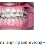 |
Figure 7: Final aligning and leveling – Frontal |
Treatment Results
After treatment, good facial harmony and balance was achieved. (Fig 8a-c). A Class I canine relationship was achieved whereas a Class III molar relationship had to be inevitably maintained. Normal overjet was achieved with effective enmasse retraction of lower anteriors and alignment of maxillary anteriors. All the rotations and abnormal axial inclinations and crowding in the maxillary and mandibular dental arches were corrected.The positive overbite that was obtained helped prevent relapse of the pre-existent severe anterior cross-bite. (Fig 8d).
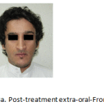 |
Figure 8a: Post-treatment extra-oral-Frontal |
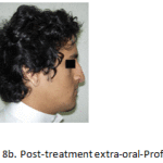 |
Figure 8b: Post-treatment extra-oral-Profile |
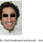 |
Figure 8c: Post treatment extra oral – Smiling |
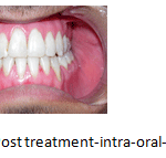 |
Figure 8d: Post treatment-intra-oral-Frontal |
The post treatment panoramic radiograph showed normal levels of alveolar bone with parallelism of the roots of the mandibular teeth well maintained with no root resorption (Fig 9). The post treatment lateral cephalometric radiograph was taken to gauge the treatment changes that was achieved (Fig 10). The lateral cephalometric measurements clearly earmarked the changes observed clinically. The Witts appraisal reading changed from -11mm to -7.5mm and the TVL to soft tissue point B reduced by 4.5mm, from -3mm to -7.5mm and the TVL to soft tissue pogonion reduced by 4mm, from -1mm to -5mm, clearly indicating the change brought about by the orthodontic camouflage with absolute anchorage and a mild clockwise rotation of the mandible. The lower anterior facial height showed a slight increase by 2mm. There was no dramatic change in the position of the maxillary incisors as they were derotated. The lower incisors were retroclined as evidenced by the reading lower incisor to A-Pog which changed from -3mm to -7.5mm. The lower lip to E-plane changed fron -2mm to -6mm in concomitance with the retroclination of the mandibular anteriors. (Table 1). The occlusion was stable post retention.
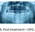 |
Figure 9: Post treatment – OPG |
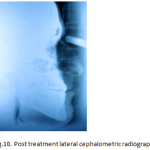 |
Figure 10: Post treatment lateral cephalometric radiograph |
Discussion
A varied number of combinations of skeletal and dental components could characterize Class III malocclusions 2 and since their etiology is multi-factorial, multiple- factor treatment is highly recommended. It is also an indubitable fact that a skeletal Class III malocclusion is often caused by some combination of maxillary deficiency and mandibular excess and the ideal treatment option of an adult skeletal Class III malocclusion with this combination is orthognathic surgery. Though the patient exhibited a skeletal Class III relationship, the almost straight profile of the patient prompted a non-surgical treatment approach and an orthodontic camouflage of the same. Camouflaging is prescribed for those patients only if there are defined indications that residual growth will not worsen the deformity after treatment; and that tooth positioning will have a favourable effect, or at least a less damaging effect on facial esthetics. The patient was an adult with no residual growth left and coupled with an almost straight profile seemed ideal for a camouflage treatment. Hence an important treatment objective in this case was to establish a natural dentoalveolar compensation orthodontically so as to establish an appropriate dentition for the patient’s skeletal pattern. The characteristics of the dentition in the patient was mesial inclination of the mandibular posterior teeth and lingual version of the mandibular incisors. Therefore, any further distal movement or lingual inclination of the incisors after mandibular premolar extractions alongwith routine orthodontic mechanics without absolute anchorage would cause unwanted complications such as root exposure and resorption of the incisors.31 It is a well acclaimed fact that the microimplants provide stationary anchorage for tooth movement in all directions.17,20,21 Bodily retraction of maxillary anterior teeth with the use of microimplants has been widely documented.16,17,19,22,23 . However the use of microimplants to bodily retract the mandibular anterior teeth has been sparsely documented. Hence the use of microimplants in the region between 35 and 36 and 45 and 46 had resulted in effective retraction of the mandibular anterior teeth and a positive overjet and overbite was achieved. The retraction of the lower anterior teeth en-masse was achieved with an archwire with crimpable hooks between the lateral incisors and canines with microimplant anchorage(MIA) sliding mechanics.17,32 With MIA sliding mechanics the enmasse retraction of the six mandibular anterior teeth was performed without anchorage loss, i.e., mesial movement of posterior teeth17,32,33 . Hence, microimplant anchorage had negated further mesial movement of the mandibular molars which could have inadvertently aggravated the already existent severe Class III molar relation. Class III elastics were not used as the microimplants provide the absolute anchorage for the enmasse distalization of the mandibular anterior teeth. Extractions were not advised in the maxillary arch as the maxilla was already retrognathic. But nevertheless, maxillary left second premolar had to be extracted as it was palatally locked and out of the arch. The maxillary anterior teeth were aligned ideally. Expansion of the maxillary arch was not very effective as the patient was an adult and the midpalatal suture was tightly interdigitated. The protruded lower lip was effectively retracted in concomitance with the effective distalization of the mandibular anteriors which improved the soft tissue profile immensely. The smile esthetics too improved dramatically. Fixed lingual retainers were bonded to prevent relapse.
Microimplants have gained widespread acceptance among patients and orthodontists because they cause less discomfort, are relatively non-invasive and pose fewer limitations in their placement.34, 35 They are also less expensive and provide absolute anchorage and cause less postoperative discomfort. Effective retraction of the mandibular incisors alongwith concurrent alveolar remodeling to correct the severe reverse overjet was adequately achieved with the use of microimplants as absolute anchorage which remained stable throughout the period of active treatment with no signs of perimplantitis and their removal post treatment too was uneventful.
Conclusion
An alternative treatment approach for correction of an adult skeletal Class III malocclusion with the use of microimplants in the mandibular arch between the second premolar and the first permanent molar had proved effective in correcting the severe Class III features of the patient, thereby restoring facial esthetics, harmony and balance.
References
- Stellzig-Eisenhauer A, Lux CJ, Schuster G. Treatment decision in adult patients with class III malocclusion: orthodontic therapy or orthognathic surgery? Am J Orthod Dentofacial Orthop 2002;122:27-38.
- Ellis E III,Mc Namara JA Jr. Components of adult class III malocclusion. J Oral Maxillofac Surg 1984;42:295-305.
- Bukhary MT. Comparative cephalometric study of Class III malocclusion in Saudi and Japanese adult females. J Oral Sci 2005;47:83-90.
- Singh GD, Mc Namara JA Jr, Lozanoff S. Craniofacial heterogeneity of pre-pubertal Korean and European-American subjects with Class III malocclusions: procustes, EDMA, and cephalometric analyses. Int J Adult Orthod Orthognath Surg 1998;13:227-40.
- Mouakeh M. Cephalometric evaluation of craniofacial pattern of Syrian children with Class III malocclusion. Am J Orthod Dentofacial Orthop 2001;119:640-9.
- Ngan P, Hagg U, Yiu C, Merwin D, Wei SH. Cephalometric comparisons of Chinese and Caucasian surgical Class III patients. Int J Adult Orthod Orthognath Surg 1997;12:177-88.
- Profitt WR, Fields HW, Sarver DM. Contemporary orthodontics.4th ed. St Louis: Mosby; 2007. p. 6-14, 300-309.
- Creekmore TD, Eklund MK. The possibility of skeletal anchorage. J Clin Orthod 1983;17:266-9.
- Roberts WE, Nelson CL, Goodacre CJ: Rigid implant anchorage to close a mandibular first molar extraction site. J Clin Orthod 28:693-704, 1994.
- Costa A, Raffainl M, Melsen B: Miniscrews as orthodontic anchorage: A preliminary report. Int J Adult Orthodon Orthognath Surg 13;201-209, 1998.
- Shapiro PA, Kokich VG: Uses of implants in orthodontics. Dent Clin North Am -32:539-550, 1998.
- Umemori M, Sugawara J, Mitani H, et al: Skeletal anchorage system for open bite correction. Am J Orthod Dentofac Orthop 115;166-174, 1999.
- Roberts WE, Engen DW, Schneider PM, et al: Implant anchored orthodontics for partially edentulous malocclusions in children and adults. Am J Orthod Dentofac Orthop 126:302-304, 2004.
- Fukunaga T, Kuroda S, Kurosaka H, et al: Skeletal anchorage for orthodontic correction of maxillary protrusion with adult periodontitis. Angle Orthod 76;165-172, 2006.
- Kanomi R. Mini-implant for orthodontic anchorage. J Clin Orthod 1997;31:763-7.
- Park HS. The skeletal cortical anchorage using titanium micro-screw implants.Korean J Orthod 1999;29:699-706.
- Park HS, Base SM, Kyung HM, Sunj JH. Micro-implant anchorage for treatment of skeletal Class I bialveolar protrusion. J Clin Orthod 2001;35:417-22.
- Park HS, Kwon DG, Sung JH. Nonextraction treatment with microscrew implant. Angle Orthod 2004;74:539-49.
- Park HS, Bae SM, Kyung HM, Sung JH. Simultaneous incisor retraction and distal molar movement with microimplant anchorage. World J Orthod 2004;5:164-71. 20. Park YC, Lee SY, Kim DH, Lee SH. Intrusion of posterior teeth using mini-screw implants. Am J Orthod Dentofacial Orthop 2003;123:690-4.
- Giancotti A, Greco M, Mampieri G, Arcuri C. The use of titanium minisrews for molar protraction in extraction treatment. Prog Orthod 2004;5:236-47.
- Park HS, Yoon Dy, Park CS, Jeoung SH. Treatment effects and anchorage potential of sliding mechanics with titanium screws in comparison with the Tweed-Merrifield technique. Am J Orthod Dentofacial Orthop. 2008;133:593-600.
- Park HS, Kwon TG, Kwon OW. The treatment of open bite with microscrew implants anchorage. Am J Orthod Dentofacial Orthop 2004;126:627-36.
- Faerovig E, Zachrisson BU. Effects of mandibular incisor extraction on anterior occlusion in adults with Class III malocclusion and reduced overbite. Am J Orthod Dentofac Orthop 115:113-124, 1999.
- Kim YH, Han UK, Lim DD, et al. Stability of anterior open bite correction with multiloop edgewise archwire therapy: A cephalometric follow-up study. Am J Orthod Dentofac Orthop 118:43-54, 2000.
- Lin J, Gu Y. Preliminary investigation of nonsurgical treatment of severe skeletal Class III malocclusion in the permanent dentition. Angle Orthod 73:401-410, 2003.
- Saito I, Yamaki M, Hanada K. Nonsurgical treatment of adult open bite using edgewise appliance combined with high–pull headgear and Class III elastics. Angle Orthod 75:277-283, 2005.
- Janson G, De Souza JE, Alves FA, et al. Extreme dentoalveolar compensation in the treatment of Class III malocclusion. Am J Orthod Dentofac Orthop 128:787-794, 2005.
- Moullas AT, Palomo JM, Gass JR, et al. Nonsurgical treatment of a patient with a class III malocclusion. Am J Orthod Dentofac Orthop 129(4 suppl): S111-S118, 2006.
- Kuroda Y, Kuroda S, Alexander RG, et al. Adult Class III treatment using a J-hook headgear to the mandibular arch. Angle Orthod 80:336-343, 2010.
- Kaley J, Phillips C. Factors related to root resorption in edgewise practice. Angle Orthod 1991;61:125-32.
- Park HS. The Use of Micro-Implant as Orthodontic Anchorage. Seoul, South Korea: Nare Pub Co; 2001:83-151.
- Lee JS, Park HS, Kyung HM. Micro-implant anchorage in lingual orthodontic treatment for a skeletal Class II malocclusion. J Clin orthod. 2001;35:643-647.
- Mah J, Bergstrand F: Temporary anchorage devices: A status report. J Clin Orthod 39:132-136, 2005.
- Kuroda S, Sugawara Y, Deguchi T, et al: Clinical use of miniscrew implant as orthodontic anchorage: Success rate and postoperative discomfort. Am J Orthod Dentofac Orthop 131:9-15, 2007.


