
K. Kumar
Department of Electronics and Instrumentation Engineering, Sathyabama University, Chennai- 600119, Tamil Nadu, India.
DOI : https://dx.doi.org/10.13005/bpj/540
Abstract
In this paper a noble model of thermograms imaging apparatus,which may have an impact towards the pattern study of a woman breast is introduced. Infrared (IR) thermography determines the surface temperature of an object or human body using thermal IR measurement camera. It is a method which is contactless and completely non-invasive. These properties make IR thermography a useful method of analysis that is used in various areas to detect, monitor and predict irregularities in many fields from engineering to medical and biological observations. System integrating passive thermal imaging with geometrical data from active scanner or an IR camera. We outline the potential benefits of this system in medical applications. In particular, we emphasize the benefits of using this system for preventive detection of breast cancer. The model deals with the potentials which are left ahead in regular diagnosis of women to determine Microcalcifications for diagnosis of abnormal growth in the breast tissues and cancer.
Keywords
Ductal Carcinoma in situ; Microcalcifications; Mammograms Infrared radiations; Digital Thermal Imaging
Download this article as:| Copy the following to cite this article: Kumar K. Micro Calcifications Detection for Breast Cancer Diagnosis using Infrared Thermal Imaging Apparatus. Biomed Pharmacol J 2014;7(2) |
| Copy the following to cite this URL: Kumar K. Micro Calcifications Detection for Breast Cancer Diagnosis using Infrared Thermal Imaging Apparatus. Biomed Pharmacol J 2014;7(2). Available from: http://biomedpharmajournal.org/?p=3255 |
Introduction
All bodies at temperatures greater than 0K emit radiation. The basic equations that link the absolute temperature of the body with the intensity and wavelength of the emitted radiation are given by the Planck, Stefan-Boltzmann, and Wein displacement law.
A major amount of thermal radiation emitted by an object emitted radiation belongs to the thermal infrared range, in a wave length between 7 and 15 mm. Infrared thermography is the technique of producing an image from infrared radiation, otherwise invisible to the human eye. Thermal cameras have sensors that capture the emitted or reflected thermal radiation from the substance. Images generated by these cameras are referred as thermograms where each image pixel corresponds to a digital value, proportional to the amount of received energy. Infrared thermography is a non-invasive (noncontact, non-destructive) imaging method. This makes infrared thermography a widely applicable technique, for fields such as building and electrical installation inspections and in industrial environments but also in biology and medicine.
Human body temperature distribution is influenced by many biological and physiological complex factors.Infrared thermography allows temperature distribution visualization of the human body and has been used in medical practice since the 1950s. The earliest medical applications of thermography was screening for sub epidermal tumour. Due to the drawback of less-sensitivity and low resolution, it was mainly used as an alternative method to detect breast tumour mass. Production of more sophisticated instruments, as well as increasing resolutions of cameras has allowed for more accurate tumour detection algorithms thus further establishing thermography as a viable alternative to standard tumour detection methods. Use of thermography in the field of medical oncology lies in the fact that tumours generally have increased temperature gradients compared to surrounding normal tissue.
The reason for this is an increase in blood supply and angiogenesis, as well as an increased metabolic rate of tumour tissue. Standard 2D thermograms are dependent on the distance and angle between the camera and the observed object. This means that it is very difficult to carry out quantitative and reproducible measurements or to correlate two different 2D thermograms of the same object. There is a need for standardising thermograms in order to solve this problem. Many studies and analyses have dealt with the problem of standardisation of thermogram.
It is low cost because it uses standard widely available electronic devices: projector and digital camera and the process acquisition is fully automated. Using the model data acquired by the scanner it is possible to define spatial data that includes shape and temperature distribution information . Also correlating thermograms is easier and can be performed automatically using point registration algorithms. This paper proposes a comprehensive conceptual model that enables combining scanning methods with thermal imaging to produce standardised thermograms, and integrating new approaches to temperature distribution data analysis and visual analytics based on thermograms. Such a system provides a new base for medical applications based on non-invasive diagnostic methods.
Need For Diagnosis-Thethermal Way
Mammography screening programs have shown to be effective in decreasing breast cancer mortality through the detection and treatment of early onset of breast cancers.
Most imaging studies and biopsies of the breast are conducted using mammography or ultrasound, in some cases magnetic resonance imaging (MRI). Although by now some improvement has been achieved, there are still some challenges and directions for future research such as developing better enhancement and segmentation algorithms are left behind. Emotional disturbances are known to occur in patient’s mind even after treatment.
Microcalcifications are tiny specks of mineral deposits (calcium) that can be scattered throughout the mammary gland, or occur in clusters. When found on a diagnosis, a radiologist will then decide whether the specks are of concern – usually, this is not the case.Generally, they indicate the presence of tiny benign cysts but can signify the presence of early breast cancer.so, it is important to attend normal checking sessions, as recommended by the health service. Microcalcifications are caused by Aging – the majority of diagnoses are made in women over 50, Benign causes, genetic causes Depending on what pattern the Microcalcifications present determines the next course of the action, whether it should be proceeded for new methods, or more regular checking.
Thermal Imaging – Architecture
System architecture defines system components and their synchronous operation. There are different approaches in creating thermography systems, depending on system usage and demands. Operation of system components is described by system workflow.
Overview
The model will determine the presence of Microcalcifications, or DCIS the stage 0 of a breast cancer. The study of thermograms is done in a pattern order. The thermograms are obtained from four IR cameras are reviewed and compared with the next and previous patterns of data’s to determine if any abnormal growth occurs in the soft tissues of the breast.
Design
The apparatus comprises a platform for supporting a female patient in front down, prone position, including an opening permitting one breast of the patient to be vertically pendant below the surface of the platform; scanning mechanism disposed below the platform to scan the breast without compression or contact. The mechanism includes a source of IR signals or thermal cameras which are four in numbers with a vacuum compressor.
The circuit for the rotation and zooming and movement of the cameras around the platform. It involves a digital camera for the display and also a computer programmed for storing and displaying images of the tissue in the breast derived in the thermal cameras. The model of the apparatus with the platform designed for is also generated by us which is shown in the fig. 1.
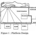 |
Figure 1: Platform Design
|
The extraction of the images or the thermograms is based on the thermal imaging technology. The only thing our model uses is that it uses n number usually four or five IR thermal cameras instead of one which gives us four different patterns of the temperature readings of the breast.
System Components
In this System we propose combines different multidimensional data sources: a VGA scanner and thermal camera, and a visualization display into a comprehensive multispectral visualisation tool suitable for medical applications .This allows the system to obtain spatial data and surface temperature distribution. The scanner used with the system uses a Digital Light Processing (DLP) video projector and a digital camera.
A video projector is used to project structured light patterns on the object to be scanned. The digital camera is used to acquire images of the object under controlled lighting. Both devices work together with the help of a PC, which generates a series of patterns, projected by the emitter and synchronizes the camera capture process.
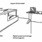 |
Figure 2
|
The thermal camera is used to capture thermograms – images showing surface temperature distribution. Based on the surface temperature distribution it is possible to isolate areas of interest for different medical conditions. The VGA scanner and the thermal camera are controlled by software components installed on standard PC for driving system workflow processes. Result of system workflow is displayed on special visualization display.
Acquisition Process
This process is used to acquire the object thermograms consisting temperature distribution of object’s surface.
Thermal camera calibration
To compute intrinsic and extrinsic parameters of the thermal camera a black and white checkerboard panel is used. The thermal camera, although it captures thermal radiation, is able to detect the calibration pattern due to the fact that black and white coloured objects have very different emissivity factors. This means that a black object emits more thermal radiation than a white object if both are illuminated using white light, even though the both objects are at the same temperature depicts thermograms of a black and white checkerboard pattern.
The temperature radiation values on pixels marked by the black line are also shown. These two figures show the difference in captured thermal radiation between black and white fields of the pattern. In order to determine the extrinsic parameters of the thermal camera, captured thermal images of the pattern are used along with images captured by the camera.
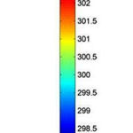 |
Figure 3: Temperature map acquisition
|
Medical Applications
Various medical applications are based on analysis and/or visualisation of data relevant for processes happening in the human body. The temperature distribution is connected to biological and physiological processes that can normally occur or are the result of an illness. Thermo grams allow modelling different models and processes based on scientific studies of medicine and biomedicine (e.g. breast cancer temperature dissipation). This means new data analysis methods have to be invented and new method of visualisation can be applied to enable medical professionals setting up a more reliable and comprehensive diagnosis.
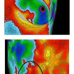 |
Figure 4
|
This process is used to acquire the object thermograms consisting temperature distribution of object’s surface. The figure shows temperature map captured by the thermo camera.
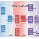 |
Figure 5
|
Conclusion
Thermal imaging alone may be successful in the diagnosis of early breast cancers and can easily differentiate malignant and benign tissues. Most important factor in the design for the model states and to that of the Indian issues. India has a tremendous age shift contrast to that of the countries where other diagnosis like mammograms is successful. But mammograms have not been much successful in directing the Indian issues of breast cancers. We hope thermal imaging in the above mentioned model does prove to be the alternative procedure.
The Detection of the cancer in its early stage should the main aspect in view of the technologies present, mammograms and thermograms pose a great value for the society and decreasing the mortality among women. Various studies conclude that when used as part of a multimodal approach (clinical examination + thermography +mammography or anatomical test), 95% of early stage cancers will be detected.
Reference
- Baker L. Breast Cancer Detection Demonstration Project: Five Year Summary Report. Ca
- Cancer J Clin. 1982;32:196-229. Beahrs Oh, Et Al. Report Of The Working Group To Review The Nci-Acs Breast Cancer
- Demonstration Project. J Natl Cancer Inst. 1979;62:639-698. Brown Sl, Silverman Bg, Berg Wa. Rupture Of Silicone-Gel Breast Implants: Causes, Sequelae, And Diagnosis. Lancet. 1997;350:1531-1537.
- Fenton Jj, Taplin Sh, Carney Pa, Et Al. Influence Of Computer-Aided Detection On Performance Of Screening Mammography. N Engl J Med. 2007;356:1399-1409.
- Barone S, Paoli A, Razionale A V 2006. A Biomedical Application Combining Visible And Thermal 3d Imaging. Xviii Congreso Internactional De Ingenieria Grafica, Sitges
- Ivanescuna, Ciupitu L 2010 Vision System For Human Body Infrared Thermography. Ieee 19th International Workshop On Robotics In Alpe-Adria-Danube Region (Raad)
- Rongqian Yang, Yazhu Chen 2011 Design Of A 3-D Infrared Imaging System Using Structured Light. Ieee Transactions On Instrumentation And Measurement
- Rocchini C, Cignoni P, Montani C, Pingi P, Scopigno R 2001 A Low Cost 3d Scanner Based On Structured

