N. Mariappan
Department of Hand Surgery, Sri Ramachandra Medical College and Research Institute, Sri Ramachandra University (deemed), Porur, Chennai, India.
Corresponding Author E-mail: drn_m@hotmail.com
DOI : https://dx.doi.org/10.13005/bpj/1739
Abstract
Nanotechnology is manipulation of matter on atomic, molecular and supramolecular scale. It has extensive range of applications in various branches of science including molecular biology, Health and medicine, materials, electronics, transportation, drugs and drug delivery, chemical sensing, space exploration, energy, environment, sensors, diagnostics, microfabrication, organic chemistry and biomaterials. Nanotechnology involves innovations in drug delivery,fabric design, reactivity and strength of material and molecular manufacturing. Nanotechnology applications are spread over almost all surgical specialties and have revolutionized treatment of various medical and surgical conditions. Clinically relevant applications of nanotechnology in surgical specialties include development of surgical instruments, suture materials, imaging, targeted drug therapy, visualization methods and wound healing techniques. Management of burn wounds and scar is an important application of nanotechnology.Prevention, diagnosis, and treatment of various orthopedic conditions are crucial aspects of technology for functional recovery of patients. Improvement in standard of patient care,clinical trials, research, and development of medical equipments for safe use are improved with nanotechnology. They have a potential for long-term good results in a variety of surgical specialties including orthopedic surgery in the years to come.
Keywords
Burns; biosensors; cancer diagnosis; carbon nanotubes; cancer treatment; drug delivery; diagnostics; implants; molecular imaging; wound care nanotechnology
Download this article as:| Copy the following to cite this article: Mariappan N. Recent trends in Nanotechnology applications in surgical specialties and orthopedic surgery. Biomed Pharmacol J 2019;12(3). |
| Copy the following to cite this URL: Mariappan N. Recent trends in Nanotechnology applications in surgical specialties and orthopedic surgery. Biomed Pharmacol J 2019;12(3). Available from: http://biomedpharmajournal.org/?p=28358 |
Introduction
Richard Feynman’s lecture in 1959 “There’s plenty of room at the bottom” led to development of nanotechnology concept.He is respected as the “Father of Nanotechnology”. Norio Taniguchi derived the term nanotechnology in year1974. He identified that materials can be manipulated precisely at nanometer level. Nano is one-billionth in the SI length unit meter (1.0 × 10⁻⁹). “Nanotechnology is the science of design, synthesis, characterization and application of materials and extremely small devices”. Extensive molecular inter-actions occur in the body on at nanometers (nm) scale in particles of less than 100nm size.The nanomaterials show drastically different behavioral properties related to melting point, conductivity, and reactivity compared to the larger particles. The Quantum wells,quantum wires and quantum dots (qdots) are nanomaterials characterized at the nanometer scale in one, two or three dimensions respectively. Fullerenes, dendrimers and semiconductor quantum dots are nanoparticles with a diameter <100 nm. Molecular proteins have sizes that range between 1 nm and larger whereas atoms are smaller than 1 nm size.
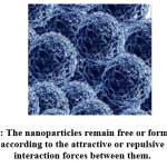 |
Figure 1: The nanoparticles remain free or form groups according to the attractive or repulsive interaction forces between them. |
Nanomedicine utilizes highly precise molecular interventions for diagnosis and treatment of diseases. Nanoscience is development of materials use at atomic, molecular and macromolecular scale and study pnenomina of altered properties due to small nature of the particles.Nanotechnology use the various design, characterization and production and applications by the shape and size at the nanometer scale.
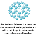 |
Figure 2: Buckminster fullerene is a round molecule of 60 carbon atoms with main application in target delivery of drugs for osteoporosis, cancer therapy and imaging. |
History
Titanium exposed to air forms an active biological field due to oxidation that promotes ingrowth of living tissues. Discovery of this phenomenon in 1950’s lead to the development of nanotechnology and a tremendous leap in bone implant applications in medical technology.
Table 1: Landmarks in the history of development of nanotechnology
| 2000 Years ago | Greeks used sulfide nanocrystals and Romans used them to prepare hair dye. |
| 1000 Years ago | Different sizes of gold NPs used in the making of stained glasses for windows and they emitted different colors |
| 1959 | Physicist Richard Feynman’s famous lecture “There’s Plenty of Room at the Bottom” – The beginning of the Concept of nanotechnology |
| 1974 | The term ‘Nanotechnology’ coined by Norio Taniguchi |
| 1977 | Drexler (MIT) introduced molecular nanotechnology concepts |
| 1981 | IBM develops Scanning Tunneling Microscope (STM). |
|
1985 |
“Bucky ball”( Fullerenes) was discovered in 1985.Harry Kroto of Sussex university, Richard Smalley and Robert Curl of Rice University won the 1996 Nobel Prize in Chemistry. |
| 1986 | K. Eric Drexler first used the term “nanotechnology” in his book titled: “Engines of Creation: The Coming Era of Nanotechnology”.
Atomic-force microscopy (AFM) was invented. |
| 1991 | MITI, Japan announced the bottom-up “atom factory” concept and Sumio Lijima discovered Carbon nanotubes. |
| 1997 | First molecular nanotechnology company (Zyvex) was founded by James R.Von Her ll. First design of Nanorobotic system introduced |
| 1999 | “Nano Medicine” published by R.Freitas, first book on nanomedicine. Nanotechnology safety guidelines were introduced. |
| 2000 | National Nanotechnological initiative (NNI) was launched. |
| 2001 | Feynman prize for developing “Theory of nanometer–scale electronic devices” and carbon nanotubes and nanowires synthesis. |
| 2002 | Feynman prize was given for making DNA enable the self-assembly utilized for new structures and to work on model molecular machine system |
| 2003 | Feynman prize was given to described electronic and molecular structures of new materials. Amalgamation of biological motors of single molecule was estwith nanoscale silicon devices. |
| 2004 | Policy conference on ‘Advanced Nanotech’ and Centre for nano- mechanical system were established. Feynman prize for Design of stable protein structures and novel enzyme with various other altered function was introduced |
| 2005-2009 | Robotics, 3-D networking and active nano products (3-D nano systems) that change their state during use were introduced. An improved walking DNA nanorobot was discovered. |
| 2011 | Era of molecular nanotechnology started. Nano processors that work with a programmable nanowire circuit were identified.
DNA molecular robots were identified to go along a branched track. Silicon Dimers were mechanically manipulated on a silicon surface. |
| 2012 | Nano sensors and Nanotechnology Knowledge Infrastructure (NKI) are the two Nanotechnology initiatives (NSIs) launched by NNI. |
| 2015 | Global market of nanotechnology reaches 1 trillion US Dollars. |
| 2016 | Graphene Nano devices used for DNA sequencing |
| 2017 | A new type of 3D computer chip that combines two cutting-edge nanotechnologies with increased memory and logic circuits. An all-women team of researchers from Indian Institute of Technology (IIT) Delhi has developed a new drug delivery platform using nanoparticles. |
| 2018 | The study of plasmon dissipation across graphene. New class of nanomaterials called metal-organic frameworks or “MOFs,” used to take carbon dioxide from atmosphere and combine it with hydrogen atoms to convert it into valuable chemicals and fuels. |
| Future 2020 | An estimated 6 Million workers will be needed worldwide by 2020.The first “molecular Assembler” to be developed |
The concept of nanotechnology
At nanoscopic size, the materials acquire altered physical and chemical properties.They have unique and advantageous surface charac-teristics compared to their original size. Nanoparticles are between 1 and 100 nm in size with a surrounding interfacial layer. Nanotechnology enables exploitation and organization of nanomaterials of one dimension below 100 nm. High surface area to volume ratio is responsible for the infinite property achieved by their smaller size. Mechanical, optical, magnetic, and chemical characteristic of nanoparticles achieved due to its particle size which enable them as ideal choice for wide applications in the fields of science, electronics, defense cosmetics and medicine. It has developed into a multibillion-dollar industry.Types of nanotechnologies described by Rice University, Houston are
Wet nanotechnology is the study of biological system in water environment.
Dry nanotechnology focuses on carbon. Silicon and inorganic materials are also parts of dry nanotechnology.
Computational nanotechnology related to modeling and stimulation of complex nanometer scale structures.
Nanotechnology perspectives are materials perspectives (larger to smaller) and molecular perspectives (simple to complex). Classification of Nanomaterials are according to their diamension, phase composition,organic or inorganic.
Dimension-based classification
Nanorods, nanowires, nanotubes, nanofibers, platelets – size ≤100 nm
Quantum dots, hollow spheres dimensions < 100 nm
Phase composition-based classification
Single phase solids – Amorphous crystalline particles and layers
multi-phase solids are matrix composites coated particles that include colloids, aerogels, ferrofluids, etc
Organic and inorganic types
The organic types of nanomaterials
Discovery of C60 was a major discovery in the field of carbon chemistry. Buckyballs are self-possessed of carbon atoms linked to three other carbon atoms by covalent bonds in a perfect sym-metrical pattern (Kroto et. al, 1985[1])This molecule was named as Buckminster fullerene after an American architect Buckminster Fuller. C60 is the cheapest and easiest to produce by vaporization of graphite into helium using laser beam.
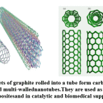 |
Figure 3: Sheets of graphite rolled into a tube form carbon nanotubes, single and multi-walled nanotubes.They are used as structural composites and in catalytic and biomedical supports. |
Carbon nanotubes (CNTs) are considered as fourth allotrope of carbon with a cylindrical nanostructure.They are elongated fullerene carbon nanotubes rolled up, with hexagonal carbon honeycomb sheets in perfect order that remain open or closed at ends. Currently carbon nanotubes are made by various solid and gaseous carbon-based production technologies like (a) electric arc release which is an electrical breakdown of a gas which gives away an ongoing plasma discharge (Ebbesen and Ajayan, 1992; Iijima, 1991) [2, 3] (b) laser ablation where a elevated level of power laser is used to vapourize carbon at high temperatures from a graphite target (Guo et al., 1995; Thess et al., 1996[4, 5]), and (c) chemical vapor deposition, the most common method of cnt production is catalyzed chemical vapor deposition of hydrocarbons (Kong et al., 1998; Li et al., 1996; Ren et al., 1998). [6, 7, 8]
Carbon nanotubes-based materials: Boron or Nitrogen doped carbon nanotubes were prepared (1994) by replacing the carbon atoms with Boron or Nitrogen elements in all types of nanotubes.
Inorganic materials
An entirely inorganic spherical fullerene-like molecules are developed by adding copper, chlorine, iron, carbon, phosphorus and nitrogen (Bai et.al.2004 [9] ). The spherical structure is three times larger of C60.(2) Inorganic nanowires have no inner cavity, and are synthesized by using metals like silver, bismuth, cadmium and semiconductors viz. zinc. (3) Quantum dots are spherical nanosized crystals of all semiconductor metals (cds, cdTe, ZnS) and their alloys. A quantum dot varies in size from 2 to 10 nm in diameter. Quantum dots have core made up of semiconductor, coated by ZnS.
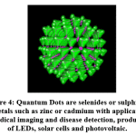 |
Figure 4: Quantum Dots are selenides or sulfides of metals such as zinc or cadmium with applications in medical imaging and disease detection, production of LEDs, solar cells and photovoltaic. |
Near-Infrared Fluorescence Imaging (NIRF) and positron emission tomography (PET) of integrin αvβ3, with quantum dots-based probes have improved tumor targeting efficacy and can detect tumors even at a lower concentration. Integrin αvβ3,is a receptor for vitronectin with two components integrin β3 and integrin αv (CD61) and is expressed by platelets. Quantum dots are suitable nanoparticles for cell tracking, reticulo-endothelial system mapping, and tumor targeting.
Dendrimers (Dendron means tree) synthesized in early 1980’s are repetitively branched, complex, synthetic and spherical molecules.They are known as arborols and cascade molecules. Poly-amidoamine (PAMAM) dendrimers are known as Starburst dendrimers.
Figure 5: Dendrimers are repetitively branched molecules as the basic structure. PAMAM is the most well-known dendrimer
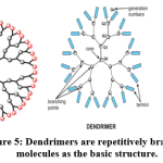 |
Figure 5: Dendrimers are repetitively branched molecules as the basic structure. PAMAM is the most well-known dendrimer |
Addition of different chemical species to the dendrimer surface enables different functionalities like detecting dye molecules,imaging agents and pharmaceutically active compounds.They also develop affinity ligands,radioligands and targeting components. An important application is Dendrimer/DNA complexes are used for gene delivery.[10]
Nanomedical applications are immensely complex and has role in multiple disciplines of science,medicine and engineering. Pre-clinical applications of nanotechnology are in vitro diagnostics: Nano-technology requires smaller samples in point of care (POC) diagnostics. There is no chance for sample deterioration and avoids intensively laborious work. It is less time consuming and achieves accurate results.
In vitro tool of diagnosis: biosensors contain biological liquid.They signal specific biological molecules in solution, a transducer convert it to a quanti-fiable signal. It can be a single biosensor or contain many integrated bio-sensors.
To measure parts of genome Gene chip devices are designed.They use DNA fragments as reusing elements for diagnostic purposes.
Table 2: Nanotechnology impact on clinical practice.
| Analytical, imaging tools |
| Nanomaterials and devices |
| Therapeutic drug delivery system |
| Targetted drug delivery, drug eluting |
| Safety, environmental, manufacturing and clinical use |
| Medical, surgical and imaging |
In vivo diagnostics
Traditional imaging techniques detect the diseased tissue and later contrast agents were injected to identify position/place of disease. Refinement by nanotechnology integrates imaging tools and contrast agents to detect disease even at single cell level. Nanotechnology achieves
Specific identification of cancerous cells.
Enable to identify locus of inflammation, developing tumor and visualization of vascular structure.
Treat the cancerous tissues early without affecting normal healthy tissues.
Multifunctional imaging programs are possible with nanotechnology due to the enormous surface area of nanoparticles. Radionuclide-based molecular imaging methods and molecular magnetic resonance imaging (mMRI) have sensitivity and are quantitative but has low resolution typically >1mm. Multimodality imaging using nanoparticles has better intrinsic and extrinsic resolution and higher rates of accuracy. Imaging labels such as super paramagnetic iron oxide (SPIO) nanoparticles for cellular imaging and drug delivery. Quantum dots, and micro bubbles are used for nuclear imaging of tumor angiogenesis and in identifying atherosclerosis and cancer. The advantages are that magnetization property is achieved in a magnetic field and they form a stable colloidal suspension. This process is important for applications in bio-medical sciences. They induce faster T2/T2 relaxation hence utilized as contrast agents for MRI. Magnetic force for clinically controlled targeting applications can be directed to desired site in the body.SPIO act as carrier vehicles that make it unique in magnetic targeting, local hyper-thermic effects and visualization with magnetic resonance imaging. SPIOs are typical tools for theragnostic application that combines diagnosis, treatment and follow up of a disease. External alternating magnetic field stimulates SPIO crystals and produce local hyperthermia-based therapies. SPIO labeled macrophage infiltration act as surrogate marker for stem cell viability.
The clinical applications of SPIO include magnetic separation process, hyperthermia therapy and stem cell tracking. In oncology they are used in MR contrast imaging of liver tumor and lymph nodes metastasis. Imaging done before therapy is to identify inflammatory involvement and is used as a prognostic biomarker. It acts as anticancer drug delivery system and used to measure pharmacodynamics response to cancer treatment program. SPIO ferumoxytol is not approved for imaging purposes. They are commercially available in the US as Feraheme® and in Europe as Rienso®.
Nanopharmaceuticals
are of two types (1) Therapeutic agent itself acts as own nanocarrier system and (2) Therapeutic agents with modified characteristics by coated nanoparticle carriers or nanoengineered drugs are used. They are functionalized or entrapped to develop new properties. Poorly soluble, poorly absorbed drugs acquire new properties suited for better drug delivery. Labile biologically active substances are converted by Nano particulate systems into stable and ideal drugs for better delivery. In commercial terms the economic life of proprietary drugs is extended and the time to market (TTM) for drugs are shortened. Recently, newer drug carriers are developed using third generation vectors. It consists of biodegradable core together with a polymer envelope (PEG) with membrane recognition ligands. Nanoparticles used for drug carriers for chemotherapeutics deliver medications directly to tumors, without any harm to the healthy tissues. [11, 12]
Bioavailability of orally administered insulin is the challenge for development of oral insulin drug for the patients with Diabetes Mellitus. Absorption of oral insulin is the main concern, selenium nanoparticles (SeNPs) used for oral insulin delivery over-come the absorption barrier. Pharmacological bioavailability improved by 9.15% compared to subcutaneous insulin. INS-SeNPs not only reduce oxidative stress and improve pancreatic islet cell function but also help promote glucose utilization. It has the disadvantage of producing significant hypoglycemic effect in both normal and diabetic animal models. Further clinical trials are needed for use in human diabetic patients.
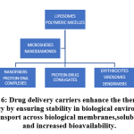 |
Figure 6: Drug delivery carriers enhance the therapeutic efficacy by ensuring stability in biological environment, transport across biological membranes, solubility and increased bioavailability. |
Precision drug targeting depends on the efficiency of drug-targeting systems on the three components namely targeting structure, drug attachment or carrying element and the drug delivered. Unique molecular feature for different diseases known as “unique address” makes it possible for site-specific targeting of drugs. It is a milestone in the improvement of drug delivery systems. The therapeutic target must be abundantly expressed by most diseased cells and absent from healthy issues. The methods used for drug targeting are (1) unique molecular structures target cancer molecules of various organs and tissues or (2) antibodies to these unique molecular structures are used as therapeutic agents. Stabilization of drug molecules by nanoscale drug delivery systems are used as gene delivery vectors and prevents rapid degradation of drugs. Nano-particles, nanowires, nanocages and dendrimers are the different types of Nano-sized carriers with clear advantages:
Drug reaches target cell without being degraded.
Enhance drug absorption in cancer cells.
Avoids side effects and
It has no interaction with normal cells.
Passive targeting works on the basis of “Enhanced permeability and retention”. Tumors with leaky blood vessels cause nanoparticles to seep to the site of damage and accumulate. This reduces the accumulation of cytotoxic drug and reduces its side effects. Active targeting: Molecules bind particular cellular receptors that are attached to nanoparticles. This brings drugs into cancerous cells as it specifically targets receptors. Destruction within is to destroy tumors thermally by Nano shells. Infrared light selectively kills tumor cells without damage to the healthy cells around the periphery of the tumor. Drug delivery devices: Nano pumps are tested for insulin delivery. Nanofabrication of ultra-small devices with storage and release of pharmaceutical ingredients directly deliver anticancer drugs to the tumor. Nanoencapsulation: Nano capsules carry vaccines, antibiotics or genes for targeted delivery and release with reduced side effects. Osaka University in Japan has developed a model for targeted delivery to human liver made of genes and drugs.
Viral nanoparticles (VNP) in medicine are bio-nanomaterials available in nature. VNP technology is crucial in chemical engineering and development of bio-medical applications. Genome-free component of VNPs are subclass of VNPs known as virus-like particles (VLP). VNPs are important tools for applications in medicine (1). Virus-based vaccines are prepared from genetically engineered VNPs (2) Chemically engineered VNPs are used in targeted drug-delivery. Their common applications are Pegylated liposomal doxorubicin, albumin bound paclitaxel and Poly (lactic-co-glycolic acid) preparations for drug delivery. In biomedical imaging nanoparticle-based contrast agents are used for magnetic resonance imaging.
Nanoscale fabrication is a significant step in nanoscience and nanotechnology and Nano scale level characterization of physical, chemical and biological properties is crucial. The idea of scaling is basic in the evolution of the micro-electronics. Scaling of devices below 10 nm size is expected to be impossible because of physical, technological and economic reasons. Heisenberg’s uncertainty or indeterminacy principle suggests that a length scale of a few nanometers is possible. Nanofabrication can be divided into 2 types. The “Bottom up” approach uses chemical synthesis or self-assembly and have applications in optics, pharmaceuticals, electronics, Nano fluids, catalyzes, Nano composites coatings and tissue engineering. They are divided into Gas-phase method and Liquid-phase methods.
Bottom Down approaches for Nano scale fabrication
Employs “self-assembly” process
Under specific conditions the atoms and molecules arrange themselves into the product.
The “top down” approach use the nanolithography and their nanoscale fabrication features include
Similar to a sculptor cutting away at a block of marble.
Begins at large scale and ends with a Nano scaled product
Used in the computer industry in creating microprocessors
Utilizes techniques such as, colloidal synthesis, epitaxy and lithography.
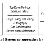 |
Figure 7: Top-down and Bottom-up approaches for nanoscale fabrication. |
Nanotechnology applications in Surgery
Nanotechnology has played a major role in all surgical specialties with fabrication of surgical implants and tissue engineering products. They have improved imaging technology, drug delivery systems and scaffolds fabrication with improved material-cell interaction that are useful in surgical practice of wound healing.
Surgical tools
Surgical blades made with diamond nanolayers has major advantages of
As diamond has a low friction coefficient, it decreases the penetration force in tissues.
It has chemical and biological inertness. It has the property of little physical adhesion to materials or tissues.
Plasma-polished blade available as Diamaze PSD-Plasma Sharpened Diamond. It has a decreased coating thickness from 5-25 μm to 0.5 μm with reduced surface roughness to 20-40 nm. They are ideal for precise surgery in the fields of Plastic, ophthalmic and neuro-surgery.
Suture needles and sutures
Nanoneedles: Suture needles made of nanosized stainless-steel crystals are available as Sandvik Bioline, RK 91TM Needles. Ideal suture needles for ophthalmic and plastic surgery should have characteristics of good ductility, strength and resist corrosion (Wilkinson2004[13]). Needles are made of stainless-steel and nanosized particles of 1-10 nm size quasi crystals by thermal ageing techniques acquire the above charecteristics. Medical wires Sandvik Bio-line (1RK91™) have very high tensile strength and hardness levels. Incorporation of silver nanocoating on silk sutures are recent approach against surgical infections. Nano tweezers will make precise cellular level surgery possible in the near future Gluing by aqueous nanoparticle solutions containing Stöber (Mesoporous) silica or iron oxide are used for Nano bridging and effectively controls bleeding and repairs damaged tissues. Liver wound edges glued by nanoparticles stopped bleeding quickly. Large numbers of nanoparticles produce millions of bonds between two surfaces for good healing. Nanoparticles are so small, and they do not have any adverse impact on wound healing process. No chemical reaction is needed for the healing process and they show aesthetic healing.
Recently, surgical sutures are made with advanced technologies. They possess adequate mechanical functions as well as drug-eluting capacities. Electro spun drug-eluting sutures with or without bupivacaine has been developed for postoperative pain management. Multifilament suture materials have the risk of infection, antibiotic eluting sutures prevent infection and achieve good healing properties. Absorbable antibiotic-eluting sutures with poly(L-lactide), PEG and levofloxacin for ophthalmic surgery are being used with good results. Surgical sutures coated with curcumin loaded Gold NPs for surgical site infections (SSI) are also available. Heparin-immobilized nanofiber for Vascular suture applications are being used.
Drugs like aceclofenac or insulin incorporated with the sutures exhibit 4% and 15% loading, release the drug for 7 and 10 days, respectively. Aceclofenac reduces epi-dermal hyperplasia and cellularity in skin inflammation and insulin shows wound healing properties with improved cellular migration. On-composite nanomaterial and nanotechnological applications in surgery are:
Nanofabricated drains
Self-assembling nanofibers for hemostasis
Conduits for focused direction in nerve repair and
Analysis of neuroprotection and neuro-modulation in real time and modifying the processes.
The surgeon can visualize the interior part of the surgery by ‘Smart ‘instruments that have sensors within the medical device or an instrument made by embedded technology method. It helps to take corrective steps at an early stage of complications.
Minimally invasive surgery
In interventional cardiology the dreaded complication is the risk of thrombus formation on the surface of intravascular catheters. Recently, catheters made of nanotube-based polymer reinforced with multi walled carbon NTs used as a filler in nylon12 (matrix) have contributed to the advances in minimally invasive surgery (Endo et al.,2005a [14]). Nucleation function of the carbon nanotubes reduces thrombogenicity by changes in electrostatic properties and dense surface changes. Advantages of the nanotechnology-based devices include enhanced mechanical properties, faster recovery to original shape and better handling properties, high resistance to fracture and their capacity to prevent and treat biofilm-associated infections on medical devices.
Optical Nano surgery
Optical tweezers (single-beam gradient force trap) is a power-full instrument with highly focused laser beams. They are used for noninvasive manipulation of micron-sized objects in the order of piconewtons, together with single living cells or organelles surrounded by cells, and viruses (Ashkin et al., 1987; Ashkin and Dziedzic, 1987 [15,16]). They are ideal for nanomanipulation of biological molecules, e.g. DNA (Bennink et al., 2001[17]). Near-infrared constant wave laser light from optical tweezers generates stress in nematodes by expressing an integrated heat shock- responsive reporter gene. (Leitz et al., 2002[18]).
Visualization
of drug distribution and metabolism can be done by tracking their movement. Cells are colored with dyes and they glow when excited by light of a certain wavelength. Quantum dots luminescent tags are attached to proteins, have the capacity to penetrate cell membranes. The dots are bioinert materials of various sizes and their movement throughout the body can be tracked. The occurrence of light worn to make an assembly of quantum dots fluorescent simultaneously make another group incandescent. Light of particular wave length will lit both groups depending on the size of the dot. “Nano cameras” are used for visualization of surgery and computers can be used to control the Nano-sized surgical instruments
Nanomedical Devices
are classified based on their level of invasiveness. Intravascular catheters, smart stents and endoscopes are are products of nanotechnology. Needles for electro-stimulation and syringes are other important applications. Gene or cell transfection systems efficiently delivers nucleic acids, proteins and siRNA into any cell types. Detection of circulating molecules at low concentration are possible by on-line monitoring sensors. Their ability to cross biological barriers like the blood-brain barriers will widen their usefulness. DNA origami is folding of DNA in range of the nanoscale size to generate non-arbitrary two and/ or three-dimensional shapes at the nanoscale. DNA Nano-scale structures act as both structural and functional element. Self-assembled DNA nanostructures are capable of being used as smart drug delivery vehicles in molecular-scale diagnostic. . Currently, nanodevices are implants used for detecting cancer and other diseases. Implanted as sensors they confirm myocardial infarction by detecting extravasation of three cardiac marker peptides namely, Cardiac troponin1 (cTn1), Myo-globin and CK-MB.Further research is on to develop efficient biological DNA-nano machines. Recent research works are on DNA nanostructures coated with virus capsid proteins. They improve transport to human cells to enhance the performance of drug delivery systems and for diagnostic applications at the molecular scale level for fabri-cation of a modular DNA-based enzymatic nanoreactor.
Nanotechnology in wound healing
Skin Wound healing is a highly coordinated, spatio-temporally regulated process with sequential and overlapping steps. Hemostasis, inflammation, proliferation, and remodeling is an ongoing continuous process.[19] Multiple cell types including fibroblasts, keratinocytes, immune, endothelial, and progenitor cells take part in the mechanism of wound healing.[20] Multiple dyanamic and continuous molecular and cellular events are controlled by growth factors, cytokines and chemokines. Final outcome of this process depends on the skin and microbiome interactions. The interleukin (IL) and growth factors signaling networks organize cell to cell and cell to extracellular matrix (ECM) interactions to fully heal a wound. Any alterations in this sequence leads to non-healing, chronic wound.[21] Antimicrobial nanobased dressings, immunomodulating antimicrobial nanoparticles, Gene modifying/silencing technologies and Growth factor-releasing nanoparticles are the different modalities available for wound care.
Typical features of a chronic wounds are unresolved inflammation and non-migratory epidermis.There is impaired fibroblast function and ECM deposition, decreased angiogenesis and increased levels of proteases. The presence of bacterial colonization and/or infection will lead to the chronicity of the wounds. [22,23,24,25,26,27] There is an amplified interest in finding more competent therapies for chronic wounds like venous, diabetic and pressure ulcers.[28]
Wound care is one of the excellent and widely used applications of nanotechnology. Nanotherapies target different phases of wound repair with good results. Nanomaterials in wound healing must have intrinsic properties beneficial for wound treatment and create an ideal microenvironment for healing and serve as delivery vehicles for thera-peutic agents.[29] Topical drug delivery in cutaneous wound healing depends on various factors, may be cell-type specific and used for a limited time only until the wound has healed. Chronic wounds are real challenge and various approved therapies are available. Bioengineered human dermal substitutes, recombinant platelet derived growth factor (rhPDGF) and skin equivalent are ideal for management of chronic wounds.[30, 31] “Smart biomaterials” like Human skin equivalent and dermal substitutes promote healthy healing process by its interaction with the wound environment. Stimulation of chemotaxis of neutrophils, macrophages, proliferation of fibroblasts, and smooth muscle cells are the beneficial effects on wound healing by PDGF.[32, 33]
Antimicrobial textile surfaces and wound care products are prepared by cotton and polyester fabrics treated with nanosized silver colloidal solutions (25-50 ppm). Another process involves melt-spinning of polypropylene and silver nanoparticles of 15 nm size.
Biomedical “smart” textiles
Passive smart textiles sense the environment only. Active smart textiles have a sensing purpose and act as movers in wound healing process.Very smart textiles have the capability of adapting their behavior to the circumstances and aid the healing process.
Woven fabrics and textiles using carbon nanotubes
Traditional textile spinning techniques are used in making of carbon nanotubes yarns.Wet spinning techniques are used for SWCNTs and dry spinning techniques are used for making MWCNTs and SWCNTs.
Wound care delivery platforms in wound care products
Nanoscale bio-degradable polymer fibers have been widely used in wound care products.They are produced by electro spinning technology and have potential applications in wound care in future. Wound care products are used as an effective and efficient hemostatic agent. Micro-dispersed oxidized cellulose technology and nanofiber technology are integrated in the process.
Nanoparticles and nanofibers for wound dressings have two layers of silver-coated,high -density polyethylene mesh. Electro spun nanofibrous membrane, polyurethane and silk fibroin nanofibers be the most commonly used in wound dressings. A rayon adsorptive polyester core used in delivery of nanocrystalline silver from a non-adherent, non-abrasive surface
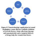 |
Figure 8: Nanotechnology application in wound healing has a nano fibrous scaffolds. Addition of Growth factors, Stem cells,Gene therapy and nanomaterials and a targeted delivery of drugs help the wounds heal faster. |
Electro spun materials have the advantages of high surface area-to-volume ratio,wide range of pore size and has high porosity. These factors favour cell attachment, cell adhesion, growth and proliferation. They also improve spreading of type I collagen. Porous structure allow fluid exudates from the wound to pass through and avoids wound desiccation, and prevents exogenous infections.
Silver-based nanoparticles
Silver and its ions forms have been used for their bactericidal properties for thousands of years.[34, 35] Introduction of anti-microbial agents containing silver bactericidal properties have revolutionized burn wound care.[36] Silver acts at multiple levels of cell cycle. It blocks respiratory enzyme pathways, alters microbial DNA and brings changes to the cell wall. Its antibacterial activity reduces the chances of developing resistance. [37] It is effective against multidrug-resistant organisms and has low systemic toxicity.[38] Pure silver nanoparticles (SNPs) markedly increases the rate of silver ion release.
Nitric oxide-delivering nanoparticles
Have a broad-spectrum antibacterial property against both Gram-positive and Gram-negative bacteria (DeRosa et al.[39]). The capability of NO in destroying MRSA biofilms was described by Miller et al.[40] and nitric oxide-releasing small molecules promote cell dispersal in P.aeruginosa biofilms (Barraud et al[41]). A silica nano-particle with NO-releasing capacity delivers the drug to pathogenic bacteria with increased bactericidal efficacy against planktonic P.aeruginosa cells (Hetrik et al.[42]). The other applications for wound healing are nanoengineered scaffolds with growth factor added to nano-materials. The future will be for nano scaffolds with added stem cell to stimulate wound healing. Nanotherapies with gene, ran interference (rnai), and small interfering rna (sirna) have infinite potential in wound care. Cell-type specificity will be achieved with nanotechnology-based targeted delivery in near future.
Gastrointestinal ulcers
Nanoparticles embedded with specific antibiotics are used in clinical applications for infections of the gastrointestinal tract. Smaller particle size and adhesiveness act against bacteria present in the mucosa make it ideal for treatment.[43] Abundant mucus production in the gastrointestinal tract, especially in the stomach,favors attachment of small particles.[44] Immune cells easily take up small particles with more adhesion property for better efficacy. Nanoparticle-bearing antibiotic treatment for H. pylori infection is with combination of two antibiotics (clarithromycin, metronidazole or tetracycline) and a proton pump inhibitor is very useful for gastro intestinal tract ulcer management.
Nanotechnology in Surgical Specialities: Surgical Oncology
Surgical Oncology
There is a paradigm shift from tissue staining to tissue imaging with nanoparticles.Their applications are wide spread in tissue visualization by advances in immune-histochemistry, profiling multiple sclerosis (MS), imaging MS, magnetic resonance imaging, detection of biomolecules, cancer cells and stem cell tracking. Surgery is the first choice and the most effective modality in treating human cancers. Complete surgical resection of cancers with a clear margin all around is the single most significant predictor of patient endurance. The challenges for surgeons during surgery are accurate identification of malignant tissues and removal of the entire tumor with adequate negative surgical margins.Total removal of the involved lymph nodes that drain the area involved by tumor disease and localization of small local residual tumor are the most important factors in implementation of adjuvant therapies.
Preservation of normal uninvolved structures is curcial for maintaining form and function. Gold Nanoparticles have a special property called the ‘Enhanced Permeability and Retention Effect’ (EPR Effect).Certain sized molecules of gold NPs accumulate in cancer cells more than in normal cells. Gold nanoparticles have a huge surface area and ‘Surface Plasmon Resonance’ properties. Gold nanoparticles restrict cancer meta-stasis.
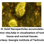 |
Figure 9: Gold Nanoparticles accumulate at the tumor site, help in visualization of tumor tissue and normal tissues (Courtesy: Georgia institute of Technology). |
Two types of gold nanoparticle shapes are more competent in converting light into heat energy.(1) Gold nanorods are solid cylinders as small as 10 nm in diameter. With different combinations of diameter and length of nanorods, the wavelength of light that the nanorod absorbs can be changed.
(2) Nanospheres have gold coating over a silica core. Changes made in the thickness of the gold coating and in the diameter of the silica core the wavelength of the light that the nanosphere absorb may be altered.
The clinical uses of gold nanoparticles (AuNPs) are
used for cancer diagnosis
Spectroscopic cancer imaging
Functionalized imaging agents for cancer detection
Treat prostate cancer with fewer side effects than chemotherapy
Nanosized particles are particularly efficient in evading the reticuloendothelial system.
AuNPs for cancer phototherapy
Photodynamic therapy (PDT)
Sonodynamic therapy (SDT) is utilized in activation of the cytotoxic effect of chemical compounds (sonosensitizers) and
focus on malignant sites situated deeply within tissues.
Difficulties experienced in AuNPs for cancer theranostics are its instability in the blood circulation, difficulties in targeting specific cells of interest and activation in response to the tumor microenvironment(TME).It poses issues for systemic AuNP delivery for cancer diagnosis and phototherapeutics.Gold NPs are successfully used for Rheumatoid arthritis,Alzheimer’s disease and cancer detection. Nanoparticles of colloidal gold, quantum dots, and polymeric liposomes have particular functional and structural properties compared to discrete molecules/bulk materials that are used in clinical applications.[45] Recently,targeting ligands (monoclonal antibodies, small molecules or peptides) are combined with nanometer-sized particles. They target malignant tumor cells and alter tumor microenvironments like tumor stroma and tumor vasculatures with high speci-ficity and affinity and help reduce the size of the tumor.[46]
Theragnostic is a treatment strategy that combines therapeutics with diagnostics. It is an important integrated diagnostic, imaging and cancer treatment protocol.[47,48] Diagnostic optical methods have problems related to tissue penetration whereas nanotechnology intra-operative imaging helps in tumor localization. It is an useful tool to assess tumor margins and for mapping of sentinel lymph nodes. They are used for identification of residual tumor cells or detect micrometastases.They help surgeon to confidently remove the tumour with better survival rates of the patients.Identification of adjacent vital structures is another advantage so that morbidity following the surgery is reduced. Nanoparticle contrast materials (sizes between 10–100 nm diameter) accumulate in solid tumors targeting both active and passive mechanisms.Their large surface area enables combining multiple diagnostic and therapeutic agents for theragnostics.
Quantum dot (QD) is a semiconductor crystal of nanometer size with distinctive condu-ctive propreties determined by its size.They form a group of fluorescent labels for use in biology and medicine.[49] Quantum dots and colloidal gold with their special optical and electronic properties of light emission with superior signal brightness are ideal for use.They resist photo bleaching with broad spectrum of absorption for excitation of multiple fluorescence colors simultaneously. QDs nanoscale scaffolds are used for designing multi-functional nanoparticles.Surface-Enhanced Raman Scattering (SERS) nanoparticles are brighter than near-infrared quantum dots and more intense than organic dyes. QDs have a high degree of sensitivity and selectivity.Intense fluo-rescent signals and multiplexing properties help the agents get concentrated in tumors. They are cleared from other normal tissues and organs. The long-term toxicity and fate of nanoparticles need to be evaluated further for authenticity of its safety. [50]
Quantum dot (QD) is a semiconductor crystal of nanometer size with distinctive condu-ctive propreties determined by its size.They form a group of fluorescent labels for use in biology and medicine.[49] Quantum dots and colloidal gold with their special optical and electronic properties of light emission with superior signal brightness are ideal for use.They resist photo bleaching with broad spectrum of absorption for excitation of multiple fluorescence colors simultaneously. QDs nanoscale scaffolds are used for designing multi-functional nanoparticles.Surface-Enhanced Raman Scattering (SERS) nanoparticles are brighter than near-infrared quantum dots and more intense than organic dyes. QDs have a high degree of sensitivity and selectivity.Intense fluo-rescent signals and multiplexing properties help the agents get concentrated in tumors. They are cleared from other normal tissues and organs. The long-term toxicity and fate of nanoparticles need to be evaluated further for authenticity of its safety. [50]
Thoracic surgery
Imaging of tumors of thorax and their intraoperative localization have always been a challenge. Nanotechnology based imaging helps intra-operative tumor localization.They also play a role in lymph node mapping and in accuracy of tumor resection.They are crucial in the adjuvant lung cancer therapy and help improve the survival rate of patients with lung cancer. Drug delivery of anticancer drugs like paclitaxel, docetaxel and doxorubicin has shown improved survival rates. Nanotech- nology has enabled monitoring of tumor microenvironment and make molecularly targeted lung cancer therapy a reality. Next frontier in lung cancer therapy involves integration of diagnosis, targeting and drug monitoring with controlled release by developing ‘Theragnostic’ multifunctional nanoparticles. [51,52]
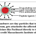 |
Figure 10: Nanoburrs are tiny particles that travel through the blood stream, gets attached to the affected arteries and deliver medicines like Paclitaxel directly to the damaged tissue.(Image credit Massachusetts Institute of Technology). |
The heart
Nanotechnology plays a role in detection and treatment of defective heart valves and arterial plaque.Nanorods nanomaterials are made up of minute gold rods alters the structure of valves made of collagen, a fibrous protein. There is a requirement for off-the-shelf implantable tissue engineered heart valves(TEHVs). A functional and viable implantable TEHV constructs may soon be realized by microfabrication and 3-D hydrogel preparation techniques. Growth enhancer (steroid) drugs incorporated in valve fabrication help produce more collagen. Nanomaterials with drugs incorporated to it are used in the treatment of aneurysm to prevent hemorrhages. Nanotechnology has improved the treatment of clogged arteries.Nanoparticles are lipid-based mole-cules and form a sphere shape called a micelle.The pectinase is attached to the surface and it is released onto the plaque. Nanoburrs are nanoparticles coated with a tiny amount of protein, which gets stuck to the target area and release drugs for several days.
Artificial Heart
Repair of damaged heart tissues and other organs is made possible by manufacturing high-functioning artificial tissues. They are durable and give adequate support for the tissues repaired with them for optimal function. Hydrogel scaffolding reinforced by carbon nanotubes are used for making cardiac tissue patches.Diamond-like carbon(DLC) are used in design of implantable human heart pump.
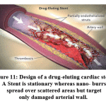 |
Figure 11: Design of a drug-eluting cardiac stent. A Stent is stationary whereas nano- burrs spread over scattered areas but target only damaged arterial wall. |
Nanotechnology applications in vascular surgery
Stents or synthetic bypass grafts for vascular intervention procedures require stent and catheters with less thrombogenicity and minimal tissue reaction.Tissue engineering and nanotechnology help in making of coated stents for making it less thrombogenic and making of implantable materials with drug-eluting capacity. Thrombus formation is the main reason for graft failure. It is due to endothelial dysfunction. An ideal graft must have biomechanical properties similar to healthy natural vessel, capacity to resist hyperplasia of the intima and possess thrombo-resistant properties. Percutaneous coronary interventions employing brachytherapy benefits from the unique electrical properties of Diamond-like carbon (DLC) and recently coronary artery stents are also made of DLC.
Polyhedral oligomeric Silsesquioxane (POSS) is a nanostructured chemical between ceramic and organic materials. Nanocomposite Polyhedral oligomeric Silsesquioxane poly (carbonate-urea) urethane (POSS-PCU) has all features of ideal graft.Its amphi-philic nature repels platelets and gives anti-thrombogenic properties. [53] Fumed silica nanoparticle releases nitric oxide, enhances the anti-thrombogenicity of the polymer. Nanopatterning of the graft luminal surface is done to promote cellular adhesion and to stimulate endothelialization.This is due to the presence of bioactive peptides associated with POSS-PCU.
Nanotechnology applications in Neurosurgery
Femtosecond laser neurosurgery:Tissue damage by use of conventional LASER is high as heat production occurs first before it cuts.Ultra-short pulse lasers are used to cut nanosized cell structures like nerve cells by an extremely precise surgery(Yanik et al.,2004[54]). “Nano scissors” produce low-energy femtosecond near-infrared laser pulses without harming normal surrounding tissues. The low energy and short laser pulses have minimal thermal damage. This is due to reduced mechanical effects like plasma extension and shock waves. Also the heat does not get accumulated and ther is no extension of thermal damage to normal surrounding tissues. (Colombelli et al.,2004[55]). Fifty percent of cut axons exhibited regrowth within 24 hours. Femto-second laser systems find applications in corneal refractive surgery in ophthal-mology (Juhasz et al.,1999[56]) and in dermatology (Kumru et al., 2005 [57])
Radiosurgery
Image-guided techniques enhance accuracy,stability and east to operate. Radiosurgery is a type of stereotactic radiotherapy. It is called as Gamma knife or cyber knife treatment. It is a very accurate targeted radiation therapy in large doses to destroy brain tumors. Femtosecond laser surgery of axotomy of neurons was performed with functional regeneration. It is now possible to cut single neurons and remove dendrites without damaging cell viability
Tissue engineering in neurosurgery
Neurite out-growth, branching, and formation of neural networks synpses are required for the refurbish or regeneration of the central nervous system subsequent to brain or spinal cord injury.[58,59,60] Single walled function-alized CNTs and multiple walled Functionalized carbon nanotubes help in regeneration of neural tissues. Carbon-nanotubes interfaced on phosphate glass fiber constructed into a 3-D, is a new class of neural scaffold that supports good cell viability and neuronal interactions.
Nanotechnology applications in ophthalmic surgery
Reactive oxygen species (ROS) are important causes of cataract and other ocular diseases. Oxidation is main feature of cataract formation.High surface area to volume ratio property of nano-materials play a crucial role in treatment of various eye conditions.Exposed position of the eye and the ease of accessibility, offers nanotechnology a wide range of applications in many ocular conditions
Measurement and continuous monitoring of intraocular pressure in glaucoma patients.
treatment of new vessels formation in choroidal theragnostics
To prevent scars following glaucoma surgery,
Treatment of oxidative stress,
Gene therapy for treatment of retinal degenerative diseases.
In age-related macular degeneration defective (retinal) pigmented epithelial cells can be replaced by a scaffold for delivery of stem cells.
Prosthetics and Regenerative nanomedicine in ophthalmic surgery.
Sight-restoring therapy
Polymer-based artificial tears contain petrolatum and lanolin.They are associated with redness, irritation and eye pain when used for treatment of dry eyes. When treated with medium-chain triglycerides as a liquid lipid and then dispersed in polyvinyl pyrrolidone solution, they form a nanoscale-dispersed eye ointment (the ointment matrix particle size about 100 nm).NDEO has no cytotoxicity to human corneal epithelial cells and is safe for ophthalmic use.It restores normal corneal and conjunctival morphology. Its improved drug delivery prevents postoperative scarring in patients with retinal degenerative diseases. Ocular drug delivery systems available at present are microemulsions, nanoparticles, nanosuspensions, dendrimers, liposomes, niosomes, and cyclodextrins. Nanomedicine, nanoimaging, and nanodiagnostics will further the frontiers of ocular diagnostics,drug delivery and therapy. Retinal nerve cell regeneration, nanoretina and nanotechnology-based eye implants are the future development that will help millions of people in need.
Nanoceria nanoparticles [Cerium oxide (CeO2) 5nm diameter] have a large surface area to volume ratio that effectively clears the reactive oxygen intermediates. Junping Chen et al demonstrated that intra-vitreal inoculation with nanoceria particles prevents light damage in rodents[61]. Macular degeneration and diabetic retinopathy related to oxidative damage can also be treated. There is no need for repetitive dosing of nano-ceria particles and they regenerate their function as scavengers of reactive radicals.
Glaucoma
Fluctuations of intraocular pressure (IOP) is difficult to monitor and its measurement is valuable for treatment of glaucoma.[62] A disposable soft silicon contact lens with sensor (CLS) embedded in it measures the corneal curvature produced by changes in IOP. This platinum-titanium strain gauge sensor has a total thickness of 175nm.The CLS is capable of measuring IOP to within 0.2mm Hg with 95% confidence interval and monitor IOP for up to 24 hours. The pressure profiles recorded were flat, fluctuating or in the form of spikes depending on diurnal versus nocturnal periods, activity and with or without prostaglandin analog treatment.
Pathological neovascularization in posterior segment of the eye is due to age-related macular degeneration and diabetic retinopathy. Treatment is a confront due to the anatomical and physiological ocular barriers. Topical, systemic, periocular, impale and intraocular are the routes for nanocarriers-based drug delivery into the posterior segment of the eye. Gold, silver and silicate nanoparticles act as inhibitors of neovascularization even without carrying the drugs.
Nanotechnology applications in field of plastic and reconstructive surgery
Soft tissue repair and healing
Nanotechnology has tremendously improved clinical care of traumatic and burn wounds.[63] Dressing materials of different composition are designed to improve wound healing by acting at all phases of healing process. Three- dimensional nanofibers scaffold resembles the native extracellular matrix (ECM) and enhance host tissue regeneration. Mechanical integrity, absorption of fluids, temperature control, and gas exchange properties are essential for ideal wound healing and tissue repair.Nanofibers provide all favourable criteria for the microenvironment for good healing to occur.
Implants and prostheses
are part of day-to-day plastic surgery practice. Implants used in breast augmentation and reconstruction following mastectomy procedures require adequate strength to withstand deformation and have the ability to prevent capsular contracture formation and must be durable.The shell of the silicone breast implants is made up of silicone rubber nanocomposite, reinforced with nanosized SiO2. Micro/macrotexurization surface modifications increase the roughness of the surface. Surface modifications available are siltex texturing, patterned surface and Biocell surface. Capsular contracture formation is prevented by halofuginone coated silicone implants that have anti-angiogenic property. Nanofiber coatings on the implants deliver tumor-specific anticancer drugs for area-specific chemotherapy to the tumor bed. The undesirable side effects of current systemic chemotherapy regimens are avoided. [64]
Tissue and organ engineering in plastic surgery
Reconstruction and repair of different tissues utilized in plastic surgery is made a reality by nanotechnology. Electro spun nanofiber matrices are urbanized for skeletal muscle regeneration.[65] It provides enough tissue and avoids the donor site morbidity of rib graft.Nasal cartilage is used for complex nasal reconstruction procedures following cancer, trauma, or congenital defects.[66] Artificial skin has been used for treatment of skin defects.[67,68] Advanced manufacturing facilities with available new biomaterials and products with good healing have enhanced aesthetic appearance.
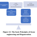 |
Figure 12: The basic Principles of tissue engineering and Regeneration. |
Nerve tubulisation
Management of nerve gaps over 5 mm requires autologous nerve graft procedures. The problems faced are the limited nerve donor sites and associated morbidity. New approaches in peripheral nerve repair have developed nanostructured tubular and porous conduits. They guide the regenerating nerves in proper alignment without loss of regerated axons. Biomaterials, embryonic stem cells, schwann cells, neural stem cells may be loaded to the conduits for enhanced regeneration. Chitosan nanofiber mesh tubes used in sciatic nerve injuries in a rat model showed partial recovery of sensory function of the nerve’s regeneration. (Wang et al [69] ). Nanophase silver-impregnated Type I collagen scaffold increase the amount of adsorbed proteins that are required for nerve healing. The time to nerve regeneration is decreased and an increase in the thickness of myelin sheaths aid better nerve conduction.
Nanotechnology in maxillofacial surgery
Maxillofacial surgery and dentistry has the potential to bring enormous advances with the applications of nanotechnology through nanorobotics, nanomaterials and biotechnology.[70] Nanorobots help clinicians in diagnostics, and various therapeutics process using natural nanomaterials which holds the potential to enhance the reconstruct a patient’s craniofacial skeleton and dentition.
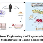 |
Figure 13: Tissue Engineering and Regenerative medicine and biomaterials for Tissue Engineering. |
Nanodentistry impression materials
Nanofillers in vinyl poly-siloxanes, produce a unique siloxane material with better flow and enhanced dental charecteristics for use as an impression material.Dentifrices made of nanosized hydroxyl apatite crystals are used as protective layer on tooth enamel in the restoration.The small dentifrobots are invisible, crawling at a slow speed and are inexpensive mechanical devices. They are safely deactivated by themselves if swallowed accidentally and are programmed with strict occlusal avoidance protocol.
Materials to induce bone growth
Hydroxyapatite nanoparticles are used to treat bone defects. Nanocrystals have modified surface with nanopores that adsorb proteins. Calcium sulphate is used to fill small spaces found in post extraction tooth sockets. It is used in periodontal bone defects and as an adjunct to the longer lasting bone graft material.
Dental Implant failure is due to insufficient bone formation around the biomaterial. Osseointegration is a bioactive method with direct physio-chemical bonding and involve the use of titanium implants.Surface roughening at the nanoscale level increases osteoblastic adhesion. Osseotite Dental Implants are titanium implants with nanosized deposits of hydroxyapatite and calcium phosphate that increases bone formation around the implant. Orthodontic wires must be corrosion resistant and have a good surface finish. Sandirk Nano flex is ultra-high strength stainless steel with good formability.This prevents loss of vitality and resorption. Tooth-straightening in horizontal and vertical position of tooth without pain is possible with the use of orthodontic nanorobots.
Nanotechnology applications in orthopedic surgery
Nanotechnology provides a multitude of new tools for applications in orthopedics. Osseointegration of implant materials, repair and regeneration of meniscus, osteochondral defects and vertebral disk are the important applications. It has an important role in targeted drug delivery in treatment of bone cancers.
Bone is made up of flexible matrix and bound minerals. Bone matrix is composed of elastic collagen fibers and ground substance. It is a biocomposite material complex of mineral mixture of calcium and phosphate in the form of hydroxylapatite, proteins with type I collagen fibrils, and water. Dimensions of the mineral and organic constituents are on the nanometer scale (Rho et al.,1998 [71]). The most important concerns with conventional orthopedic and dental implants are failure and limited life-time especially for young patients. Bone substitutes developed by nanotechnology have improved strength and longevity of the implants.Technological advances made in implant manufacture have made possible the development and applications of biosensors, sensitive diagnostic systems, and controlled drug delivery systems. [72]
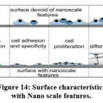 |
Figure 14: Surface characteristics with Nano scale features. |
Osseointegration is stimulation of rapid new bone formation that make the implants firmly fixed within the bone. Material properties of the implant and mechanical characteristics of the surrounding bone tissue should be matched.They must be in direct chemical and physical bonding with adjacent bone surfaces to achieve a good osseointegration.There should be no fibrous tissue interface formation. Stainless steel and cobalt chrome alloys are used for their good mechanical properties but stiffness of solid materials resulted in stress-shielding and bone resorption. Osseointegration minimizes stress and strain at the tissue-implant interface for better performance and the longevity of implants.
Applications of nanomaterial in orthopedic surgery are related to improving implant longevity,treatment of osteoporotic vertebral fractures, control of infection, treatment of orthopedic oncology, orthopedic tissue engineering and in stem cell regenerative medicine.Nanomaterials develop superior physio-chemical properties due to modifi-cations in physical charecteristics and resultant energetics of original materials. The major implant applications are (i) Reconstructive joint replacement (ii)Spinal implants (iii) orthobiologics and (iv) trauma implants. The most important mechanisms in the process of osseointegration are
Greater adsorption and interactions of selected proteins is enhanced
New bone growth is stimulated by creating nanoscale roughness of bone.
Regeneration of defective bone tissue occurs by a combination of living cells and growth factors with a biomaterial nanoscaffold.
Nanomaterial implants reduces infection rates due to the larger surface area, a healthy environment,with antibiotics incorporated and a better wound healing.
Extracellular adhesion proteins interact with nanophase implant scaffolds and have osteoblast adhesion. There is good new bone formation better fusion between implant and bone occurs.
Alumina nanocomposites have shown improved mechanical properties compared to monolithic alumina. Silicon carbide nanoparticles,Yttrium aluminium garnet and Zirconium Oxide NP are the examples. Alumina-zirconia nanocomposites are also known as Zirconia toughened alumina (ZTA). Fine-grained alumina matrix reinforced with Zircona particles, gives the toughness to the alumina matrix. The risk of aseptic loosening is a major cause of failure in total joint replacements (TJRs). Nanotextured materials improves osteoblast adhesion, osteointegration and is also effective in the treatment for bone defects.The use of nanocomposite implants in the treatment of osteochondral knee defects was demonstrated by Kon et al.[73] Gelatin, bioactive ceramics, biodegradable polymers, and polysaccharides such as agarose are the materials used. Their physical properties and nanoscale features stimulates cell growth and tissue regeneration in the human body. Methods available to reduce the implant failure rates, avoid complications and prolong the life of the implants are by
Implant coating materials
Surface charecteristics modifications
Bone replacement materials and
Tissue Engineering techniques.
Nanostructured implant coatings prevent wear,erosion and corrosion and provide thermal insulation. Nano-structured diamond, hydroxyapatite, and metalloceramic coatings are the most common coating materials.They are classified as
bioceramics of calcium phosphate
coatings with metal ion
Peptides and ECM components
Titanium nanotubes
Coatings materials for sustained delivery devices for osteogenic growth factor and drugs.
Nanostructured diamond coatings are ultra-smooth nanostructured diamond (NSD and USND) for titanium and cobalt-based alloy metal implant surfaces.They provide good adhesion properties to titanium alloys and poor adhesion to cobalt-chrome and steel substrates (Catledge et al., 2002a [74]) and the lifetime implants is increased by 30 to 40 years (Catledge et al., 2002b [75]). Nanostructured hydroxyapatite coatings are used extensively in orthopedic and dental implants. Hydroxyapatite biocoatings and tita-nium coatings connect the implants structurally and functionally with the human bones. This is achieved by an increase in osteoblast function that promotes bone formation around the implant.Recently, the next generation nanostructured hydroxy-apatite coatings are prepared by electrophoretic deposition technique.
Diamond-like carbon(DLC) has properties similar to diamond and it exists in seven different forms. Its main property is reducing abrasive wear of the implant surfaces. They are commonly used in implants for hip and knee joints replacement surgeries.
Nanostructured metalloceramic coatings
The main drawback of ceramics is their hard coatings on metallic substrate that has less adhesion properties.The presence of Chromium in Cr-Ti-N ternary system has an influence on the corrosion resistant properties of the system. They act as functionally graded metalloceramics. Ti-Cr-N coatings impart good wear-resistance, high hardness and excellent high-temperature resistance to implant surface.
Nanoporous ceramic implant coatings
Titanium made alloy implant surfaces are coated with nanoporous alumina layer by iodization of aluminum (Briggs et al., 2004; Karlsson et al., 2003[76, 77] ). This results in a highly adherent coating that can withstand stresses, shear and tension, similar to the bone it will be implanted.
Implant coating nanomaterials
Titanium and their alloys have good osseointegration properties and they are ideal implant materials. New bone growth is stimulated by surface roughening techniques like sandblasting and hydroxyapatite coating (Ducheyne et al., 1986[78]) or by formation of titanium dioxide or titania (Uchida et al., 2003[79]). Helical rosette nanotubes (Chun et al., 2004[80]) and Titania nanotubes are the newer class of nanomaterials that have been used with good long-term results(Oh et al., 2005[81]). Helical rosette nanotubes are organic nanomaterials with two basic DNA components, guanine and cytosine (Fenniri et al., 2001[82] Functionalized helical rosette nanotubes are ideal for next generation of orthopedic implants as they enhance osteoblast function and implant longevity.
Titania nanotubes are prepared on the titanium substrate using anodization process. “Nano-inspired nanostructure” is sodium titanate made from either alkaline treatment or sol-gel method. It is coarser than its precursor sodium titanate.The smallest is the nanofiber hydroxyapatite. HA-based nanofiber is prepared from Gelatin-calcium phosphate (caP) sol. Gelatin stabilized gold nanoparticles supported by titania nano-tubes are used in medical implants.Gelatin is the denatured product of collagen used as an immobilization matrix for biosensor production.
Surface modifications
Failures of implants are due to decreased osseointegration and fibrous encapsulation at the tissue-implant interface(Kaplan et al.,1994[83]) Identification and creation of nanostructured surfaces using nanofibers has led to an enhanced implant performance. Nanofiber alumina have an increased cytocompati-bility properties.Alumina nanofibers in both delta and theta crystalline phases increase osteoblast cell activity and also calcium deposition.This is due to enhanced adsorption of vitronectin as a result of decreased adsorption of apolipoprotein A-1 and due to increased adsorption of calcium on nanophase alumina.
Surface roughness modification of implants at Nanometerscale enhance bone growth towards the implant surface. Innumerable mechanical, thermal, chemical, electro-chemical and laser methods are available to improve orthopedic and dental implants. Nanophase alumina (Webster et al., 2001[84]) and poly (lactic-co glycolic acid) (PLGA) cast of carbon nanofibers (Price et al., 2004[85]) are commonly used due to their increased osteoblast function and orthoclastic response. Anaphase alumina(Webster et al.,2001[86]), copolymer mixtures of polystyrene and polybromostyrene {PS/PBrS} (Dalby et al., 2002; Dalby et al., 2003[87, 88]), PLGA (Vance et al., 2004[89]), and ceramics (Mustafa et al., 2005 [90] are the various materials used for surface roughness modification.
Bone replacement materials
Artificial bone is a laboratory created bone-like material used as bone graft. Hydroxyapatite and collagen fibers are the major components of bone. Chondroitin sulfate, keratan sulfate and lipid are also present. Organic polysaccharides (chitin,chitosan,alginate) and minerals (hydroxyapatite) are the material types prepared in bone grafting procedures. Nanoclay fillers reinforced bone cement has enhanced mechanical properties. Selenium, nanoceramics, alumina, titania,carbon,nanometals, Ti6AlV, cobalt chrome alloys and nanocrystalline,diamond display nanophase characteristics.[91,92] Bone replacement materials are used to treat fractures of the bones, periprosthetic fractures during hip revision surgery, acetabular reconstruction, filling cages in spinal column surgery, osteotomies and for filling bone defects in children.[93]
Charecteristics of applications of bone replacement materials
An Injectable bone matrix with 100% synthetic nanoparticular hydroxyapatite in paste form. This is completely absorbed after a few months.[94]
Engineered synthetic bone product loaded with bone void filler composed of hydroxyapatite nano crystals, have structure similsr to native bone crystals.
Synthetic replacement material for cancellous bone grafts with β tricalcium phosphate nanoparticles has been developed. Compared to conventional tricalcium phosphate they possess larger surface area, higher porosity, enhanced bioresorption and vascular invasion (Szpalski and Gunzburg, 2002[95]).
Nanocomposite scaffold implants composed of Type I collagen and nanostructured hydroxyapatites are used for the treatment of osteochondral defects of the knee joint.
Tissue engineering
involves scaffolds,signals and cells used to generate tissues in a limitless number of arrangements. In orthopedics restoration of pathologically altered tissues by cell transplantation in suitable scaffolds and biomolecules lead to tissue regeneration. Scaffolds can be metals, ceramics and polymers. Biological signals commonly used are rhBMP-2 and platelet rich plasma (PRP), osteogenic and angio-genic growth factors. Most commonly used cell types are mesenchymal stem cell (MSCs). Sources of MSCs are amniotic fluid-derieved, skin,periosteum and umbilical cord blood.
Cartilage engineering in orthopedic surgery for repair of cartilage defects or loss in an articular joint or meniscus in order to restore function has been used for many years.
Chitosan is the deacetylated form of chitin, an abundantly available poly-saccharide.Chitosan has minimal foreign body reaction with an intrinsic antibacterial property, can be moulded to various shapes and it forms porous structures ideal for cell in-growth and osteoconduction.Applications of chitosan are as bone graft substitutes, chitosan based hydrogels and wound healing bandages, drug delivery and for gene delivery strategies.Gene therapies with chitosan will be a reality in the coming years.
Nanotechnology for prevention and treatment of osteoporosis
Reduced bone density and micro architectural bone damages are charecteristics features of osteo-poresis. It can be primary or secondary. Fractures of the spine are more common than other bone fractures due to osteoporosis. Aims of treatment of osteoporetic vertebral fracture (OVF) are relief of pain, to restore height and functional stability of the vertebral body involved. Nanotechnology enhances bioavailability of nanosized calcium carbonate and calcium citrate, reduces the risk of osteoporosis and are used for treating osteoporotic vertebral fractures. Vertebroplasty and kyphoplasty are possible with bone fillers, injectable nanomaterials or Polymethyl-methacrylate (PMMA) bone cement. Development of calcium phosphate cement (CPC). [96] and calcium sulfate cement (CSC) with improvement in charecteristics of bone cements have better clinical applications. Injectable hydrogels are new tools for bone healing and regeneration.
An ideal Injectable nanomaterials for vertebroplasty and kyphoplasty treatment must have high injectability and homogeneity during injection. It should have setting properties with sufficient handling times. It must give adequate mechanical strength with appropriate stiffness to match neighboring vertebral bodies. They must have optimal stimulus for new bone ingrowth and provide porous structures for osseo-integration and angiogenesis. It should have low risk of necrosis or infection and radiopacity for imaging during surgery. Conventional materials have monomer toxicity, high temperatures causing damage to tissues, a failure to integrate into bone and presence of excessive stiffness that causes fracture. New nanomaterial incorpo-rated bone cements overcome these complications. PMMA bone cements with nano- phase MgO and BaSO4 shows higher osteoblast adhesion densities than pure PMMA cement. (Ricker et al[97]).
Calcium phosphate cements (CPCs) are used in bone defects and in tissue engineering. It has chemical and biological similarities to natural bone and has molding capability upon mixing and is an alternative to PMMA bone cement. CPCs are classified into brushite (dicalcium phosphate dihydrate) CPC or apatite CPC.There is an increase in the fracture resistance with CPC ultrafine nanofibers, similar to cortical bone. Degradation of the fibers makes pores and inter-connective channels for bone in-growth.[98] The elastic modulus and yield strength of PMMA decreases when mixed with 2% aqueous solution of sodium hyaluronate gel.[99] Addition of CNTs into CPC increase the strength, and bio-mineralized CNTs also increase the strength of CPC (Wang et al [100]). Multi-walled CNTs and bovine serum albumin composite result in a high-strength CPC. (Chew et al[101]). CPC/multi-walled CNT/ bovine serum albumin composite improves strength and leads to strong interface bonding with CPC and improves reactivity and wettability. Bovine Serum Albumin (BSA) and MWCNT also help improve the mechanical properties, resulting in stronger CPC composites and promote HA growth. Recent applications of CPC are 3-D printing,injectability, stem cell, growth factor or drug delivery.Their applications include pre-fabricated CPC scaffold, 3-D printing, injectable CPC scaffolds and CPC scaffold construct for bone tissue engineering.
Injectable hydrogels are similar to extra-cellular matrix and are carriers of drugs,cells, or growth factors for bone repair or regeneration. Hydrogels are used to fill gaps in bone defects and treat injuries in non-weight bearing areas.They also deliver growth factors, bioactives and drug molecules, are ideal platform for bone tissue engineering. Biodegradable poly(3- hydroxybutyrate-co-3 hydroxy-hexanoate)nanoparticles added to chitosan produce an injectable, thermo- reversible chitosan/β glycerol-phosphate hydrogel system.[102] It is suited for a long term sustained and controlled drug delivery of vancomycin for the treatment of orthopedic infections (Abdel-Bar et al[103]) Hydrogels alone cannot be applied for treatment of load-bearing bones. Injectable composite hydrogel of poly (N-isopropylacrylamide) reinforced by super para-magnetic iron oxide nanoparticles, has high elasticity (Campbell et al [104; [105]
Gelatin-based hydrogels modified by nanoparticles
Gelatin is a natural biopoly-mer widely used in pharmaceutical industry.Gelatin nanoparticles are used in anti-cancer drug delivery (Methotrexate, doxorubicin,paclitaxel, cisplatin), Protein and vaccine delivery,gene delivery ,pulmonary and ocular drug delivery and nutraceutical delivery. Biodegradable gelatin hydrogel with gold NPs has been developed.[106,107] The gel-gold nanoparticles have potential for treating fractured bones. Nanomaterials are widely used for manufacture of implants, as scaffolding for faster recovery. Safety of their applications in treatment for bone defects need further evaluation for long term benefits.
Prevention of Infection
Antibiotic-resistant infection is the main concern in joint replacement surgeries. Multiple drug-resistant staphylococcus aureus (MRSA) infection has increased recently. Biofilm matrix formation aurond the implant makes it difficult for the infection to be controlled and may nacessiate removal of implant. Treatment with nanophase silver incorporated as a power source into the implant design stimulates wound healing.The body fluids act as conducting medium between battery and silver, creates a low-level charge to enable the medicines to kill even antibiotic-resistant bacteria such as methicillin-resistant staphylococcus aereus. Titanium orthopedic implants with nanophase silver incorporated onto the surface shows strong, immediate anti-adhesive and bactericidal effects lasting for up to 30 days. “Drug Eluting nanostructured coating” materials deliver antibiotics and other
drugs on implant surface to prevent infection.
Nanotechnology applications in orthopedic oncology involve both diagnostic and treatment modalities. Diagnostic:Nanoparticles carry ligands that bind to specific molecules on the targeted cell. Loading the NP with a contrast,the imaging of the tumor is done at cellular level.The mutated p15 gene is tumor marker for osteosarcoma.They are used for identification of pulmonary metastasis also.Superparamagnetic iron oxide NP or quantom dots are good contrast agents for targeted MRI.They help identification of normal tissues, vital structures near the surgical field and identification of residual cancer tissues.Hollow nanoshells, gold nanocages, carbon and titanium nanotubes are used in diagnostic applications. Treatment applications (1) Passive or active targeting of drug delivery (2) Drug loaded nanostructure against tumor cells.(3) Nanovehicles interacting with molecular pathways(4)Hydroxyapatite NP in different osteosarcoma cell lines and (5)Gene therapy.(4) Nanophase (5) Selenium is a potentiator of chemo-therapeutic agents. It inhibits malignant osteoblastic growth at the implant-tissue interface.[108]
Bone regeneration
Functionalized SWCNTs are similar to collagen and form scaffolds for bone therapy (Zhao et al., 2005 [109]. Functionalized SWCNTs are ideal materials for artificial bone and to promote growth of bone. They take the role of collagen scaffold for the nucleation and growth of hydroxyapatite in bone when implanted as solutions or substratum as support scaffold. (Zhao et al., 2005 [110])
Human mesenchymal stem cells (hMSC) cultured on TiO2, smaller nanotubes capture local proteins easily and create an extracellular matrix-like for easy adhesion of hMSC. Larger nanotubes make hMSCs elongated and induce differentiation into osteoblastic cell-lines (Oh et al.[111]) Larger nanotubes have less capture of local proteins and hMSCs develop filopodia to cover larger surface area for adequate adhesion.This technique improves osteoinduction that involve gene therapy.[112,113]
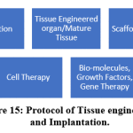 |
Figure 15: Protocol of Tissue engineering and Implantation. |
s
Stem Cell Regenerative Medicine of Orthopedic Surgery
Age, disease, damage or some genetic defects cause loss of tissues or organs. Regenerative medicine is the process of regenerating tissues that have lost their fractions. Regenerative medicine allow to grow tissues in laboratory, safely implant them and help regeneration. Stem cells exhibit enormous self repair potential and have many applications in regenerative medicine. Another advance in regenerative medicine is development of biomaterials. Nanotechnology provides nanoparticles and scaffolds for tissue engineering, and surface nanopatterning modifies specific biological responses from host tissues.[114]
Nanotechnology in gene therapy
Gene therapy has beneficial effects for treatment genetic disorders like Diabetes mellitus.[115,116] cystic fibrosis [117,118] and alpha 1 anti-trypsin deficiency. RNA interference (RNAi) is the method of treatment with good results. Interfering RNAs (iRNA) regulate specific gene sequencing, silencing and down regulating processes. Non-coding RNA leads to the development of therapeutic agents with potential for different diseases including viral diseases and cancers. The methods to guarantee targeted delivery and high stability are the challenges in this technique.The delivered material should not elicit undesirable immune response.
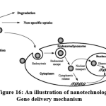 |
Figure 16: An illustration of nanotechnology Gene delivery mechanism |
Nanoparticles in gene delivery
Polymer nanoparticles (PNPs) deliver genes, therapeutic proteins or drugs. They are dissolved as a nanoparticle or encapsulated within as nanocapsule. PNPs are synthesized from non-toxic biodegradable, biocom-patible polymers. Chitosan, cyclodextrin, polyethylenimine (PEI), poly (lactic-co -glycolic acid) (PLGA), and dendrimers are the best suited materials. Polycations such as polyline help overcome the DNA size barrier as it “can condense DNA into toroidal nanostructures” to sizes less than 150 nm that can be carried into the cell.
Dendrimers for gene delivery Mono dispersity, specialized structure and surface functional groups are the properties that make dendrimers valuable tools in gene delivery.
Liposomes are minute vesicle-like structures created by self-assembly in the course of lipids energetic interactions and are commonly used for gene delivery
Magnetic nanoparticles: Paramagnetic NPs are used as drug carriers.They target tissues using strong magnetic fields, and are used in cancer treatment. The novel technique of magnetofecion overcomes the drawbacks of in vivo gene therapy.
Gold nanoparticles: The optical and physicochemical properties of Gold Nano-particles (AuNPs), allow easy transfection into cells and have unique biocompatibility that make them non-toxic.
Quantum dots for labeling genetic material: QDs are used in applications of catalysis, phosphors, photovoltaic, light emitting diodes (LEDs) and biological labeling. QDs bind to proteins and receptors to check with which molecules they interact. Hence useful in labelling techniques
Viral vectors used for gene transfer have safety concerns. Production of antibodies against viral vectors occurs due to stimulation of immune system and naked DNA can not cross the negatively charged cell membrane as these are also negatively charged. [119,120] Nanoparticle based gene therapy has evolved to over-come the problems as a mode to transfer genetic material. “Hybrid Nanomedicine” consists of TiO2 Semi-conductor nanoparticle of size 4.5 nm linked covalently to oligonucleotide DNA. [121,122,123,124]. Nanomedicine applications help in cure of HIV, cancer, in monitoring, diagnosing and treating a variety of diseases. It restores lost tissue at cellular level and monitor neuro-electric signals and stimulate bodily systems.
Nanorobots
“There is Room at the bottom -NO There is Plenty of Room at the Bottom” indicate the phenomenal expansion of nanotechnology-related applications. Nanorobots are designed to perform precise intracellular surgery.
 |
Figure 17: Nanotech medical robots (“nanomedibots”) perform functions beyond imagination. (NANOBOT-Image Credit: The Center for Responsible Nanotechnology) |
Nanorobot propulsion function happens with glucosea and body sugars and oxygen. Nanomanipulation is by using coded ultrasound signals, controlled by computer under supervision of surgeon. Search for pathology, diagnosis and removal of the lesion are the main areas of its applications of this technology. The exterior carbon atoms of nano robot has inert properties. The super-smooth surface of the nanorobots with minimal immune response helps in targeted function.
Nanorobotics is the technology of manufacture of ‘smart’ nanodevices that can be used for diagnostic and therapeutic purposes. It involves creating machines or robots at microscopic scale of a nanometer (10−9 meters). Nanorobots are essentially nano- electromechanical systems (NEMS) which are comparable to biological cells and organelles in size and this technology is known as Nanorobotics[125,126]. Types of nanorobots designed by Robert A. Freitas Jr as artificial blood are: i. Respirocytes. ii. Microbivores and iii. Clottocytes.
Applications of nanorobotics
They enhance human health and functioning by repair of damaged tissues,monitoring body function at the molecular level,removal of cancer tissues and pathological plaques.Composition of nanorobots can be biochip, bacteria-based, positional nanoassembly or Nubots (nucleic acid robots).The various fields of applications of nanorobotics are microrobotics,drug delivery, biomedical, treatment of brain aneurysm, cancer therapy, and communication systems.Its diamondoid structure form gives its strength. Miniature robots are designed so that it can be introduced into the body through the vascular system.They can pass through catheters also to under-take basic surgical procedures.Motion, force or a signal created at cellular level power the robot functional. Precise intracellular surgery can be performed through external guidance and monitoring
Clinical applications of nanorobots are in monitoring, diagnosis, treating diseases and targeted delivery of specific drugs.Treatment and diagnosis of Diabetes: Glucose monitoring nanorobots use the chemosensor, determine the time and dose of insulin to be injected. Dentistry: Dentifrobots functions in oral analgesia, to desensitise tooth and in straightening of irregular set of teeth. Pharmacytes nanorobots are loaded with drugs into its payload and used for drug delivery. Surgical nanorobots programmed by surgeons, act as semi-autonomous onsite surgeon by nanomanipulation. These miniature robots can be introduced into the body through vascular access or through catheters. The surgeon can perform precise intracellular surgery, through external guidance and monitoring.Cancer Detection and treatment: Transferrin, a polymer component of nanorobots is capable of detecting tumour cells.They kill cancer cells without damaging the healthy cells.They do not have complications such as hair loss, nausea etc.associated with conventional treatment modalities.GeneTherapy is done by comparing the molecular structure of both proteins and DNA found in the cell.Genetic diseases are treated by nanorobots with accuracy and with much comfort to the patient.
Minimally invasive surgical techniques and 3-D imaging data have contributed to the development of robotic surgery that provide stability of the system and its ability to work at small scales.“HybriDot” surgical robot has both automatic and manual functions for drilling and tapping before the introduction of screws into a bone.[127,128] They have are classified as Class II Medical devices. Femur preparation in total Hip Arthroplasty and Percutaneous screw fixation for pelvi-acetabular fracture, Acetabular cup placement and distal locking of intramedullary nails are tasks completed by the robot.[129,130] In total knee replacement surgery (TKR) it helps in preoperative planning, bone cutting and accuracy of the prosthetic alignment. In spine surgery, percutaneous insertion of screws with perfection is possible.
Minimally invasive microrobots are used to remove and repair atherosclerotic plaque inside arteries. Nanoparticle-assisted surgery illuminates cancerous tissues for early identification and its complete removal. They are useful tool to scan the body for metastasis. Local drug delivery system helps in diagnosis and treatment of cancers in a more cell-to-cell fashion. Nanobot is set to over-turn the basic paradigm of today’s medicine. Cancer-specific nanoparticle vehicles enhanced by personalized genomics is now possible. This will pave way for individualized therapies in near future.
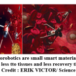 |
Figure 18: Nanorobotics are small smart materials can function with less tto tissues and less recovery time. (Nanorobotics: Credit : ERIK VICTOR/ Science Photo Library) |
An artificial flagella that mimics natural bacteria in size is developed,with swimming-enabled technology on nanobots.They are being used for retinal surgery.There is a shift from a treatment model to a prevention model.Nanotechnology provides body sensors to check and kill pathogens, before the patient has any symptoms. Advantages of nanorobot are that it helps the patients to get rid of the disease without any side effects and operate at specific site with a minimal tissue destruction.
Disadvantages of nanorobot are
The initial design cost is very high and very complicated.
Creation of stray fields affecting bioelectric-based molecular recognition systems.
Radiofrequency, electric fields, electromagnetic pulses affect nanorobots function due to electrical interference
Difficulties encountered are complex nature to design, customize and hard to interface
Its capability to destruct human body at the molecular level, have brutal risk in the field of terrorism
Privacy is a potential risk and more eavesdropping may occurs
Harmful effects may occur if the nanorobot is not very accurate
Field of Medical Engineering
combines both nanotechnology and micromachining technology.They find applications in pacemakers, hearing aids and in vision reha-bilitation. Recent applications include neuro-engineered systems for motor control, microchip based drug delivery systems and in prosthetic knee systems. Applications in bionics also have helped millions of sufferes.
Artificial internal organs for digestive Tracts:
An implantable artificial sphincter for patients with stoma following surgery for colon cancer who cannot have control over defecation has been designed and has improved Quality of Life (QOL) of such patients.
Artificial esophagus using a 3D-printed scaffold coated with mesenchymal stem cells and seeded in fibrin has been developed. Reconstruction of esophagus following cancer surgery is now made simpler with the availability of an artificial esophagus.
Stents with peristalsis and hyperthermia function are used in patients with inoperable and terminal esophageal cancer as an alternative treatment. It is completely non-invasive, has hyperthermia effect on the carcinoma tissue and maintains peristalsis function of esophagus.
Implantable type undulation pump ventricular assist device and undulation pump total artificial heart are in developemental stages. An undulation pump is a small Rotary blood pump (RP), that produces pulsation. The Eva heart is the rotary blood pump tested for endurance and anti-thrombogenicity. Rotary blood pumps are the most desirable ventricular assist system (VAD) for the small children.
Artificial Myocardium using nanotechnology: Nano sensor and nanocontrol chips are being developed as a first step towards the design for artificial myocardium.
Anatomical Modeling for cardiomyoplasty and/or cardiac transplantation is the gold standard of treatment at advanced end-stage heart failure patients.
Brain Function Control Units: Patients with warning sign of an attack of epilepsy are instructed to press a control switch implanted under their skin. The focus of epilepsy is cooled and epilepsy is aborted.
Following total artificial heart implantation, hypertension was observed in chronic experimental studies. Hypertension is a baroreflex function. As the blood pressure increases, there is decrease in heart rate and peripheral arterial dilation.Decreasing the cardiac output and peripheral arterial resistances,blood pressure returns to normal. Diagnosis of baroreflex sensitivity is by a hardware that measures the responses of the artery.
Nanotechnology and stem cell research
Magnetic nanoparticles (MNPs), quantum dots are being used for molecular imaging, delivery of gene and/or drugs into stem cells and tracing of stem cells.
Carbon nanotubes, fluorescent CNTs and fluorescent MNPs are the common nano-materials used in regenerative medicine for regulation of proliferation and differen-tiation of stem cells. It is also possible to track and image stem cells, to identify specific cell lineage and study their biology. Stem cells are modulated by mixing of nanocarriers with biological molecules. Translational medicine is “an interdisciplinary branch of the biomedical disciplines supported by three main pillars: benchside, bedside and community”
Stem cell-based therapeutics have found its place in prevention, diagnosis and treat-ment of various diseases.Intracellular access is a reality by nanodevices.They are also used for intelligent delivery and sensing of biomolecules.The available technologies have a great role to play in biomedical applications, stem cell microenvironment and in tissue engineering.
Future of nanotechnology
Gene editing technology is required for advancing the functional abilities of MSC. Gene editing MSCs have the risk of tumor formation that is prevented by an advanced CRISPR/Cas9 (Clustered Regularly Interspaced Short Palindromic Repeats) technology. Only one Cas9 protein is required for gene silencing process. This system consists of a nuclease that cut genomic DNA, and a guide RNA (gRNA) that recruits the nuclease to the target site. Alterated sequence of the gRNA makes Cas9 nucleases directed toward the DNA target .[131, [132] The system has properties of gene knockout, genome correction, and cassette knockin and gene deletion. CRISPR/Cas9 regulates TGF-b signalling through precise SMAD protein knockdown.[133] CRISPR/Cas9 can be activated in a reversible manner to avoid permanent genome editing.[134] and block the gene expression in other cell types but not MSCs.[135] The advantage of the CRISPR/Cas9 system is the very low effect in gene editing.[136] First clinical trial using CRISPR/Cas9 technology in human was conducted in China in 2016 to treat patients with metastatic non-small cell lung cancer.[137. 138,139,140]
Gene modulation is gaining grounds with a combination of EV application and CRISPR/ Cas9 technology. Inhibiting a drug’s metabolism enhances its effective dose. Most drugs are metabolized in the body by P450 3A4(CYP3A4). Micellar nano-sized cytochromeP450 inhibitors block hepatic metabolism of docetaxel. Theragnostic Liposome-nanoparticle hybrids for drug delivery and Bioimaging: Liposome–quantum dot (L–QD) hybrid vesicles are nanoconstructs for cell imaging. Liposomal-topotecan (L-TPT) enhances the efficiency of TPT by preventing systemic clearance. It allows extended time to accumulate in tumors.Hydrophobic CdSe/ZnS QD and TPT were located in the bilayer membrane and inner core of liposomes, respectively.
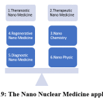 |
Figure 19: The Nano Nuclear Medicine applications. |
Nanonuclear Medicine: Radioisotopes are used extensively in the development of nanoparticle based therapeutics. Radioisotopes are an integral part for purposes of drug development, diagnostic and therapeutic applications. Assessment of bio-distribution, circulation half-life and pharmacokinetics of nanoparticles has been done by radiolabeling. Radio-isotope is ideal for the diagnostic component of the Dual diagnostic and therapeutic functionalities (theranostics). Treatment of cancers are achieved by delivery of nanoparticles by therapeutic radioisotopes. NPs properties are wide and flexible and enables use of radioisotopes in newer applications. Radio-isotopes versatility and potential with a promise to develop wide range of nanoapplications in the future.
Nanotechnology in India
The Initiative Division in the Department of Electronics and Information Technology (DeitY) has taken several major steps for the promotion of Nanoelectronics research and innovation in the country. Indian Nanoelectronics Users Programme (INUP) initiated by DeitY is being implemented at Centers of Excellence in Nanoelectronics (CEN) at Institute of Science (IISc) and IIT Bombay. This has provided a great opportunity for Research and Development community all over the country. The art of nanofabrication facilities, research and skill development in Nanoelectronics has taken a giant leap with this initiative.
Nanotechnology initiatives in India
Keeping abreast with the development of nanotechnology and its applications in various fields including defence the Government of India has taken initiatives for the new advances
The 9th Five-Year Plan (1998-2002)– Research programs in super conductivity, robotics, neuro-sciences, carbon and nanomaterials were included for the first time in Science and Technology research.
2000: Launch of “Programme on Nanomaterials: Science and Devices” by the Department of Science and Technology (DST).
In 2001-2002: “Nanomaterials: Science and Devices” Expert Group on DST was set up.
The 10th Five Year Plan (2002-07): Nanomaterials Science and Technology Mission (NSTM) initiated by Government of India.
On 3 May 2007: DST launched Mission on NanoScience and Technology (Nano Mission) to foster, promote and develop all aspects of nanoscicence and nanotechnology for the benefit of the country.
The Eleventh Five-Year Plan (2007-2012): Nano material and Nano devices in health and disease projects with high value and large impact on socio-economic delivery were announced. A Budget allocation of Rs. 1000 crore was earmarked for the Nano Mission in 2007.
Twelfth Five Year Plan (2012-2017): Approval for continuation of Mission on Nano Science and Technology (Nano Mission) in its Phase-II at a total cost of Rs. 650 crore was obtained.
Conclusion
Nanotechnology has improved all the spheres of Medicine and Health care. Advances in the fields of drug and gene delivery,biomedical imaging and diagnostic biosensors has enhanced patient care. It plays a great role in all surgical specialties especially with regard to cancer diagnosis, imaging and treatment.Complex and innovative hybrid technologies have widened their applications have potential for development in future.High-efficiency delivery transporters for biomolecules into cell are produced using single-walled carbon nano-tubes.Nanotechnology manipulation of genetic material and discovery of new biological components are possible. Wound healing and care of burn injuries are most beneficial applications that helps heal wounds and reduce the sufferings of patients. The development of Smart bandages in the near future will change the concept of wound care.Further research will widen the horizons in medicine, regenerative medicine, stem cell research and nutraceuticals for many useful applications. All discoveries made, new innovations described and useful advances made will ultimately help serve the mankind and improve human life to end their sufferings.
Conflict of Interest
The authors declare that they have no conflict of interest.
References
- Kroto HW, Heath JR, O’Brien SC, Curl RF and Smalley RE (1985). C60: Buckminster fullerene. Nature 318, 162-163.
- Ebbesen TW and Ajayan PM (1992). Large-scale synthesis of carbon nanotubes. Nature 358, 220-222.
- Iijima S (1991). Helical microtubules of graphitic carbon. Nature 354, 56-58.
- Guo T, Nikolaev P, Thess A, Colbert DT and Smalley RE (1995). Catalytic growth of single-walled nanotubes by laser vaporization. Chem. Phys. Lett. 243, 49-54.
- Thess A, Lee R, Nikolaev P, Dai H, Petit P, Robert J, Xu C, Lee YH, Kim SG, Colbert DT, Scuseria G, Tomanek D, Fisher JE and Smalley RE (1996). Crystalline ropes of metallic carbon nanotubes. Science 273, 487-493
- Kong J, Soh HT, Cassell AM, Quate CF and Dai H (1998). Synthesis of individual single-walled carbon nanotubes on patterned silicon wafers. Nature 395, 878-881.
- Li YL, Kinloch IA and Windle AH (2004). Direct spinning of carbon nanotube fibers from chemical vapor deposition synthesis. Science 304, 276-278.
- Ren ZF, Huang ZP, Xu JW, Wang JH, Bush P, Siegal MP and Provencio PN (1998). Synthesis of large arrays of well-aligned carbon nanotubes on glass. Science 282, 1105-1107.
- Bai J, Virovets AV and Scheer M (2004). Synthesis of inorganic fullerene-like molecules. Science 300, 781-783.
- Li S, Szalai ML, Kevwitch RM and McGrath DV (2003c). Dendrimer disassembly by benzyl ether depolymerization. J. Am. Chem. Soc. 125, 10516-10517.
- Dubin CH (2004). Special delivery: pharmaceutical companies aim to target their drugs with nano precision, Mech. Eng. Nanotechnol., 126(Suppl.): 10-12.
- Dass CR and Su T (2001). Particle-mediated intravascular delivery of oligonucleotides to tumors: associated biology and lessons from genotherapy. Drug Delivery, 8: 191- 213.
- Wilkinson JM (2004). Micro- and nanotechnology: Fabrication processes for metals. Med. Device Technol. 15, 21-23.
- Endo M, Koyama S, Matsuda Y, Hayashi T and Kim Y-A (2005a). Thrombogenicity and blood coagulation of a microcatheter prepared from carbon nanotube-nylon-based composite. Nano Lett. 5, 101-105.
- Ashkin A and Dziedzic JM (1987). Optical trapping and manipulation of viruses and bacteria. Science 235, 1517-1520.
- Ashkin A, Dziedzic JM and Yamane T (1987). Optical trapping and manipulation of single cells using infrared laser beams. Nature 330, 769-771.
- Bennink ML, Leuba SH, Leno GH, Zlatanova J, de Grooth BG and Greve J (2001). Unfolding individual nucleosomes by stretching single chromatin fibers with optical tweezers. Nature Struct. Biol. 8, 606-610.
- Leitz G, Fallman E, Tuck S and Axner O (2002). Stress response in Caenorhabditis elegans caused by optical tweezers: Wavelength, power, and time dependence. Biophys. J. 82, 2224-2231.
- Eming, S. A.; Martin, P.; Tomic-Canic, M. Wound repair and regeneration: mechanisms, signaling, and translation. Sci. Transl. Med. 2014, 6, 265sr266−265sr266.
- Pastar, I.; Stojadinovic, O.; Yin, N. C.; Ramirez, H.; Nusbaum, A. G.; Sawaya, A.; Patel, S. B.; Khalid, L.; Isseroff, R. R.; Tomic-Canic, M. Epithelialization in wound healing: a comprehensive review. Adv. Wound Care 2014, 3, 445−464.
- Midwood, K. S.; Williams, L.; Schwarzbauer, J. E. Tissue repair and the dynamics of the extra-cellular matrix. Int. J. Biochem. Cell Biol. 2004, 36, 1031−1037.
- Lindley, L. E.; Stojadinovic, O.; Pastar, I.; Tomic-Canic, M. Biology and Biomarkers for Wound Healing. Plast. Reconstr. Surg. 2016, 138, 18S−28S.
- Brem, H.; Tomic-Canic, M. Cellular and molecular basis of wound healing in diabetes. J. Clin. Invest. 2007, 117, 1219−1222.
- Kalashnikova, I.; Das, S.; Seal, S. Nanomaterials for wound healing: scope and advancement. Nanomedicine 2015, 10, 2593−2612.
- Tocco, I.; Zavan, B.; Bassetto, F.; Vindigni, V. Nanotechnology- Based Therapies for Skin Wound Regeneration. J. Nanomater. 2012, 2012, 1−11.
- Wei, E. X.; Kirsner, R. S.; Eaglstein, W. H. End points in dermatologic clinical trials: A review for clinicians. J. Am. Acad. Dermatol. 2016, 75, 203−209.
- Barrientos, S.; Brem, H.; Stojadinovic, O.; Tomic-Canic, M. Clinical application of growth factors and cytokines in wound healing. Wound Repair Regen. 2014, 22, 569−578.
- Richmond, N. A.; Vivas, A. C.; Kirsner, R. S. Topical and biologic therapies for diabetic foot ulcers. Med. Clin. North Am. 2013, 97, 883−898.
- Lo SF, Hayter M, Chang CJ, Hu WY, Lee LL: A systematic review of silver-releasing dressings in the management of infected chronic wounds. J. Clin. Nurs. 17(15), 1973–1985 (2008).
- Breitbart, A.S., Grande, D.A., Laser, J. et al. Treatment of ischemic wounds using cultured dermal fibroblasts transduced retrovirally with PDGF-B and VEGF121 genes. Ann Plast Surg 2001, 46: 555-61.
- Koveker, G.B. Growth factors in clinical practice. Int J ClinPract 2000, 54: 590-3.
- Herndon, D.N., Nguyen, T.T., Gilpin, D.A. Growth factors. Local and systemic. Arch Surg 1993, 128: 1227-33.
- Rudkin, G.H., Miller, T.A. Growth factors in surgery. PlastReconstrSurg 1996, 97: 469-76.
- Monteiro DR, Gorup LF, Takamiya AS, Ruvollo-Filho AC, de Camargo ER, Barbosa DB: The growing importance of materials that prevent microbial adhesion: antimicrobial effect of medical devices containing silver. Int. J. Antimicrob. Agents 34(2), 103–110 (2009).
- Melaiye A, Youngs WJ: Silver and its application as an antimicrobial agent. Expert Opin. Ther. Pat. 15, 125–130 (2005).
- Atiyeh BS, Costagliola M, Hayek SN, Dibo SA: Effect of silver on burn wound infection control and healing: review of the literature. Burns 33(2), 139–148 (2007).
- Neal AL: What can be inferred from bacterium-nanoparticle interactions about the potential consequences of environmental exposure to nanoparticles? Ecotoxicology 17(5), 362–371 (2008).
- Jain J, Arora S, Rajwade JM, Omray P, Khandelwal S, Paknikar KM: Silver nanoparticles in therapeutics: development of an antimicrobial gel formulation for topical use. Mol. Pharm. 6(5), 1388–1401 (2009).
- DeRosa F, Kibbe MR, Najjar SF, Citro ML, Keefer LK, Hrabie JA: Nitric oxide-releasing fabrics and other acrylonitrile-based diazeniumdiolates. J. Am. Chem. Soc. 129(13), 3786–3787 (2007).
- Miller C, McMullin B, Ghaffari A et al.: Gaseous nitric oxide bactericidal activity retained during intermittent high-dose short duration exposure. Nitric Oxide 20(1), 16–23 (2009).
- Barraud N, Schleheck D, Klebensberger J et al.: Nitric oxide signaling in Pseudomonas aeruginosa biofilms mediates phosphodiesterase activity, decreased cyclic di-GMP levels, and enhanced dispersal. J. Bacteriol. 191(23), 7333–7342 (2009).
- Hetrick EM, Shin JH, Paul HS, Schoenfisch MH: Anti-biofilm efficacy of nitric oxide-releasing silica nanoparticles. Biomaterials 30(14), 2782–2789 (2009).
- Lai SK, Wang YY, Hanes J: Mucuspenetrating nanoparticles for drug and gene delivery to mucosal tissues. Adv. Drug Deliv. Rev. 61(2), 158–171 (2009).
- Behrens I, Pena AI, Alonso MJ, Kissel T: Comparative uptake studies of bioadhesive and non-bioadhesive nanoparticles in human intestinal cell lines and rats: the effect of mucus on particle adsorption and trans 11. Alivisatos P. The use of nanocrystals in biological detection. Nat Biotechnol 2004;22:47–52. [PubMed: 14704706]
- Ferrari M. Cancer nanotechnology: opportunities and challenges. Nat Rev Cancer 2005;5:161–71. [PubMed: 15738981]
- Nie SM, Xing Y, Kim GJ, et al. Nanotechnology applications in cancer. Annu Rev Biomed Eng 2007;9:257–88. [PubMed: 17439359]
- Liu Z, Cai W, He L, et al. In vivo biodistribution and highly efficient tumor targeting of carbon nanotubes in mice. Nat Nanotechnol2007;2:47–52. [PubMed: 18654207]
- Heath JR, Davis ME. Nanotechnology and cancer. Annu Rev Med 2008;59:251–65. [PubMed: 17937588]
- Gao XH, Yang L, Petros JA, et al. In-vivo molecular and cellular imaging with quantum dots. Curr Opin Biotechnol 2005;16:63–72. [PubMed: 15722017].
- Davis ME, Chen Z, Shin DM. Nanoparticle therapeutics: an emerging treatment modality for cancer. Nat Rev Drug Disc 2008;7:771–82.
- Babapulle MN, Joseph L, Bélisle P, Brophy JM and Eisenberg MJ (2004). A hierarchical Bayesian metaanalysis of randomised clinical trials of drug-eluting stents. Lancet 364, 583-591.
- Liu X, Huang Y, Dens J, De Scheerder I, Hanet C, Vandormael M, Legrand V, Vandenbossche JL, Missault L and Vrints C (2003b). Study of antirestenosis with the biodivYsio dexamethasone-eluting stent (STRIDE): A first-in-human multicenter pilot trial. Catheter. Cardiovasc. Interv. 60, 172-178.
- Endo M, Koyama S, Matsuda Y, Hayashi T and Kim Y-A (2005a). Thrombogenicity and blood coagulation of a microcatheter prepared from carbon nanotube-nylon-based composite. Nano Lett. 5, 101-105.
- Yanik MF, Cinar H, Cinar HN, Chisholm AD, Jin Y and Ben Yakar A (2004). Neurosurgery: Functional regeneration after laser axotomy. Nature 432, 822-822.
- Colombelli J, Stelzer EHK and Grill SW (2004). Ultraviolet diffraction limited nanosurgery of live biological tissues. Rev. Sci. Instrum. 75, 472-478.
- Juhasz T, Loesel FH, Kurtz RM, Horvath C, Bille JF and Mourou G (1999). Corneal refractive surgery with femtosecond lasers. IEEE J. Select. Topics Quantum Electr. 5, 902-910.
- Kumru SS, Cain CP, Noojin GD, Cooper MF, Imholte ML, Stolarski DJ, Cox DD, Crane CC and Rockwell BA (2005). ED50 study of femtosecond terawatt laser pulses on porcine skin. Lasers Surg. Med. 37, 59-63.
- Mattson MP, Haddon RC and Rao AM (2000). Molecular functionalization of carbon nanotubes and use as substrates for neuronal growth. Journal of Molecular Neuroscience 14, 175-182.
- Zhang X, Morgan A, Ozkan CS, Prasad S, Ozkan M and Niyogi S (2005). Guided neurite growth on patterned carbon nanotubes. Sens. Actuators, B 106, 843-850.
- Hu H, Ni Y, Mandal SK, Montana V, Zhao B, Haddon RC and Parpura V (2005). Polyethyleneimine functionalized single-walled carbon nanotubes as a substrate for neuronal growth. J. Phys. Chem. B 109, 4285-4289.
- Chen J, Patil S, Seal S, McGinnis JF. Rare earth nanoparticles prevent retinal degeneration induced by intracellular peroxides. Nature Nanotechnology. 2006 Nov;1(2):142-50.
- Pajic B, Pajic-Eggspuchler B, Haefliger I. Continuous IOP fluctuation recording in normal tension glaucoma patients. Curr Eye Res. 2011 Oct 6.
- Hromadka M, Collins JB, Reed C, Han L, Kolappa KK, Cairns BA, Andrady T, van Aalst JA. Nanofiber applications for burn care. J Burn Care Res 2008;29:695‑703.
- Puskas JE, Luebbers MT. Breast implants: the good, the bad and the ugly. Can nanotechnology improve implants? Wiley Interdiscip Rev Nanomed Nanobiotechnol 2012;4:153‑68
- Klumpp D, Horch RE, Kneser U, Beier JP. Engineering skeletal muscle tissue – new perspectives in vitro and in vivo. J Cell Mol Med 2010;14:2622‑9.
- Oseni A, Crowley C, Lowdell M, Birchall M, Butler PE, Seifalian AM. Advancing nasal reconstructive surgery: the application of tissue engineering technology. J Tissue Eng Regen Med 2012;6:757‑68.
- Gerstle TL, Ibrahim AM, Kim PS, Lee BT, Lin SJ. A plastic surgery application in evolution: three‑dimensional printing. Plast Reconstr Surg 2014;133:446‑51.
- Hu MS, Maan ZN, Wu JC, Rennert RC, Hong WX, Lai TS, Cheung AT, Walmsley GG, Chung MT, McArdle A, Longaker MT, Lorenz HP. Tissue engineering and regenerative repair in wound healing. Ann Biomed Eng 2014;42:1494‑507.)
- Wang W, Itoh S, Konno K, Kikkawa T, Ichinose S, Sakai K, Ohkuma T, Watabe K. Effects of Schwann cell alignment along the oriented electrospun chitosan nanofibers on nerve regeneration. J Biomed Mater Res A 2009;91:994‑1005.
- Bhardwaj A, Bhardwaj A, Misuriya A, Maroli S, Manjula S, Singh AK. Nanotechnology in dentistry: present and future. J Int Oral Health 2014;6:121‑6.
- Rho JY, Kuhn-Spearing L and Zioupos P (1998). Mechanical properties and the hierarchical structure of bone. Med. Eng. Phys. 20, 92-102.
- Kon E, Delcogliano M, Filardo G, et al. Novel nano-composite multi-layered biomaterial for the treatment of multifocal degenerative cartilage lesions. Knee Surg Sports Traumatol Arthrosc 2009;17:1312-5.
- Kon E, Delcogliano M, Filardo G, et al. A novel nano-composite multi-layered biomaterial for treatment of osteochondral lesions: technique note and an early stability pilot clinical trial. Injury 2010;41:693-701.
- Catledge SA, Borham J, Vohra YK, Lacefield WR and Lemons JE (2002a). Nanoindentation hardness, and adhesion investigations of vapor deposited nanostructured diamond films. J. Appl. Phys. 91, 5347.
- Catledge SA, Fries MD, Vohra YK, Lacefield WR, Lemons JE, Woodard S and Venugopalan R (2002b). Nanostructured ceramics for biomedical implants. J. Nanosci. Nanotechnol. 2, 293-312.
- Briggs EP, Walpole AR, Wilshaw PR, Karlsson M and Pålsgård E (2004). Formation of highly adherent nanoporous alumina on Ti-based substrates: a novel bone implant coating. J. Mater. Sci. Mater. Med. 15, 1021- 1029
- Karlsson M, Pålsgård E, Wilshaw PR and Di Silvio L (2003). Initial in vitro interaction of osteoblasts with nano-porous alumina. Biomaterials 24, 3039-3046.
- Ducheyne P, Van Raemdonck W, Heughebaert JC and Heughbaert M (1986). Structural analysis of hydroxyapatite coatings on titanium. Biomaterials 7, 97-103.
- Uchida M, Kim HM, Kokubo T, Fujibayashi S and Nakamura T (2003). Structural dependence of apatite formation on titania gels in a simulated body fluid. J. Biomed. Mater. Res. 64, 164-170.
- Chun AL, Moralez JG, Fenneri H and Webster TJ (2004). Helical rosette nanotubes: A more effective orthopaedic implant material. Nanotechnology 15, S234-S239.
- Oh SH, Finõnes RR, Daraio C, Chen LH and Jin S (2005). Growth of nano-scale hydroxyapatite using chemically treated titanium oxide nanotubes. Biomaterials 26, 4938-4943.
- Fenniri H, Mathivanan P, Vidale KL, Sherman DM, Hallenga K, Wood KV and Stowell JG (2001). Helical\ rosette nanotubes: Design, self-assembly, and characterization. J. Am. Chem. Soc. 123, 3854-3855.
- Kaplan FS, Hayes WC, Keaveny TM, Boskey A, Einhorn TA and Ianotti JP (1994). Form and function of bone.In: Orthopaedic Basic Science, edited by Simon SR. American Acadamy of Orthopaedic Surgeons, Rosemont, Illinois, p. 127-185.
- Webster TJ, Schadler LS, Siegel RW, et al. Mechanisms of enhanced osteoblast adhesion on nanophase alumina involve vitronectin. Tissue Eng 2001;7:291-301.
- Price RL, Waid MC, Haberstroh KM, et al. Selective bone cell adhesion on formulations containing carbon nanofibers. Biomaterials 2003;24:1877-87.
- Webster TJ, Ejiofor JU. Increased osteoblast adhesion on nanophase metals: Ti, Ti6Al4V, and CoCrMo. Biomaterials 2004;25:4731-9.
- Dalby MJ, Childs S, Riehle MO, Johnstone HJ, Affrossman S and Curtis AS (2003). Fibroblast reaction to island topography: changes in cytoskeleton and morphology with time. Biomaterials 24, 927-935.
- Dalby MJ, Riehle MO, Johnstone H, Affrossman S and Curtis AS (2002). In vitro reaction of endothelial cells to polymer demixed nanotopography. Biomaterials 23, 2945-2954.
- Vance RJ, Miller DC, Thapa A, Haberstroh KM and Webster TJ (2004). Decreased fibroblast cell density on chemically degraded poly-lactic-co-glycolic acid, polyurethane, and polycaprolactone. Biomaterials 25, 2095-2103.
- Mustafa K, Oden A, Wennerberg A, Hultenby K and Arvidson K (2005). The influence of surface topography of ceramic abutments on the attachment and proliferation of human oral fibroblasts. Biomaterials 26, 373- 381.
- Sato M, Webster TJ. Nanobiotechnology: implications for the future of nanotechnology in orthopedic applications. Expert Rev Med Devices 2004;1:105-14.
- Durmus NG, Webster TJ. Nanostructured titanium: the ideal material for improving orthopedic implant efficacy? Nanomedicine 2012;7:791-3.
- Chris Arts JJ, Verdonschot N, Schreurs BW, et al. The use of a bioresorbable nano-crystalline hydroxyapatite paste in acetabular bone impaction grafting. Biomaterials 2006;27:1110-8.
- Appleford MR, Oh S, Oh N, et al. In vivo study on hydroxyapatite scaffolds with trabacular architecture for bone repair. J Biomed Mater Res A 2009;9:1019-27.
- Szpalski M and Gunzburg R (2002). Applications of calcium phosphate-based cancellous bone void fillers in trauma surgery. Orthopedics 25, s601-s609.
- Barinov SM, Komlev VS. Calcium phosphate bone cements. Inorg Mater. 2011;47:1470–1485.
- Ricker A, Liu-Snyder P, Webster TJ. The influence of nano MgO and BaSO4 particle size additives on properties of PMMA bone cement. Int J Nanomedicine. 2008;3:125–132.
- Canal C, Ginebra MP. Fibre-reinforced calcium phosphate cements: a review. J Mech Behav Biomed Mater. 2011;4:1658–1671.
- Boger A, Bohner M, Heini P. Properties of an injectable low modulus PMMA bone cement for osteoporotic bone. J Biomed Mater Res B Appl Biomater. 2008;86:474–482.
- Wang X, Ye J, Wang Y, Chen L. Reinforcement of calcium phosphate cement by bio-mineralized carbon nanotube. J Am Ceram Soc. 2007;90: 962–964.
- Chew KK, Low KL, Sharif Zein SH, et al. Reinforcement of calcium phosphate cement with multi-walled carbon nanotubes and bovine serum albumin for injectable bone substitute applications. J Mech Behav Biomed Mater. 2011;4:331–33964.
- Peng Q, Sun X, Gong T, et al. Injectable and biodegradable thermosensitive hydrogels loaded with PHBHHx nanoparticles for the sustained and controlled release of insulin. Acta Biomater. 2013;9:5063–5069.
- Abdel-Bar HM, Abdel-Reheem AY, Osman R. Optimized formulation of vancomycin loaded thermoreversible hydrogel for treatment of orthopedic infections. J Pharm Sci. 2014;5:2936–2946.
- Campbell SB, Patenaude M, Hoare T. Injectable superparamagnets: highly elastic and degradable poly(N isopropylacrylamide)-superparamagnetic iron oxide nanoparticle (SPION) composite hydrogels. Biomacromolecules. 2013;14:644–653.
- Cao L, Werkmeuster JA, Wang J, Glattauer V, McLean KM, Liu C. Bone regeneration using photocrosslinked hydrogel incorporating rhBMP- 2 loaded 2-N, 6-O-sulfated chitosan nanoparticles. Biomaterials. 2014;35:2730–2742.
- Kim HH, Park JB, Kang MJ, Park YH. Surface-modified silk hydrogel containing hydroxyapatite nanoparticle with hyaluronic acid-dopamine conjugate. Int J Biol Macromol. 2014;70:516–522.
- Reddy NN, Varaprasad K, Ravindra S, et al. Evaluation of blood compatibility and drug release studies of gelatin based magnetic hydrogel nanocomposites. Colloids Surf A Physicochem Eng Asp. 2011; 385(1):20–27.
- Tran P, Sarin L, Hurt RH, et al. Titanium surfaces with adherent selenium nanoclusters as a novel anticancer orthopedic material. J Biomed Mater Res A 2010;93:1417-28.
- Zhao L, Wang H, Huo K, et al. Antibacterial nano-structured titania coating incorporated with silver nanoparticles. Biomaterials 2011;32:5706-16.
- Zhao B, Hu H, Mandal SK and Haddon RC (2005). A bone mimic based on the self-assembly of hydroxyapatite on chemically functionalized single-walled carbon nanotubes. Chem. Mater. 17, 3235-3241.
- Oh S, Brammer KS, Li YS, Teng D, Engler AJ, Chien S, Jin S. Stem cell fate dictated solely by altered nanotube dimension. Proc Natl Acad Sci U S A 2009;106:2130‑5.)
- Bhakta G, Mitra S and Maitra A (2005). DNA encapsulated magnesium and manganous phosphate nanoparticles: Potential non-viral vectors for gene delivery. Biomaterials 26, 2157-2163.
- Polini A, Pisignano D, Parodi M, Quarto R, Scaglione S. Osteoinduction of human mesenchymal stem cells by bioactive composite scaffolds without supplemental osteogenic growth factors. PLoS One 2011;6:e26211.
- Pal N, Quah B, Smith PN, et al. Nano-osteo-immunology as an important consideration in the design of future implants. ActaBiomater 2011;7:2926–2934.
- Cash, K. J., & Clark, H. A. (2010).Nanosensors and nanomaterials for monitoring glucose in diabetes.Trends Mol Med, 16(12), 584-593.doi:10.1016/ j.molmed .2010 .08.002
- Pickup, J. C., Zhi, Z.-L., Khan, F., Saxl, T. and Birch, D. J. S. Nanomedicine and its potential in diabetes research and practice. Diabetes/Metabolism Research and Reviews, 24,604. (2008) doi: 10.1002/dmrr.893.
- Drumm ML et al.Correction of the cystic fibrosis defect in vitro by retrovirus-mediated gene transfer. Cell 1990; 62: 1227–1233. | Article | PubMed | ISI | ChemPort |
- Engelhardt JF et al.Expression of the cystic fibrosis gene in adult human lung. J Clin Invest 1994; 93: 737–749. | PubMed | ISI | ChemPort |
- Nowrouzi A, Dittrich M, Klanke C et al. 2006. genome-wide mapping of foamy virus vector integrations into human cell line. J Gen Virol, 87, 1339-1347.
- Paulus W, Baur I, Boyce MF, Breakefield XO and Reeves SA.1996. Self-contained, tetracycline-regulated retroviral vector system for gene delivery to mammalian cells. J Virol, 70, 62-67.
- Pinheiro AV, Han D, William M, Shih WM, Yan H (2011) Challenges and opportunies for structural DNA nanotechnology. Nature Nanotechnol 6: 763-772.
- Schulz M, Binder WH (2015) Mixed hybrid lipid/polymer vesicles as a novel membrane platform. Macromol Rapid Commun 36(23):2031–2041.
- Kowal J, Wu D, Mikhalevich V, Palivan CG, Meier W (2015) Hybrid polymer-lipid films as platforms for directed membrane protein insertion. Langmuir 31(17): 4868–4877.
- Ruysschaert T, et al. (2005) Hybrid nanocapsules: Interactions of ABA block copoly- mers with liposomes. J Am ChemSoc 127(17):6242–6247.
- A.G.Requicha,Nanorobots,NEMS and Nanoassembly. proceedings of the IEEE,2003,9,1922-1926.
- A.Weir, D.P.Sierra, J.F.Jones, A Review of Research in the Field of Nanorobotics; Sandia National Corporation:Albuquerque, New Mexico,2005.
- Leung KS, Tang N, Cheung WH, Ng WK. Robotic arm in ortho- paedic trauma surgeryeearly clinical experience and a re- view. Punjab J Orthop 2008;10:5e9.
- Kuang SL, Leung KS, Wang TM, Chui LH, Liu WY, Wang Y. A novel passive/active hybrid robot for orthopaedic trauma surgery.Int J Med Robotics Comput Assist Surg 2012;8: 458e67.
- Devito DP, Kaplan L, Dietl R. Clinical acceptance and accuracy assessment of spinal implants guided with spine assist surgical robot: retrospective study. Spine 2010;35:2109e15
- Karia M, Masjedi M, Andrews B, Jaffry Z, Cobb J. Robotic assistance enables inexperienced surgeons to perform uni- compartmental knee arthroplasties on dry bone models with accuracy superior to conventional methods. AdvOrthop 2013; 2013:481039. http://dx.doi.org/10.1155/2013/481039. Epub 2013 Jun 19.
- Cho SW, Kim S, Kim JM, Kim JS. Targeted genome engineering in human cells with the Cas9 RNA-guided endonuclease. Nat Biotechnol 2013;31(3):230e2.
- Mout R, Ray M, Lee YW, Scaletti F, Rotello VM. In vivo de- livery of CRISPR/Cas9 for therapeutic gene editing: progress and challenges. BioconjugChem 2017.
- Lee JS, Grav LM, Lewis NE, FaustrupKildegaard H. CRISPR/Cas9-mediated genome engineering of CHO cell factories: application and perspectives. Biotechnol J 2015; 10(7):979e94.
- Cheng AW, Wang H, Yang H, Shi L, Katz Y, Theunissen TW, et al. Multiplexed activation of endogenous genes by CRISPR- on, an RNA-guided transcriptional activator system. Cell Res 2013;23(10):1163e71.
- Qi LS, Larson MH, Gilbert LA, Doudna JA, Weissman JS, Arkin AP, et al. Repurposing CRISPR as an RNA-guided platform for sequence-specific control of gene expression. Cell 2013;152(5):1173e83.
- Kleinstiver BP, Pattanayak V, Prew MS, Tsai SQ, Nguyen NT, Zheng Z, et al. High-fidelity CRISPReCas9 nucleases with no detectable genome-wide off-target effects. Nature 2016.
- Cyranoski D. Chinese scientists to pioneer first human CRISPR trial. Nat News 2016;535(7613):476.
- Gibson G, Yang M. Gene editing in chondrocytes using CRISPR/CAS9. OsteoarthrCartil 2016;24:S2e3.
- van den Akker G, van Beuningen H, Davidson EB, van der Kraan P. CRISPR/CAS9 mediated genome engineering of human mesenchymal stem cells. OsteoarthrCartil 2016;24:S231.
- Yang M, Zhang L, Stevens J, Gibson G. CRISPR/Cas9 mediated generation of stable chondrocyte cell lines with targeted gene knockouts; analysis of an aggrecan knockout cell line. Bone 2014;69:118e25.


