
Gulzhan Yermukhanova, S. D. Asfendiyarov, G. Zaitenova, G. Ksetayeva and K. Sebitova
Professor, Doctor of Medical Science, The Kazakh National Medical University, Kazakhstan.
DOI : https://dx.doi.org/10.13005/bpj/413
Abstract
We created an experimental model of hemangioma in the dog penis and studied the effectiveness of sclerotherapy using a magnesium-based metal rod in treating this tumor. We found that the magnesium rod possessed thrombogenic and sclerotic properties.
Keywords
Cavernous Hemangioma; Therapeutic
Download this article as:| Copy the following to cite this article: Yermukhanova G, Asfendiyarov S. D, Zaitenova G, Ksetayeva G, Sebitova K. The Therapeutic Effects of a Magnesium-Based Metal Rod in an Experimental Model of Cavernous Hemangioma in the Dog Penis. Biomed Pharmacol J 2013;6(2) |
| Copy the following to cite this URL: Yermukhanova G, Asfendiyarov S. D, Zaitenova G, Ksetayeva G, Sebitova K. The Therapeutic Effects of a Magnesium-Based Metal Rod in an Experimental Model of Cavernous Hemangioma in the Dog Penis. Biomed Pharmacol J 2013;6(2). Available from: http://biomedpharmajournal.org/?p=2717 |
Intrduction
Experimental models of hemangioma to clinically evaluate the therapeutic effects of pharmacological and chemical agents on angiomatosis tissue are available in the field of experimental medicine. One of these models involves the use of barium sulfate to track capillaries within hemangiomas in rabbit femur skin [1]. Cavernous bodies in the penis are composed of fibrinopurulent elastic bundles that create a network of crossbeams. Among the dense plexus of crossbeams, endothelial-lined caves filled with blood are present [2]. Currently recognized models of cavernous hemangioma in the dog penis involve treatment with sclerotherapy using 70% ethyl alcohol, a method widely used in clinical practice. We established a new experimental model of cavernous hemangioma in the dog penis that more or less simulated the natural clinical appearance of cavernous hemangiomas [2] and involved the use of sclerotherapy with a magnesium-based metal rod (magnesium rod) composed of a mixture of biogenic metals, with magnesium (95%) being the major constituent. This magnesium rod was approved for use in sclerotherapy in experimental models of hemangioma. The objective of this study was to investigate the therapeutic effects of this magnesium rod using our newly developed experimental model.
Materials and Methods
This study was conducted at the scientific and educational laboratory at the Kazakh National Medical University.
Prior to the procedure, the magnesium rod, which had a diameter of 1 mm, was divided into 1.5-cm rods containing 30 mg of magnesium. One end of the rods was pointed to facilitate insertion into the corpus cavernosum. The membranula, which was formed by oxidation upon contact with air, was removed from the surface of the metal by dipping each rod into 10% acetic acid before introduction into the penis.
Fifteen purebred male dogs aged 2–5 years and weighing 4–13 kg were used in this study. All dogs passed quarantine requirements and were culled by the veterinary clinic [3]. The dogs were divided into 2 groups: 7 animals were studied 14 days after introduction of the magnesium rod (group 1) and 8were studied 28 days after introduction of the magnesium rod (group 2). The experiment was conducted under general anesthesia, which was induced by intravenous administration of 2% Rometar at a dosage of 0.1 mL/kg body weight and intramuscular administration of Kalipsol at a dosage of 0.2 mL/kg body weight. After establishing a deep narcotic sleep, one magnesium rod was introduced into the penis of each dog using the technique described as follows. The skin of the penis was pulled back to expose the base and the magnesium rod was inserted completely into the corpus cavernosum, approximately 1 cm to the right of the head. Minor bleeding was stopped by mechanical tamponade. The skin was repositioned and animals were transferred to a vivarium where they received a normal diet and water. All animals were stable during the entire experiment and during the course of the 14- and 28-days observations.
Results and Discussion
The group 1 dogs (n = 7) were examined 14 days after introduction of the magnesium rod. Following the induction of general anesthesia as described above, the lower third of the penis was examined, and regions of a uniform, soft, elastic consistency were identified by palpation in the region where the magnesium rod was inserted. Thereafter, the bottom third of the penis was amputated.
The amputated penis from each dog was divided into three parts: a central portion corresponding to the site where the magnesium rod was inserted and two lateral portions corresponding to sites around the central portion. The tissues were processed for routine histology at the diagnostic center.
The group 2 dogs (n = 8) were examined in a similar fashion 28 days after introduction of the magnesium rod.
Sections of the amputated penis were fixed in 10% buffered formalin within a week after surgery and subsequently embedded in paraffin. Tissue blocks were sectioned with a microtome and cross sections were stained with hematoxylin and eosin or van Gieson’s picro fuchsin. These cross sections were examined under a DMLS binocular microscope and images were captured by a Leica DFC-280 digital camera at magnifications of 100´, 200´, and 1000´ [4, 5, 6].
Morphological examination of cavernous tissue cross sections from the penis of a healthy dog showed vascular cavities separated from each other by trabecular partitions consisting of fibroelastic bundles and smooth muscular fibers. These vascular cavities were lined with endothelial cells and filled with blood (Figure 1).
In the group 1 dogs, 14 days after insertion of the magnesium rod, an integumentary multilayered flat epithelium with moderate basal cell hyperplasia was observed in the central portion corresponding to the site where the rod was inserted.
Parakeratosis was noted in the surface layers and lymphoplasmacytic infiltration was observed in the subepithelial layer (Figure 2).
Figure 1. A hematoxylin and eosin-stained cross section of cavernous bodies in the dog penis
Magnification, 100´
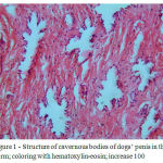 |
Figure 1: Structure of cavernous bodies of dogs’ penis in the norm; coloring with hematoxylin-eosin; increase 100
|
Figure 2. A hematoxylin and eosin-stained cross section of cavernous bodies in the dog penis prepared 14 days after introduction of a magnesium rod
Edema and sparse lymphohistiocytic and perivascular lymphoid infiltration are observed.
Magnification, 100´
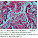 |
Figure 2: Cavernous body of a dog’ penis in 14 days after introduction of a magnesium rod; edema and scanty lymphohistiocytic and perivascular lymphoid infiltration; coloring with hematoxylin-eosin; increase 100
|
Figure 3. A hematoxylin and eosin-stained cross section of cavernous bodies in the dog penis prepared 14 days after introduction of a magnesium rod
Edema has separated the fibers in the tissues beneath the multilayered flat keratinizing epithelium to separate. Magnification, 100´
Thickening of cavernous tissue between the trabecular partitions was observed in one animal. Vascular cavities, some expanded and some elongated, without an endothelial layer were found in the same tissue (Figure 3).
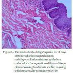 |
Figure 3: Cavernous body of dogs’ a penis in 14 days after introduction magnesium rod; multilayered flat keratinizing epithelium under which the separation of fibres of tissue elements owing to edema is visible; coloring with hematoxylin-eosin; increase 100 thickened fibrosised partitions. In a lumen of separate cavities there are parietal calcifications (Figure 7).
|
Adjacent to the central portion of the penis, multilayered flat epithelium with acanthosis, parakeratosis, and focal hypoplasia was observed. In the cavernous tissue, many vascular cavities of various diameters, some enlarged and some shrunken, were visible. Occasionally, erythrocytes were also observed. Spaces in between the trabecular partitions were wide and fibrosed with small hemorrhagic foci (Figure 4).
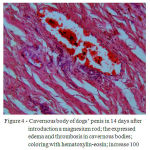 |
Figure 4: Cavernous body of dogs’ penis in 14 days after introduction a magnesium rod; the expressed edema and thrombosis in cavernous bodies; coloring with hematoxylin-eosin; increase 100
|
Perivascular edema surrounded the stromal vessels, with a lining of peripheral erythrocytes in the lumina. Small parietal red thrombi were visible within individual caves (Figure 5).
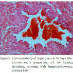 |
Figure 5: Cavernous body of dogs’ penis in 14 days after introduction a magnesium rod; the forming thrombus; coloring with hematoxylin-eosin; increase 100
|
In the group 2 dogs, 28 days after insertion of the magnesium rod, a multilayered flat epithelium with moderate basal cell hyperplasia and parakeratosis was observed in the central portion of the penis corresponding to the site of rod insertion. The connective tissue stroma was also fibrotic with thick fibers (Figure 6).
The cavernous tissue consisted of round vascular cavities divided by thick fibrotic partitions. The lumina of these cavities contained parietal calcifications (Figure 7).
Figure 4. A hematoxylin and eosin-stained cross section of cavernous bodies in the dog penis prepared 14 d after introduction of a magnesium rod
Edema and thrombosis are observed within the cavernous bodies.
Magnification, 100´
Figure 5. A hematoxylin and eosin-stained cross section of cavernous bodies in the dog penis prepared 14 d after introduction of a magnesium rod
Developing thrombi are observed within the cavernous bodies. Magnification, 100´
Figure 6. A hematoxylin and eosin-stained cross-section of cavernous bodies in the dog penis prepared 28 days after introduction of a magnesium rod.
The walls of the cavernous bodies have thickened by the growth of coarse connective tissue fibers.
Magnification, 100´
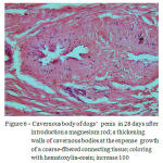 |
Figure 6: Cavernous body of dogs’ penis in 28 days after introduction a magnesium rod; a thickening walls of cavernous bodies at the expense growth of a coarse-fibered connecting tissue; coloring with hematoxylin-eosin; increase 100
|
Figure 7. A hematoxylin and eosin-stained cross section of cavernous bodies in the dog penis prepared 28 days after introduction of a magnesium rod
A dystrophic calcification is observed in the wall of a cavity in the region where the rod was introduced. Magnification, 100´
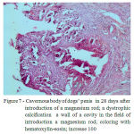 |
Figure 7: Cavernous body of dogs’ penis in 28 days after introduction of a magnesium rod; a dystrophic calcification a wall of a cavity in the field of introduction a magnesium rod; coloring with hematoxylin-eosin; increase 100
|
Conclusions
On the basis of our results, we conclude that magnesium rods cause sclerosis of cavernous bodies in experimental models of hemangioma in the dog penis. Nonspecific inflammation, including fibrinoid swelling (homogenous focal changes), occasional stromal edema, and loosening of connective tissue fibers, was observed 14 days after introduction of the magnesium rod into the corpus carvenosum. Vascular cavities of various diameters, some enlarged and some shrunken, were observed, with a few containing erythrocytes. Spaces between the trabecular partitions were wide and fibrosed, with small hemorrhagic foci. Developed and developing thrombi were noted in the majority of cavities. Twenty-eight days after introduction of the metal rod, a fibrosed connective tissue stroma with infiltration of fibrocytes and fibroblasts was observed. The cavernous tissue also contained round vascular cavities divided by thick fibrotic partitions. The lumina of individuals cavities contained parietal calcifications indicative of sclerosis in the cavernous bodies. These findings indicate the effectiveness of sclerotherapy using magnesium-based metal rods in the treatment of cavernous hemangiomas in the dog penis.
References
- Safina G.G. Surgicaltattoo of the capillary (planar) hemangiomas; – Kazan, 1982. – p. 17
- Yermuhanova G.T., Solovyev M.M., Supiev T.K., Colycheva NI., Galyapin A.S. way of modeling hemangioma. Author’s certificate # 1638720 from December 1, 1990.
- Zapadnyuk I.P., Zapadnyuk V.I., Zahariya K.A. Laboratory animals, their breeding, maintenance and use in the experiment. – Kiev: Vishcha school, 1983. p. 349
- General pathology of human: Guide for physicians / edited by A.I. Strukova, V.V. Serova, D.S. Sarkisova: V2 – 2nd edition, revised and updated – AMS of USSR. – M: Medicine, 1990. – p. 416
- Pathoanatomical diagnosis of human tumors: V1 / edited by N.A. Krayevski, A.V. Smolyannikova, D.S. Sarkisova – 4th edition, revised and updated – M: Medicine, 1993. – p. 560
- Berlov G.A. Histological diagnosis of major human tumors (Practical Histology). Minsk, “Belarus”, 1970. – p. 328

