
J. Seetha1 and S. Selvakumar Raja2
1Department of Computer Science and Engineering, Sathyabama University, Chennai, India.
2Kakatiya Institute of Tech and Science for Women, Nizamabad-503 003. Telangana, India.
Corresponding Author E-mail: selvakumarraja1968@gmail.com
DOI : https://dx.doi.org/10.13005/bpj/1511
Abstract
The brain tumors, are the most common and aggressive disease, leading to a very short life expectancy in their highest grade. Thus, treatment planning is a key stage to improve the quality of life of patients. Generally, various image techniques such as Computed Tomography (CT), Magnetic Resonance Imaging (MRI) and ultrasound image are used to evaluate the tumor in a brain, lung, liver, breast, prostate…etc. Especially, in this work MRI images are used to diagnose tumor in the brain. However the huge amount of data generated by MRI scan thwarts manual classification of tumor vs non-tumor in a particular time. But it having some limitation (i.e) accurate quantitative measurements is provided for limited number of images. Hence trusted and automatic classification scheme are essential to prevent the death rate of human. The automatic brain tumor classification is very challenging task in large spatial and structural variability of surrounding region of brain tumor. In this work, automatic brain tumor detection is proposed by using Convolutional Neural Networks (CNN) classification. The deeper architecture design is performed by using small kernels. The weight of the neuron is given as small. Experimental results show that the CNN archives rate of 97.5% accuracy with low complexity and compared with the all other state of arts methods.
Keywords
Brain Tumors; Computed Tomography; Magnetic Resonance
Download this article as:| Copy the following to cite this article: Seetha J, Raja S. S. Brain Tumor Classification Using Convolutional Neural Networks. Biomed Pharmacol J 2018;11(3). |
| Copy the following to cite this URL: Seetha J, Raja S. S. Brain Tumor Classification Using Convolutional Neural Networks. Biomed Pharmacol J 2018;11(3). Available from: http://biomedpharmajournal.org/?p=22844 |
Introduction
Brain tumor is one of the vital organs in the human body, which consists of billions of cells. The abnormal group of cell is formed from the uncontrolled division of cells, which is also called as tumor. Brain tumor are divided into two types such low grade (grade1 and grade2) and high grade (grade3 and grade4) tumor. Low grade brain tumor is called as benign. Similarly, the high grade tumor is also called as malignant. Benign tumor is not cancerous tumor. Hence it doesn’t spread other parts of the brains. However the malignant tumor is a cancerous tumor. So it spreads rapidly with indefinite boundaries to other region of the body easily. It leads to immediate death.12
Brain MRI image is mainly used to detect the tumor and tumor progress modeling process. This information is mainly used for tumor detection and treatment processes.MRI image gives more information about givenmedical image than the CT or ultrasound image. MRI image provides detailed information about brain structureand anomaly detection in brain tissue.Actually, Scholars offered unlike automated methods for brain tumors finding and typecataloging using brain MRI images from the time when it became possible to scan and freight medical imagesto the computer. Conversely, Neural Networks (NN) and Support Vector Machine (SVM) are theusually used methods for their good enactment over the most recent few years.11 However freshly, Deep Learning (DL) models fixed a stirring trend in machine learning as the subterranean architecturecan efficiently represent complex relationships without needing a large number of nodes likein the superficial architectures e.g. K-Nearest Neighbor (KNN)and Support Vector Machine (SVM).Consequently, theygrew quickly to become the state of the art in unlike health informatics areas for examplemedical image analysis, medical informatics andbioinformatics.
Related works
In,1 the Fuzzy C-Means (FCM) segmentation is applied to separate the tumor and non-tumor region of brain. Also wavelet feature are extracted by using multilevel Discrete Wavelet Transform (DWT). Finally, Deep Neural Network (DNN) is incorporated for brain tumor classification with high accuracy. This technique is compared with KNN, Linear Discriminant Analysis (LDA) and Sequential Minimal Optimization (SMO) classification methods. An accuracy rate of 96.97% in the analysis of DNN based brain tumor classificationBut the complexity is very high and performance is very poor.
In,2 a novel bio-physiomechanical tumor growth modeling is presented to analyze the step by steps tumor growth of patients. It will be applied for gliomas and solid tumor with individual margins to seizure the significant tumor mass effect. The discrete and continuous methods are combined to make a tumor growth modeling. The proposed schemeprovides the likelihood to tacitly segmenttumor-bearing brain images based on atlas-based registration. This technique is mainly used for brain tissue segmentation.But the computation time is high.
In,3 new multi-fractal (MultiFD) featureextraction and improved AdaBoost classification schemes are used to detect and segment the brain tumor. The texture of brain tumor tissue is extracted by using MultiFD feature extraction scheme. The improved AdaBoost classification methods are used to find the given brain tissue is tumor or non-tumor tissue. Complexity is high.In,4 local independent projection-based classification (LIPC) method is used to classify the voxel of the brain. Also path feature is extracted in this method. Hence no need to perform explicit regularization in LIPC. The accuracy is low. In,5 a seeded tumor segmentation method with new CellularAutomata (CA) technique is presented, which is compared with graph cut based segmentation method. The seed selection and Volume OfInterest (VOI) is calculated for efficient brain tumor segmentation. Also tumor cut segmentation is incorporated into this work. The complexity is low. But the accuracy is low.
In,6 new brain tumor segmentation is introduced, which is also called multimodal brain tumor segmentation scheme. Also combing different segmentation algorithm in order to achieve high performance than the existing method. But the complexity is high. In,7 the survey of brain tumor segmentation is presented. Discuss about Various segmentation methods such as Region based segmentation, threshold based segmentation, fuzzy C Means segmentation, Atlas based segmentation, Margo Random Field (MRF) segmentation, deformable model, geometric deformable model, The accuracy, robustness, validity are analyzed for all the methods. In,8 hybrid feature selection with ensemble classification is applied for brain tumor diagnosis process. The GANNIGMAC, decision Tree, Bagging C based wrapper approach is used to obtain the decision rules. Also simplify the decision rules by using hybrid feature selection, which contains the combination of (GANNIGMAC + MRMR C+ Bagging C + Decision Tree).
In,9 the fuzzy based control theory is used for brain tumor segmentation and classification method. The Fuzzy Interference System (FIS) is a one special technique, which is mainly used for brain segmentation. Supervised classification is used to create a membership function of fuzzy controller. The performance is high and accuracy is low. In,10 the adaptive histogram equalization is used to improve the contrast of the image. Then Fuzzy CMeans (FCM) based segmentation is performed to separate the tumor from the whole brain image. After that Gabor feature are extracted to filter the abnormal cells of brain. Finally, the fuzzy with K Nearest Neighbor (KNN) classification is applied to find the abnormality of brain MRI image. The complexity is high. But the accuracy is low. In this work, a novel automatic brain tumor classification is performed by convolutions neural network.
Proposed System
The human brain is modeled by using design and implementation of neural network. The neural network is mainly used for vector quantization, approximation, data clustering, pattern matching, optimization functions and classification techniques. The neural network is divided into three types based on their interconnections.Three type neural networks are feedback, feed forward and recurrent network. The Feed Forward Neural network is further divided into single layer network and multilayer network. In the single layer network, the hidden layer is not presented. But it contains only input and output layer. However, the multilayer consists of input layer, hidden layer and output layer. The closed loop based feedback network is called as recurrent network.
 |
Figure 1: Block diagram of proposed brain tumor classification using CNN.
|
In the normal neural network, image cannot scalable. But in convolution neural network, image can scalable (i.e) it will take 3D input volume to 3D output volume (length, width, height).The Convolution Neural Network (CNN) consists of input layer, convolution layer, Rectified Linear Unit (ReLU) layer, pooling layer and fully connected layer. In the convolution layer, the given input image is separated into various small regions. Element wise activation function is carried out in ReLU layer. Pooling layer is optional. We can use or skip. However the pooling layer ismainly used for down sampling. In the final layer (i.e) fully connected layer is used to generate the class score or label score value based on the probability in-between 0 to 1.
The block diagram of brain tumor classification based on convolution neural network is shown in fig.1. The CNN based brain tumor classification is divided into two phases such as training and testing phases. The number of images is divided into different category by using labels name such as tumor and non-tumor brain image…etc. In the training phase, preprocessing, feature exaction and classification with Loss function is performed to make a prediction model. Initially, label the training image set. In the preprocessing image resizing is applied to change size of the image.
Finally, the convolution neural network is used for automatic brain tumor classification. The brain image dataset is taken from image net. Image net is a one of the pre-trained model. If you want to train from the starting layer, we have to train the entire layer (i.e) up to ending layer. So time consumption is very high. It will affect the performance. To avoid this kind of problem, pre-trained model based brain dataset is used for classification steps. In the proposed CNN, we will train only last layer in python implementation. We don’t want to train all the layers. So computation time is low meanwhile the performance is high in the proposed automatic brain tumor classification scheme.
The loss function is calculated by using gradient descent algorithm. The raw imagepixel is mapping with class scores by using a score function. The quality of particular set of parameters is measured by loss function. It is based on how well the induced scores approved with the ground truth labels in the training data. The loss function calculation is very important to improve the accuracy. If the loss function is high, when the accuracy is low. Similarly, the accuracy is high, when the loss function is low. The gradient value is calculated for loss function to compute gradient descent algorithm. Repeatly evaluate the gradient value to compute the gradient of loss function.
Algorithm for CNN based Classification
Apply convolution filter in first layer
The sensitivity of filter is reduced by smoothing the convolution filter (i.e) subsampling
The signal transfers from one layer to another layer is controlled by activation layer
Fasten the training period by using rectified linear unit (RELU)
The neurons in proceeding layer is connected to every neuron in subsequent layer
During training Loss layer is added at the end to give a feedback to neural network
Results and Discussion
Our Dataset contains tumor and non-tumor MRI images and collected from different online resources. Radiopaedia13 contains real cases of patients, tumor images were obtained from Radiopaedia and Brain Tumor Image Segmentation Benchmark (BRATS) 2015 testing dataset.14
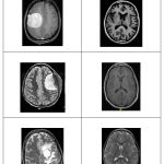 |
Figure 2: CNN based classified results
|
In this work, efficient automatic brain tumor detection is performed by using convolution neural network. Simulation is performed by using python language. The accuracy is calculated and compared with the all other state of arts methods. The training accuracy, validation accuracy and validation loss are calculated to find the efficiency of proposed brain tumor classification scheme. In the existing technique, the Support Vector Machine (SVM) based classification is performed for brain tumor detection. It needs feature extraction output. Based on feature value, the classification output is generated and accuracy is calculated. The computation time is high and accuracy is low in SVM based tumor and non-tumor detection.
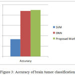 |
Figure 3: Accuracy of brain tumor classification.
|
In the proposed CNN based classification doesn’t require feature extraction steps separately. The feature value is taken from CNN itself. In fig.2. shows the classified result of Tumor and Non-tumor brain image. Hence the complexity and computation time is low and accuracy is high. The output of brain tumor classification accuracy is given in fig.3. Finally, the classification results as Tumor brain or non-tumor brain based on the probability score value. The normal brain image has the lowest probability score. Tumor brain has highest probability score value, when compared to normal and tumor brain.
Conclusion
The main goal of this research work is to design efficient automatic brain tumor classification with high accuracy, performance and low complexity. In the conventional brain tumor classification is performed by using Fuzzy C Means (FCM) based segmentation, texture and shape feature extraction and SVM and DNN based classification are carried out. The complexity is low. But the computation time is high meanwhile accuracy is low. Further to improve the accuracy and to reduce the computation time, a convolution neural network based classification is introduced in the proposed scheme. Also the classification results are given as tumor or normal brain images. CNN is one of the deep learning methods, which contains sequence of feed forward layers. Also python language is used for implementation. Image net database is used for classification. It is one of the pre-trained models. So the training is performed for only final layer. Also raw pixel value with depth, width and height feature value are extracted from CNN. Finally, the Gradient decent based loss function is applied to achieve high accuracy. The training accuracy, validation accuracy and validation loss are calculated. The training accuracy is 97.5%. Similarly, the validation accuracy is high and validation loss is very low.
References
- Mohsen H et al. Classification using Deep Learning Neural Networks for Brain Tumors. Future Computing and Informatics. 2017:1-4.
- Bauer S. et al. Multi scale Modeling for Image Analysis of Brain Tumor Studies. IEEE Transactions on Biomedical Engineering. 2012;59:1.
CrossRef - Islam A. et al. Multi-fractal Texture Estimation for Detection and Segmentation of Brain Tumors. IEEE.
- Huang M et al. Brain Tumor Segmentation Based on Local Independent Projection-based Classification. IEEE Transactions on Biomedical Engineering. 2013.IEEE.
- Hamamci A et al. Tumor-Cut Segmentation of Brain Tumors on Contrast Enhanced MR Images for Radiosurgery Applications. IEEE Transactions on Medical Imaging. 2012;31:3.
CrossRef - Bjoern H. M et al. The Multi modal Brain Tumor Image Segmentation Benchmark (BRATS). IEEE Transactions on Medical Imaging.
- Liu J et al. A Survey of MRI-Based Brain Tumor Segmentation Methods. TSINGHUA Science and Technology. 2011;19;6.
- Huda S et al. A Hybrid Feature Selection with Ensemble Classification for Imbalanced Healthcare Data A Case Study for Brain Tumor Diagnosis. IEEE Access. 2017;4.
- Karuppathal and Palanisamy V. Fuzzy based automatic detection and classification approach for MRI-brain tumor. ARPN Journal of Engineering and Applied Sciences. 2014;9;12.
- Janani and Meena P. image segmentation for tumor detection using fuzzy inference system. International Journal of Computer Science and Mobile Computing. 2013;2(5):244–248.
- Pereira S et al. Brain Tumor Segmentation using Convolutional Neural Networks in MRI Images. IEEE Transactions on Medical Imaging.
- Zhang J et al. Brain Tumor Segmentation Based on Refined Fully Convolutional Neural Networks with A Hierarchical Dice Loss, Cornell university library computer vision and pattern recognition. 2018.
- [Radiopaedia] http:// radiopedia.org.
- [BRATS 2015] https://www.smir.ch/BRATS/Start 2015.

