
Priyadarshini Choudhury1, N. Gnanasundaram2 and Atul Anand Bajoria3
1Department of Oral Medicine and Radiology, Kalinga Institute of Dental Sciences, Kalinga Institute of Industrial Technology deemed to be University.
2Sri Ramachandra Medical College and Research Institute.
3Department of Oral Medicine and Radiology Kalinga Institute of Industrial Technology deemed to be University.
Corresponding Author E-mail: priyajuly14@gmail.com
DOI : https://dx.doi.org/10.13005/bpj/1393
Abstract
Endemic fluorosis is a disease of toxic manifestation of fluoride affecting teeth, bone and other soft tissue structure. Literature reviews voluminous knowledge of dental fluorosis and skeletal fluorosis but dearth of knowledge is available regarding non-skeletal fluorosis. The aim of this research is to establish the magnitude of toxic problems of dental, skeletal and non-skeletal fluorosis and whether calcium reduces the fluoride toxicity. Methodology: A total of eight colony bred rabbits (Oryctolagus Cuniculus) were divided into three groups; group I – control group, group II – rabbits treated with 10mg/kg body wt of sodium fluoride and group III – rabbits treated with 10 mg/kg body wt of sodium fluoride and 250 mg of calcium. Histopathological study of tooth revealed core tissue with central capillaries which were calcified and outer enamel showed concentric deposits of calcium in the form of ring like bluish and black granules. Bone in fluoride fed animal showed amorphous, sprinkling of blackish blue granular deposits with in the osteoids, occupied 20-0% of bone area. The liver tissue of animals fed with 10mg/kg body wt showed periportal inflammation with lymphocytic infiltration, ballooning degeneration with lysosomal granules in cytoplasmic cells. In this research, it is found that mere fluoride ingestion causes bone changes, tooth changes and liver changes but ingestion of fluoride 10mg/kg body weight and 250 mg of calcium do not produce any change in the tissues. This shows that calcium is beneficial and nullifies the action of fluoride.
Keywords
Fluoride; Rabbits; Calcium and Dental Fluorosis
Download this article as:| Copy the following to cite this article: Choudhury P, Gnanasundaram N, Bajoria A. A. Fluoride Toxicity in Rabbits and The Role of Calcium in Prevention of Fluoride Toxicity. Biomed Pharmacol J 2018;11(1). |
| Copy the following to cite this URL: Choudhury P, Gnanasundaram N, Bajoria A. A. Fluoride Toxicity in Rabbits and The Role of Calcium in Prevention of Fluoride Toxicity. Biomed Pharmacol J 2018;11(1). Available from: http://biomedpharmajournal.org/?p=19249 |
Introduction
Fluoride exerts its effect on caries and on dental fluorosis by distinct mechanisms. The action of fluoride is not only local in the oral cavity but also systemic. It is the blood fluoride concentration that influences the metabolic variables and vice versa.1,2 For example, enamel formation is a continuous process and that the clinically visualized defects are dependent on the thickness of enamel formed under exposure to a given fluoride dose. Thus, the plasma fluoride concentration moderates the fluorosis in developing dentition.3,4,5 The systemic effect of fluoride and the average time of exposure of ameloblasts to fluoride determines dental fluorosis.6,7 The aim of this research is to evaluate the magnitude of dental, skeletal and non-skeletal fluorosis with the action of calcium and fluoride to reduce fluoride toxicity.
Materials and Method
A total number of eight colony bred rabbits (Oryctolagus Cuniculus) from different mothers were used of which 6 were males and 2 were females. The weights of the rabbits ranged from 500 grams to 1000 grams and were 6 to 8 months of age.
These rabbits were divided into 3 groups.
Group I: Control group – two animals were given prescribed diet.
Group II: Study group – four animals were fed with prescribed diet and oral administration of 10mg /kg body weight of sodium fluoride (NaF) daily for 6 months.
Group III: Study group – two animals were fed with prescribed diet and oral administration of 10mg/kg body weight of NaF with 250mg of calcium daily for 6 months.
Rabbit’s Diet
Rabbits were fed with lab animal feed for rabbits (ration computed and supplied by TANUVAS – Veterinary University, Chennai-35) in the mornings. Green grass CO-3 was fed in the evenings. Additionally fresh carrots and cabbage leaves were fed 2-3times a weak.
Fluoride and Calcium
The fluoride used in the research for ingestion in to the rabbits to find out fluoride toxicity in different organs is sodium fluoride powder prepared by S.D fine chem limited, Mumbai- 30. Calcium that is used in the research for ingestion in to the rabbits was a calcium syrup prepared by Elders pharmaceuticals.
Methodology
Animals were preserved in the cage and normal diet was given daily. Fluoride 10mg/kg body wt to the animals and fluoride 10mg/kg body wt with 250 mg calcium were given orally with syringe. Sodium fluoride stock solution was taken in a 2ml syringe according to the weight of the animal and fed by inserting the syringe in the lateral diastema of rabbit’s mouth. Calcium was fed with a 5 ml syringe by inserting the syringe in the lateral diastema of rabbit’s mouth. This procedure was performed for 180 days.
At the end of 180 days animals were clinically examined for structural and functional changes. Blood was drawn by heart puncture for estimation of Hemoglobin, RBC and WBC count. Then animals were sacrificed by using an overdose of ketamin and xylazene. Vertical incisions were made in the ventral aspect of the control and experimental animals to expose the thorax and abdomen. Then the skeletal and non-skeletal tissues and teeth were studied macroscopically.
After studying the macroscopical appearance of teeth, bone and other organs, soft tissues of stomach, intestine, liver, kidney, lung, artery, vein, ligament, muscle and teeth were collected for histopathological study with eosin and hematoxillin.
Results
Rabbits were used as an animal model to evaluate the fluoride toxicity in tooth, bone, and non-skeletal soft tissue was carried out for a period of 180 days continuously giving 10mg/kg body wt of oral ingestion of fluoride daily to the animal. Observations were also made among the experimental animals fed with 10 mg/kg body wt of NaF with 250mg of calcium. The observation of animals fed with NaF and animals fed with fluoride and calcium were made comparing the control animals. At the end of 180 days the animals were examined for toxic manifestation both clinically and histopathologically.
Clinical Observation
The weight of the control animal and experimental animal were recorded at the end of the experiment. It was found that weight of the each experimental animal with 10mg/kg body wt was slightly less than the control group of animals (Table 1). Animal activities like walking, moving the head, moving the limbs were slightly restricted than compared to the control animals. On examination of teeth, there was no brown discoloration in the experimental animal fed with NaF but mild opaque color was noticed with area of destruction in few teeth. All the above recorded changes were found in those animals fed with 10 mg NaF / kg body weight fluoride, daily. On the contrary, examination of experimental animals fed with 10mg/kg body wtNaF and 250mg calcium revealed no changes in loss of weight, restricted movement and tooth changes.
Table 1: Weight of the animals in kilograms.
| Before Experiment | After Experiment | |
| Control | 1.045 | 1.734 |
| Experimental animals fed with NaF-10mg/Kg body weight | A-1.039
B-0.833 C-1.387 D-1.341 |
A-1.531
B-1.261 C-1.552 D-1.709 |
| Experimental animals fed with NaF-10mg/Kg body weight and calcium | M-1.007
N-1.149 |
M-1.673
N-1.775 |
Histological Changes
Control and experimental animals were sacrificed by injecting xylazene and ketamin. On exposure to thorax and abdomen morphological examination of organs were made. No change with reference to size, shape, color in any of the organs of the animals fed with 10mg/kg body wt NaF and 10mg / kg body wt NaF and 250mg calcium were seen.(Figure 1& 2).
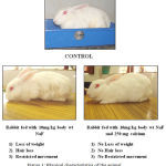 |
Figure 1: Physical characteristics of the animal
|
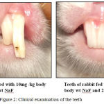 |
Figure 2: Clinical examination of the teeth
|
Tissues were collected from bone, teeth, gastric mucosa, intestinal mucosa, liver, kidney, artery, vein, lung, ligament, muscle along with bone and teeth. The structural changes in the gastric mucosa were observed with a magnifying glass. (Figure 3 to 6) The gastric mucosa in experimental animals fed with 10 mg /kg body wt showed mucosal congestion, slight erythema and mild erosion. Cut section of artery and vein showed no changes in the wall of the vessel. Intestinal mucosa had no alteration in the lumen on examination. The collected specimens from soft tissues were subjected to histopathological examination with eosin and hematoxilin stain and the observation were seen under microscope.
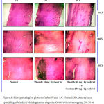 |
Figure 3: Histopathological picture of rabbit bone. 3A. Normal 3B. Amorphous sprinkling of blackish bluish granular deposits. Osteiod tissue occupying 20- 30 % of bone. 3C. No changes seen in animal fed with Fluoride and Calcium
|
Major changes were seen in the liver. The tissues of control animal showed normal architecture and normal cytological appearance. In the experimental animals fed with 10mg/kg body wt NaF, periportal inflammation sparse lymphocytes and hepatocytes, moderate lysosomal granules, ballooning degeneration were seen in the liver. On the other hand, animals fed with sodium fluoride 10mg/kg body weight and 250 mg calcium showed normal architecture with no periportal inflammation, scanty cytoplasm granules and absence of ballooning degeneration of hepatocytes. (Figure 4)
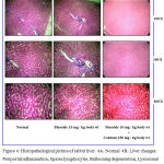 |
Figure 4: Histopathological picture of rabbit liver. 4A. Normal 4B. Liver changes: Periportal inflammation, Sparse lymphocytes, Ballooning degeneration, Lysosomal granules are seen in Fluoride fed animal. 4C. No changes seen in animal fed with Fluoride and Calcium
|
Bone Changes
Control animal showed no deposits in the bone and the bone structure was normal. In animals fed With Sodium Fluoride-10mg/Kg Body Weight, the Section of bone showed amorphous sprinkling of blackish blue granular deposits within the osteoid. These deposits were patchy and occupied 20% to 30% of the bone area. The marrow spaces were normal. These findings clearly indicates that the structure of bone reacts abnormally with the fluoride action. The presence of 20 to 30% of osteoid tissue in the structure of bone suggests that the mineralization of the bone was affected in presence of excess fluoride in the body. These changes in the structure of bone were not seen in those animals fed with NaF 10mg/kg body wt along with 250mg of calcium. (Figure 3)
Tooth Changes
In control animals, the tooth was normal. However, in animals fed with sodium fluoride-10mg/kg body weight, the central capillaries in the teeth were calcified. The outer enamel showed concentric deposits of calcium in the form ring of bluish black granules. From the above observation it is clear that fluoride disturbs the calcified area making irregular calcifications and calcified capillaries. But these type of alterations were not recorded in the tooth of animals fed with 10mg /kg body wt of sodium fluoride and calcium. (Figure 5)
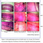 |
Figure 5: Histopathological picture of rabbit teeth. 5A. Normal 5B. Presence of concentric deposits of calcium in the form of ring like bluish black granules. Core shows central capillaries are calcified. 5C. No changes seen in animal fed with Fluoride and Calcium
|
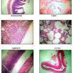 |
Figure 6: Histopathological picture of other organs of the rabbit.
|
Discussion
In the present study, the clinical parameters such as weight loss, reduction of movement, hair loss and changes in the teeth were present in those animals fed with 10 mg NaF / kg body weight fluoride. On the contrary, the experimental animals fed with NaF and calcium showed no changes in the above mentioned parameters. Paul et al8 in their study found inhibitory action of NaF on motor activity along with diminished activity of AchE in the central nervous system. This could be attributed to the lack of motor movement in NaF treated animals. Mullenix et al9 found similar results in their study as well.
Opit et al10 found out that in NaF treated animals, protein synthesis was inhibited by fluoride by blocking the Na-K activated ATPase pump. Also the activity of the AchE was inhibited by fluoride leading to muscle weakness and poor activity at the neuromuscular junction. Susheela et al11 in their study found substantial increase in the serum fluoride level on oral intake of NaF which could be due to absorption of fluoride in the gastrointestinal tract. On the other hand Schiffl et al12 in their study found elevated fluoride serum levels resulting from decreased urinary excretion of fluoride.
In the present study, the gastric mucosa of the animals fed with 10 mg /kg body wt NaF showed mucosal congestion, slight erythema and mild erosion. While those fed with NaF and Calcium showed no changes in the gastric mucosa. Das et al13 in their study found atrophy of the gastric mucosa in NaF treated animals.
In the present study animals fed with NaF only showed significant histopathological changes in the liver and bone tissues when compared to the control animals. In the present study, the animals fed with NaF only showed reduction in Hemoglobin values and the total WBC count. While those fed with NaF and Calcium showed no changes in the haematological parameters.
Boink et al14 and Farley et al15 in their study found significant hypocalcemia in NaF treated animals. Fluorapatite is poorly soluble form of calcium causing hypocalcaemic states. Calcium prevents absorption of fluoride from the gastrointestinal tract. Thus if calcium supplements are given along with NaF, hypocalcaemic state can be countered.
Chinoy and Sequeria16 in their study have concluded that in animals treated with NaF, there is depletion of body protein along with accumulation of glycogen in the body. This could be an indication for the changes in the locomotory activity of NaF treated animals. However, in animals treated with calcium supplements and NaF, no depletion of locomotor behaviour or any other parameter was observed. This proves the fact that calcium supplements help to decrease the fluoride burden in the body thus counteracting the toxicity of excess fluoride in the body.
In the present study, the animals fed with NaF only demonstrated concentric deposits of calcium in the form of ring like bluish black granules in the outer enamel layer. However these findings were not consistent with the animals fed with NaF and calcium.
Ekstrand & Oliveby17 in their study found out that low salivary calcium levels occurring due to NaF consumption caused dental lesions in NaF treated animals. Shupe et al18 concluded that the dental lesions may be the reason for animals to inadequately masticate and swallow the food causing weight loss. It has been proved that the biochemical, behavioural and the dental impairments can be reverted back significantly after 2 months of fluoride exposure.
Trivedi et al19 have reported reversal of endemic fluorosis after changing fluoride containing drinking water. Catani et al20 in their study found that animals chronically exposed to symmetrically oscillating fluoride doses can cause dental fluorosis.
Conclusion
In conclusion, it is found that adding calcium along with fluoride is beneficial to nullify the action of fluoride toxicity as evidenced by the fact of non involvement of toxic manifestations in the tissues in the animals fed with calcium and fluoride. The present study gives support to the epidemiological observation that increased fluoride concentration in the body causes deposition of fluoride within the tooth which may lead to dental fluorosis.
Acknowledgement
First and foremost I thank the Almighty for all His mercy and blessings bestowed throughout my life and career.
It is with supreme humility, I express my sincere thanks and heartfelt gratitude to my most respected Professor and Head of the Department, Dr. N.Gnanasundaram, M.D.S., for his constant encouragement, unrelenting support and valuable guidance rendered in the preparation of this Dissertation.
My sincere and heartfelt thanks to the Director Dr.B.Jayachandran Dare, MVSc,Ph.D, and Mr.Praveen M.Sc, of Biomedical Research unit and Laboratory Animal Center Saveetha University, Chennai for their guidance and cooperation throughout the study.
I also express my sincere thanks to my teacher Dr. M Arvind & my dear friends Dr.Sangeetha D and Dr. Atul Anand Bajoria for their constant cooperation, support and encouragement.
I am thankful to my Madam Dr. Meera Govindarajan, M.D., Pathologist and the other staff members for the guidance, cooperation in providing laboratory facilities for me.
Conflict of Interest
There is no conflict of interest
References
- Cury J. A., Tenuta L. M. How to maintain a cariostatic fluoride concentration in the oral environment. Adv Dent Res. 2008;20:13-16.
CrossRef - Bronckers A. L., Lyaruu D. M., DenBesten P. K. The impact of fluoride on ameloblasts and the mechanisms of enamel fluorosis. J Dent Res. 2009;88:877-893.
CrossRef - Ekstrand J. Fluorides metabolism. In: Fluoride in dentistry. Fejerskov O., Ekstrand J., Burt B (Editors). Copenhagen: Munksgaard. 1996;55-68.
- Angmar-Månsson B., Whitford G. M. Plasma fluoride levels and enamel fluorosis in the rat. Caries Res. 1982;16:334-339.
CrossRef - Aoba T., Fejerskov O. Dental fluorosis chemistry and biology. Crit Rev Oral Biol Med. 2002;13:155-170.
CrossRef - Angmar-Månsson B., Whitford G. M. Enamel fluorosis related to plasma F levels in the rat. Caries Res. 1984;18:25-32.
CrossRef - Catani D. B., Hugo F. N., Cypriano S., Sousa M. L. R., Cury J. A. Relationship between fluoride levels in the public water supply and dental fluorosis. Rev Saúde Pública. 2007;41:732-739.
CrossRef - Paul V.,Ekambaram P & Jayakumar A. R. Effects of NaF on locomotor behaviour and a few biochemical parameters in rats. Env. Toxicol. Pharmacol. 1998;6:187–191.
CrossRef - Mullenix P. J., Denbesten P. K. , Schunior A. M & Kernan W. J. Neurotoxicity of NaF in rats. Neurotoxicol. Teratol. 1995;17:169–177.
CrossRef - Opit L. J., Potter H & Charnock J. S. The effect of anions on Naπ Kπ activated ATPase. Biochim. Biophys. Acta. 1966;120:159–162.
CrossRef - Susheela A. K. & Bhatnagar M. Fluoride toxicity: a biochemical and scanning electron microscopic study of enamel surface of rabbit teeth. Arch. Toxicol. 1993;67:573–579.
CrossRef - Schiffl H. H &Binswanger U. Human urinary fluoride excretion as influenced by renal functional impairment. Nephron. 1980;26:69–72.
CrossRef - Das T. K.,Susheela A. K.,Gupta I. P., Dasarathy S & Tandon R. K. Toxic effects of chronic fluoride ingestion on upper gastrointestinal tract. J. Clin. Gastroenterol. 1994;18:194–199.
CrossRef - Boink A. B., Wemer J., Meulenbelt J., Vaessen H. A & De D. J. W. The mechanism of fluoride-induced hypocalcemia. Hum. Exp. Toxicol. 1994;13:149–155.
CrossRef - Farley J. R., Wergedal J. E & Baylink D. J. Fluoride directly stimulates proliferation and alkaline phosphatase activity on bone forming cells. Science. 1983;222:330–332.
CrossRef - Chinoy N. J. & Sequeira E. Effects of fluoride on the histoarchitecture of reproductive organs of male mouse. Reprod. Toxicol. 1989;3:261–267.
CrossRef - Ekstrand J & Oliveby A. Fluoride in oral environment. Acta odont. scand. 1999;57:330–333.
- Shupe J. L., Olson A. E., Peterson H. B & Low J. B. Fluoride toxicosis in wild ungulates. J. Amer. Vet. Assoc. 1984;185:1295–1300.
- Trivedi N., Mithal A., Gupta S. K & Godbole M. M. Reversible impairment of glucose tolerance in patients with endemic fluorosis. Diabetologia. 1993;36:826–828.
CrossRef - Catani D. B., Tenuta L. M. A., Andaló F.. A and Cury J. A. Fluorosis in Rats Exposed to Oscillating Chronic Fluoride Doses. Braz Dent J. 2010;21(1):32-37.
CrossRef

