
Malinee Pongsavee
Department of Medical Technology, Faculty of Allied Health Sciences, Thammasat University, Rangsit Campus, Patumthani 12121, Thailand.
Corresponding Author E-mail: malineep@tu.ac.th
DOI : https://dx.doi.org/10.13005/bpj/1285
Abstract
Curcumin is a compound in Curcuma longa Linn. It has shown antioxidant and anti-inflammatory properties. Formaldehyde is the chemical compound with the formula H2CO. It is the toxic chemical substance and causes many types of cancer in human. The protective effects of curcumin in formaldehyde treated lymphocytes and cancer cells were investigated. The antimutagenic effect of curcumin was investigated in the formaldehyde treated lymphocytes. The antiproliferative effect of curcumin was investigated in cancer cells (MCF-7 cells, A549 cells and CRL-2724 cells). Role of curcumin in formaldehyde treated lymphocytes for SOD activity change was also studied. The formaldehyde concentrations of 0.036, 0.072, 0.15 mg/ml, 10 and 20 µM curcumin, Trypan blue exclusion assay, MTT assay, Giemsa staining, G-banding technique for chromosome investigation and SOD activity assay were used in this study. The results showed that curcumin has the protective effects of antimutagenicity and antiproliferation in formaldehyde treated lymphocytes and cancer cells. Curcumin effects SOD activity change in formaldehyde treated lymphocytes. Curcumin may apply in the therapy of diseases caused by the chemical substance toxicity.
Keywords
Curcumin; Cancer Cells; Formaldehyde Protective Effects; Lymphocytes; SOD Activity;
Download this article as:| Copy the following to cite this article: Pongsavee M. Protective Effects of Curcumin in Formaldehyde Treated Lymphocytes and Cancer Cells and its Role in Superoxide Dismutase Activity. Biomed Pharmacol J 2017;10(4). |
| Copy the following to cite this URL: Pongsavee M. Protective Effects of Curcumin in Formaldehyde Treated Lymphocytes and Cancer Cells and its Role in Superoxide Dismutase Activity. Biomed Pharmacol J 2017;10(4). Available from: http://biomedpharmajournal.org/?p=17860 |
Introduction
Curcumin is a natural, non-toxic compound of a plant. The Curcuma longa Linn has been traditionally used as a seasoning spice in Indian cuisine and also for the treatment of diseases because of its anti-inflammatory and antioxidant properties. It has been explained as a potent inducer of apoptosis and inhibitor of cell growth or proliferation in a variety of tumor cells. It serves as inhibitor of epidermal growth factor receptor signaling, angiogenesis, cellular migration, invasion and metastasis.1 It down regulates expression of several genes including protein kinase C.2 It inhibits progression of MCF-7 cells by inhibiting telomerase function.3
Formaldehyde is a chemical compound (H2CO) which exists as gas at room temperature. It is an intermediate in the oxidation of methane as well as other carbon compounds. It is sold as a saturated aqueous solution with concentration of 37 % formaldehyde, stabilized with 10 – 15 % methanol. It is found in exhausts of automobile and in smoke of tobacco. It has also been found useful as a disinfectant, production of polymers and other chemical substances. The formaldehyde solutions are applied topically to dry the skin, such as in the treatment of warts. It preserves or fixes tissue or cells by irreversibly cross-linking primary amine groups in proteins with other nearby nitrogen atoms in protein or DNA through a –CH2-linkage. It gets converted to formic acid in the body, leading to rise in blood acidity (acidosis). When combined with phenol, urea, or melamine, formaldehyde produces a hard thermoset resin and used in carpeting. The sign of short-term exposure to formaldehyde in humans are irritation of the eyes, nose and throat, together with concentration-dependent discomfort, lacrimation, sneezing, coughing, nausea, dyspnoea and finally death.4-5 Several studies of pathologist and other professionals exposed to formaldehyde have observed an increase risk of leukemia, with myeloid leukemia being most prominent in some studies, as well as modest associations with lymphoma. It affects hematotoxicity and alters the counts of different types of blood cells. It increases B cells but decreases CD3 and CD8.6
SODs are antioxidant enzymes that catalyze the dismutation of two superoxide radicals into hydrogen peroxide and oxygen. The products of these genes are thought to protect the brain, lungs, and other tissues from oxidative stress. Limitations on the abundance of tissue samples necessitate the use of animal models and cell lines in the studies of tumor-related phenomena. Cancer cell lines have been extensively used in screening studies involving drug sensitivity, effectiveness of anticancer drugs, migration capacity and ability to induce angiogenesis.
MCF-7 cell line is the hormone-responsive breast cancer cell line. It also has differential sensitivities to estrogens and anti-estrogens, differential expression of estrogen receptor (ER), ER mRNA, and progesterone receptor, and differences in tumorigenicity and proliferation rates. The characteristics of MCF-7 cells like the estradiol-dependence for growth and low metastatic potential has lead to the assumption that they represent an early epithelial adenocarcinoma of breast. A549 cells are adenocarcinomic human alveolar basal epithelial cells fall under the squamous subdivision of epithelial cells. CRL-2724 cell line is the leukemic cancer cell line. The MCF-7, A549 and CRL-2724 cell lines were used for study the antiproliferative effect of curcumin in this study.
Cancer causes significant morbidity and mortality and is a major public health problem worldwide. Toxic chemical substance such as formaldehyde and food preservative are involved in cancer progression.7 Plant derived compounds are known to have curative potential. The antimutagenic and antiproliferative effects of curcumin in normal cell induced by formaldehyde and cancer cells were the aim of this study. The alterations in the activity of SOD was studied in formaldehyde treated lymphocytes. This study indicated that curcumin has the protective effects and the potential for the development of modern medicine. It may be applied in the treatment of many diseases such as cancer caused by chemical substance toxic.
Materials and Methods
Blood Samples
Heparinized blood from 30 consenting healthy volunteers were collected. Thirty human lymphocyte cultures were cultured in RPMI medium (Gibco BRL Life Technologies, San Diego, CA, USA) plus 10% fetal calf serum (Invitrogen, Carlsbad, CA, USA) and penicillin-streptomycin antibiotic. Phytohemagglutinin was added in RPMI medium as the mitogen and incubated for 72 hrs. in 5% CO2 incubator.
The participants were given consent to donate blood for study the antimutagenic effect of curcumin (in Curcuma longa Linn.) on the formaldehyde treated lymphocyte research. This study was approved by Thammasat University ethic committee.
Cell lines
Human breast cancer cells (MCF-7), Human lung adenocarcinoma cells (A549) and Leukemic cancer cells (CRL-2724) were cultured in DMEM/F12 supplemented with 10% fetal calf serum, 2mM L-glutamine and antibiotics (100 µg/ml streptomycin and 50 U/ml penicillin) at 37°C in a humidified incubator containing 5% CO2 . Cells were trypsinized and sub-cultured at 1: 3 ratio for routine maintenance and experiments.
Studies on Antimutagenic Effect of Curcumin in Lymphocytes Induced by Formaldehyde
This study was divided into three groups. The first human lymphocyte culture group was the control group and did not add formaldehyde and curcumin in this group. The formaldehyde concentrations of 0.036, 0.072 and 0.15 mg/ml were added for 72 hrs. in the second human lymphocyte culture group. After 72 hrs. incubation, the first group and second group were harvested and did Giemsa staining and G-banding technique for chromosome observation. The formaldehyde concentrations of 0.036, 0.072 and 0.15 mg/ml were added for 72 hrs. in the third human lymphocyte culture group following 10 µM curcumin for 24 hrs. After 24 hrs. incubation, the third group was harvested and did Giemsa staining and G-banding technique for chromosome observation.
The antimutagenic effect of Curcumin (in Curcuma longa Linn.) on the formaldehyde treated lymphocytes was studied by ANOVA test. P<0.05 was considered to be statistically significant.
Studies on the Effect of Formaldehyde on Cancer cell Proliferation and the Antiproliferative Effect of Curcumin in Cancer Cells
Study the Effect of Formaldehyde on Cancer cell Proliferation
Cells (1x 10⁶ cells/ ml) of MCF-7, A549 and CRL-2724 cell lines were plated in the 12-well plates and incubated for 24 hrs. at 37°C in a humidified incubator containing 5% CO2. After incubation, the formaldehyde concentrations of 0.036, 0.072 and 0.15 mg/ml were added in each types of the cancer cell suspensions and incubated for 24 hrs. After incubation, cell proliferation assay was studied on the formaldehyde treated cancer cells by using Trypan blue exclusion assay and count the living cells with hemocytometer.
Study the Antiproliferative Effect of Curcumin in Cancer Cells
Cells (1x 10⁶ cell/ ml) of MCF-7, A549 and CRL-2724 cell lines were treated with the curcumin concentration of 10 µM for 24 hrs. After 24 hrs. incubation, the viability of each types of cancer cells was assessed by MTT assay. The correlation between formaldehyde effect and cancer cell proliferation was studied by ANOVA test. P<0.01 was considered to be statistically significant. % Cytotoxicity Index (%CI) of curcumin was calculated for cancer cell anti-proliferation.
Study the Role of Curcumin About SOD Activity Change in Lymphocytes Induced by Formaldehyde
The formaldehyde concentrations of 0.036, 0.072 and 0.15 mg/ml were added for 72 hrs. in the two human lymphocyte culture groups. After 72 hrs. incubation, the first human lymphocyte culture group was added with 10 µM curcumin and the second group was added with 20 µM curcumin. Both groups were incubated for 24 hrs. and studied SOD activity change by SOD activity assay kit (Biovision, USA).
Results
Antimutagenic Effect of Curcumin in Lymphocytes Induced by Formaldehyde
The results showed that formaldehyde effected lymphocyte genotoxicity at chromosome level. Comparing between the first lymphocyte culture group (the control group) and the second lymphocyte culture group (added only formaldehyde), the numbers of metaphase chromosome were reduced when the concentration of formaldehyde was increased (P<0.05) (Table 1). Loss of chromosome was found in the second lymphocyte culture group comparing with the first lymphocyte culture group (Figure 1 – 3).
Table 1: Average the numbers of metaphase chromosome in the first group and the second group.
| Formaldehyde
concentrations (mg/ml) |
The numbers of metaphase chromosome (Each metaphase chromosome contains 46 chromosomes.) (metaphase) |
| The first group (control) | 1058 ± 0.3 |
| The second group
(added only formaldehyde) |
|
| 0.036 | 735 ±0.1 |
| 0.072 | 640 ± 0.4 |
| 0.15 | 0 |
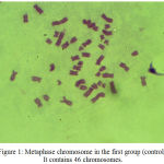 |
Figure 1: Metaphase chromosome in the first group (control). It contains 46 chromosomes.Click here to View figure |
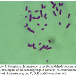 |
Figure 2: Metaphase chromosome in the formaldehyde concentration of 0.036 mg/ml of the second group. It contains 35 chromosomes. Loss of chromosome group C, D, F and G were observed.
|
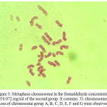 |
Figure 3: Metaphase chromosome in the formaldehyde concentration of 0.072 mg/ml of the second group. It contains 31 chromosomes. Loss of chromosome group A, B, C, D, E, F and G were observed.
|
The curcumin concentration of 10 µM exerted antimutagenic activity (at chromosome level) by inhibiting the aberrant cells proliferation in formaldehyde induced chromosomal aberration in human lymphocytes. Comparing between the first lymphocyte culture group (the control group) and the third lymphocyte culture group (added formaldehyde and curcumin), no metaphase chromosomes were found in the third group (Table 2). Curcumin effected the antimutagenic activity on the formaldehyde treated lymphocytes (P<0.05).
Table 2: Average the numbers of metaphase chromosome in the first group and the third group.
| Formaldehyde concentrations (mg/ml) | The numbers of metaphase chromosome (Each metaphase chromosome contains 46 chromosomes.) (metaphase) |
| The first group (control) | 1058 ± 0.3 |
| The third group
(added formaldehyde and 10 µM curcumin) 0.036 0.072 0.15 |
0
0 0
|
Effect of Formaldehyde on Cancer Cell Proliferation
MCF-7, A549 and CRL-2724 cells were treated with the formaldehyde concentrations of 0.036, 0.072 and 0.15 mg/ml for 24 hrs. The cancer cells were stain with 0.4% trypan blue and count the living cells with hemocytometer for detecting the effect of formaldehyde on cancer cell proliferation. The result showed that the formaldehyde concentration increasing did not affect MCF-7, A549 and CRL-2724 cell proliferation (P> 0.01) (Table 3).
Table 3 : Average the numbers of cancer cells (living cells) for the formaldehyde concentrations of 0.036, 0.072 and 0.15 mg/ml at 24 hrs
| Formaldehyde concentration (mg/ml) | Average the numbers of MCF – 7 cells ( x106 ) at 24 hrs. ± SD | Average the numbers of A549 cells ( x106 ) at 24 hrs. ± SD | Average the numbers of CRL-2724 cells ( x106 ) at 24 hrs. ± SD |
| Control | 3.5 ± 0.4 | 3.5 ± 0.3 | 3.5 ± 0.3 |
| 0.036 | 3.6 ± 0.8 | 3.6 ± 0.5 | 3.6 ± 0.4 |
| 0.072 | 3.7 ± 0.4 | 3.7 ± 0.3 | 3.7 ± 0.2 |
| 0.150 | 3.7 ± 0.6 | 3.7 ± 0.5 | 3.7 ± 0.4 |
Antiproliferative Effect of Curcumin in Cancer Cells
MCF-7, A549 and CRL-2724 cells were treated with 10 µM curcumin and incubated for 24 hrs. MTT assay was done for detecting the antiproliferative effect of curcumin. At 24 hrs., the curcumin concentration of 10 µM could inhibit all types of cancer cell proliferation (Table 4) and % Cytotoxicity Index (%CI ) was shown in Table 5.
Table 4: Average the optical density (OD) of cancer cells treated with 10 µM curcumin at 24 hrs.
| Types of cancer cells
treated with 10 µM curcumin |
OD ± SD of
cancer cells treated with 10 µM curcumin at 24 hrs. |
| MCF – 7 cells | |
| • Control group | 1.25 ± 0.42 |
| • MCF – 7 cells treated
with 10 µM curcumin |
0.62 ± 0.38 |
| A549 cells | |
| • Control group | 1.15 ± 0.87 |
| • A549 cells treated with
10 µM curcumin |
0.76 ± 0.54 |
| CRL-2724 cells | |
| • Control group | 1.08 ± 0.26 |
| • CRL-2724 cells treated
with 10 µM curcumin |
0.55 ± 0.22 |
Table 5: Percent of Cytotoxicity index (%CI) of 10 µM curcumin treated in cancer cells at 24 hrs.
| Types of cancer cells
treated with 10 µM curcumin |
% CI of 10 µM curcumin treated in cancer cells at
24 hrs. |
| MCF – 7 cells
– Control group – MCF – 7 cells treated with 10 µM curcumin |
0
50.4 |
| A549 cells
– Control group – A549 cells treated with 10 µM curcumin |
0
40 |
| CRL-2724 cells
– Control group – CRL-2724 cells treated with 10 µM curcumin |
0
49 |
Role of Curcumin on SOD Activity in Lymphocytes Induced by Formaldehyde
The activities of SOD were increased in lymphocytes induced by formaldehyde when curcumin concentrations were increased (Table 6).
Table 6: Average % SOD inhibition activities in formaldehyde treated lymphocytes at 24 hrs.
| Curcumin concentration (µM) | Formaldehyde treated lymphocytes concentration (mg/ml) | |||
| 0 | 0.036 | 0.072 | 0.15 | |
| 10 | 52.53 | 48.73 | 41.30 | 36.14 |
| 20 | 52.34 | 45.17 | 38.27 | 32.25 |
Discussion
Szende reported that the suitable formaldehyde concentration can activate cancer cell proliferation for in vitro study.8 However, the formaldehyde concentrations of 0.036, 0.072 and 0.15 mg/ml could not increase the cancer cell proliferation. These concentrations may not be the suitable for MCF-7, A549 and CRL-2724 cell proliferation. At 24 hrs. incubation, the curcumin concentration of 10 µM affected cancer cell proliferation and formaldehyde treated lymphocytes mutagenicity. Formaldehyde is one of the toxic substance that can promote cancer cell growth.9-11 Curcumin can arrest the growth of cancer cells in the G2 stage of cell cycle. This study demonstrated that the suitable curcumin concentration could induce antiproliferative effect on cancer cells (MCF-7, A549 and CRL-2724 cells) in vitro. Curcumin exerts an antiproliferation effect in human cancer cell lines.
Increased levels of cytogenetic damage by formaldehyde have been reported in the bone marrow of exposed mice and rats.12 It also has been reported about genetic toxicology in the formaldehyde exposed workers.13 Formaldehyde induces DNA-protein 14 crosslinks and inhibit p53 expression.9 For this study, the formaldehyde concentrations of 0.036, 0.072 and 0.15 mg/ml could induce lymphocyte mutagenic effect at chromosome level. It effected the numbers of metaphase chromosomes which contains 46 chromosomes. Formaldehyde increased chromosomal damage in the form of numerical aberration. At 24 hrs. incubation, the curcumin concentration of 10 µM could induce antimutagenic effect on the formaldehyde treated lymphocytes and no chromosome was observed in the third group. Curcumin effected the chromosome change in lymphocytes induced by formaldehyde. It may be suggested that curcumin protects the cell damage induced by formaldehyde by protection of oxidative stress mechanism.
Curcumin has many pharmacological effects such as antioxidation, decreasing blood lipid. Researches in vitro demonstrated that curcumin could inhibit proliferation and induce apoptosis of tumor cells at levels of DNA, mRNA, and protein.14 The tumor cell growth inhibition is associated with the promotion of apoptosis. Signal pathway of cell apoptosis includes mitochondria-mediated endogenous pathway and death receptor-mediated exogenous pathway. Caspase activation is the last common pathway. Caspase can be divided into two groups : initiative enzyme including Caspase-8, Caspase-9, Caspase-2 and Caspase-10 which can activate caspase cascade at levels of death receptor, mitochondria and endoplasmic reticulum, and does its work upstream death signal transduction, and effective enzyme including Caspase-3, Caspase-6 and Caspase-7 which is executor of cell apoptosis, and does its work at downstream death signal transduction.15 Curcumin can inhibit the NF-κB activation. Impaired proteasome function inhibit the degradation of IκB and, thereby block the nuclear translocation and transcriptional activity of NF-kappaB. NF-kappaB activity is very essential for cell survival. Its down regulation would definitely promote cell death 16. Curcumin upregulates different pro-apoptotic genes and at the same time downregulates some of the anti-apoptotic genes.17-19
Curcumin has been targeted for therapeutic application in several diseases, including glioblastoma multiforme (GBM).20 In this study, curcumin might be therapeutic application in cancer which caused by the chemical toxic substance such as formaldehyde.
Conclusions
This study demonstrated that curcumin had the antimutagenic effect in the formaldehyde treated lymphocytes and had the antiproliferative effect in cancer cells in vitro. It protects cell damage from oxidative stress. Biological effect of curcumin is multi-functional and involves multiple pathways. It may be useful in the therapy of diseases caused by the chemical substance toxicity.
Conflict of interest
The author has no conflicts of interest to declare.
Acknowledgements
This work was supported by Thammasat University research grant, 2010 and 2011. It was supported by the National Research University Project of Thailand, Office of Higher Education Commission, 2011 and 2013.
References
- Cui S., Qu X. J., Xie Y. Y., Zhou L., Nakata M., Makuuchi M,. Tang W. Curcumin inhibits telomerase activity in human cancer cell line. Int J Mol Med. 2006;18:227-31.
CrossRef - Lin J. K. Suppression of protein kinase C and nuclear oncogene expression as possible action mechanisms of cancer chemo-prevention by curcumin. Arch Pharm Res. 2004;27:683-92.
CrossRef - Pongsavee M. Effects of 744ins20 – ter240 BRCA1 mutation on breast/ovarian carcinogenesis and the role of curcumin in telomerase inhibition. Arch Med Sci Civil Dis. 2017;2:e125–9.
- Main D. M., Hogan T. J. Health effects of low-level exposure to formaldehyde. J Occup Med. 1983;25:896-900.
CrossRef - Vargova M., Wagnerova J., Liskova A., Jakubovsky J., Gajdova M., Stolcova E et.al. Subacute immunotoxicity study of formaldehyde in male rats. Drug Chem Toxicol. 1993;16:255–75.
CrossRef - Levine R. J., Andjelkovich D. A., Shaw L. K. The mortality of Ontario undertakers and a review of formaldehyde-related mortality studies. J Occup Med. 1984;26:740-46.
CrossRef - Marcsek Z. L., Kocsis Z. S., Szende B., Tompa A. Effect of formaldehyde and resveratrol on the viability of Vero, HepG2 and MCF-cells. Cell Bio Inter. 2007;31:1214-19.
CrossRef - Szende B., Tyihak E. Effect of formaldehyde on cell proliferation and death. Cell Biol Int . 2010;34:1273–82.
CrossRef - Shaham J., Bomstein Y., Melzer A., Ribak J. DNA-protein crosslinks and sister chromatid exchanges as biomarkers of exposure to formaldehyde. Int J Occup Environ Health. 1997;3:95-104.
CrossRef - Orsiere T., Sari-Minodier I., Iarmarcovai G., Botta A. Genotoxic risk assessment of pathology and anatomy laboratory workers exposed to formaldehyde by use of personal air sampling and analysis of DNA damage in peripheral lymphocytes. Mutat Res. 2006;605:30–41.
CrossRef - Zhang L., Steinmaus C., Eastmond D. A. Formaldehyde exposure and leukemia: A new meta-analysis and potential mechanisms. Mutat Res. 2009;681:150-68.
CrossRef - Kitaeva L. V., Kitaev E. M., Pimenova M. N. The cytopathic and cytogenetic sequelae of chronic inhalational exposure to formaldehyde on female germ cells and bone marrow cells in rats. Tistologiia. 1990;32:1212-16.
- Ye X., Yan W. Cytogenetic analysis of nasal mucosa cells and lymphocytes from high- level long term formaldehyde exposed workers and low level short term exposed waiters. Mutat Res. 2005;7: 22-7.
CrossRef - Lin J. K., Lin-Shiau S. Y. Mechanisms of cancer chemoprevention by curcumin. Proc Natl Sci Counc Repub China B. 2001;25:59-66.
- Maclachlan T. K., EI-Deiry W. S. Apoptotic threshold is lowered by p53 transactivation of caspase-6. Proc Natl Acad Sci USA. 2002;99:9492-97.
CrossRef - Jana N., Dikshit P., Goswami A., Nukina N. Inhibition of proteasomal function by curcumin induces apoptosis through mitochondrial pathway. J Biol Chem. 2004;279:11680-85.
CrossRef - Mukhopadhyay A ., Bueso-Ramos C., Chatterjee D., Pantazis P., Aggarwal B. Curcumin downregulates cell survival mechanisms in human prostate cancer cell lines. Oncogene. 2001;20: 7597–7609.
CrossRef - Chen A., Xu J., Johnson A. C. Curcumin inhibits human colon cancer cell growth by suppressing gene expression of epidermal growth factor receptor through reducing the activity of the transcription factor Egr-1. Oncogene. 2006;25:278-87.
CrossRef - Glienke W., Maute L., Bauer N., Bergmann L. Curcumin inhibits constitutive STAT3 phosphorylation in human pancreatic cancer cell lines and down- regulates surviving / BIRC5 gene expression. J Clin Oncol. 2007;25:15030–35.
- Luthra P. M., Lal N. Prospective of curcumin a pleiotropic signalling molecule from Curcuma longa in the treatment of glioblastoma. European Journal of Medicinal Chemistry. 2016;109:23–35.
CrossRef

