
K. Anuradha1 and K. P. Uma2
1Department of MCA, Karpagam College of Engineering, Coimbatore.
2Department of Mathematics, Hindusthan College of Engineering and Technology, Coimbatore.
Corresponding Author E-mail: k_anur@yahoo.com
DOI : https://dx.doi.org/10.13005/bpj/1281
Abstract
Oral Tumor grading can be performed in many ways. To find the depth of the tumor, the conventional TNM (Tumor, Node, Metastatis) staging has been performed by experts for several years. But this staging system is not adequate for optimal prognostication and must be supplemented by different recent methods. This study uses Fuzzy Cognitive Maps (FCM) to grade oral tumors. Eight histopathological features were used to develop Fuzzy Cognitive Map model. Active Hebbian Learning (AHL), the supervised learning algorithm is used to train and improve the FCM’s grading. 123 cases containing 85 normal and 38 abnormal cases of oral tumor were used for testing. The proposed model (FCM and AHL) achieved an accuracy of 90.58% for oral tumors of low grade and 89.47% of high grade.
Keywords
Active Hebbian Learning Fuzzy Cognitive Map; Oral Tumor; Supervised Learning; TNM;
Download this article as:| Copy the following to cite this article: Anuradha K, Uma K, P. Histological Grading of Oral Tumors using Fuzzy Cognitive Map. Biomed Pharmacol J 2017;10(4). |
| Copy the following to cite this URL: Anuradha K, Uma K, P. Histological Grading of Oral Tumors using Fuzzy Cognitive Map. Biomed Pharmacol J 2017;10(4). Available from: http://biomedpharmajournal.org/?p=17355 |
Introduction
Many attempts have been made to improve the diagnostic accuracy of the oral tumor characterization by using various types of Computer aided Technologies. Several experiments were conducted by the researchers for the classification of the tumor as benign or malignant.1 Techniques like Dental X – Rays, Positron Emission Tomography (PET), Computed Tomography (CT), Biopsy and Magnetic Resonance Imaging (MRI) are used to test the presence/absence of Tumor. Classification of tumors is an important aspect for its respective diagnosis. However, the presence of cancer can only be detected through the process of biopsy. Among all other cancers, oral cancer has the highest death rate and has witnessed no improvements over the past 40 years. World health organization (WHO) suggested a grading system for the classification of tumors. According to WHO, tumors are classified into low – grade tumors and high grade tumors. In low grade tumors, there is no invasion of tissues or metastasis and there may be less risk of further progression. However, high grade tumors are characterized by a much higher risk of progression. Premalignant tumors may progress into cancer. The correct accuracy of the diagnosis depends on the expert’s experience and knowledge. Several histopathological features were reviewed by various researchers for cancer classification.2,3 Experts combine the features to determine the final grade of tumor. In this work, histopathological features such as Differentiation, Nuclear polymorphism, Mitoses, Stroma, Mode, Stage, Vascular and Inflammatory response were taken. The proposed method is based on FCM with the implementation of Active Hebbian Learning algorithm which improves the classification accuracy of FCM.
This paper is structured as follows: Section 2 presents the related literature study. Section 3 and 4 describes the methodology of FCM and AHL respectively. The development of FCM model for grading tumors is shown in section 4. Section 5 presents the experimental results and conclusion is shown in section 6.
Literature Study
Classification and grading of cancers have been always been an epic area of interest for researchers. Muthu Rama Krishnan et al.,proposed a wavelet based texture classification for oral histopathological sections. As the conventional method involves in stain intensity, inter and intra observer variations leading to higher misclassification error, a new method is proposed. The proposed method, involves feature extraction using wavelet transform, feature selection using Kullback – Leibler (KL).4
Anuradha. K and Sankaranarayanan.K (2013) classified oral cancers using Feature Extraction Techniques. Dental radiographs were taken as input images. The tumor area is segmented using Watershed algorithm. Gray Level Co-occurrence Matrix (GLCM), Gray Level Run Length Matrix and Intensity Histogram feature extraction methods were used to extract features from the segmented image. Further, a supervised classifier, Support Vector Machine is used to classify the features as benign or malignant. Among the feature extraction methods, GLCM with SVM classifier achieved an accuracy of 96%.5
Few researchers used Fuzzy Cognitive Maps to stage cancers.
Papageorgiou. E. I et al (2003) developed a FCM model to grade Urinary Bladder Tumors using Unsupervised Hebbian Algorithm. Eight Concepts were considered for grading. The classification accuracy was 93.18% for low grade tumors and 90.59% for high grade tumors.6
Roopa Chandrika et al.,(2016) used Fuzzy Cognitive maps for grading breast tumors from Digital Mammograms. The textural features were obtained using Gray Level Co-occurrence Matrix (GLCM) and Laws Energy. These textural values are fed as input to Fuzzy Cognitive Map to classify the severity of abnormality present in digital mammograms.7
Thuthi Sarabai and Arthi.K (2016) improved Fuzzy Cognitive Map with Cat Swarm Optimization Algorithm to classify breast cancers. GLCM features were extracted from the input preprocessed image. The performance of the system is evaluated with Mean Square Error, Sensitivity and Specificity.8
Materials and Methods
123 cases of oral tumor patients with the age groups 21 to 67 were collected. 85 are normal cases and 38 are abnormal cases.
Fuzzy Cognitive Map
Fuzzy Cognitive Map was first enhanced by Kosko. B.9 These are fuzzy – graph structures which represents causal reasoning. The proposed work used Fuzzy Cognitive Maps with Active Hebbian Learning. A Fuzzy Cognitive Map integrates the accumulated experience and knowledge on the causal relationship between factors/ characteristics/ components of any system; due to the way it is constructed, i.e., using human experts that know the system and its behaviour under different circumstances.10 It is one of the unsupervised learning algorithms which classify data by using concepts. Weights are calculated between the concepts. The value of the weight indicates the strong influence between concepts. The value calculated between the interconnection of concepts lies between 0 and 1. The Fuzzy Cognitive Map represents knowledge and relates states, processes and inputs. When compared with neural networks, it is relatively easy to represent knowledge. The sample FCM is shown in Fig 1.
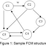 |
Figure 1: Sample FCM structure
|
The w indicates how strongly C1 influences C2. If w >0, there exists a positive causality between C1 and C2. i.e., An increase in C1 will also cause to increase C2. Negative causality is present if w < 0. If there is a decrease in C1, there will be a decrease in C2 also.
Here, in this work, the FCM model is constructed using eight concepts shown in Table 1.
Development of FCM
123 cases of oral tumor patients with the age groups 21 to 67 were collected. Using various diagnostic tools available today, experts diagnosed 85 as normal cases and 38 as abnormal cases. The abnormal cases were mostly Squamous cell carcinoma in the neck regions and the normal cases were mostly Lichen Planus. To construct a FCM, eight histopathological criteria (Table 1) were used. Different grading systems were reviewed in.2 As the database contains more cases of Squamous Cell Carcinoma, Fisher classification is used. Each criteria contains 2 – 5 possible values. These possible values are the factors of the grading system.
The initial process is to decide the number of concepts and the relation between them. These concepts encode the level of malignancy. As these concepts are interrelated to one another, the dependency matrix can be easily developed by using FCM map.
Table 1: FCM grading for Oral Tumor
| Concept | Histological Feature | Possible Assessment (Tumor scores) |
| C1 | Differentiation | Much keratin, Some keratin, Squamous, Anaplastic |
| C2 | Nuclear polymorphism | Few aniso, Moderate aniso, Many aniso, Bizarre |
| C3 | Mitoses | Occasional, Few, Moderate, Many |
| C4 | Stroma | Abundant, Dense, Delicate, None |
| C5 | Mode | Pushing, Bands, Cords, Diffuse |
| C6 | Stage | No invasion, Microinvasion, In connective tissue, Deep |
| C7 | Vascular | None, Possible, Few, Many |
| C8 | Inflammatory response | Marked, Moderate, Slight, None |
| C9 | Degree of Tumor grade | Low, high |
The FCM grading model was developed using the eight concepts (C1 to C8): The eight concepts represents the eight variable of the tumor grading system. The Ninth concept (C9) represents the degree of tumor grade. Concept C1 represents the differentiation, C2 represents Nuclear polymorphism, C3 represents Mitoses, C4 represents Stoma, C5 represents Mode, C6 represents Stage, C7 represents Vascular and C8 represents Inflammatory response. The ninth concept represents the degree of tumor grade. All the values are in the interval (0, 1). The threshold (0.5) decides which event is stimulated.
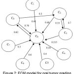 |
Figure 2: FCM model for oral tumor grading
|
The fuzzy rule for each interconnection was evaluated using Fuzzy reasoning and from that fuzzy weights (Equation 1) are defuzzified. A fuzzy set is modelled {positive very high, positive high, positive medium, positive weak, Zero, negative weak, negative medium, negative low and negative very low} by using the degree of influence of concepts. These fuzzified values are converted into numerical values using defuzzification method.
The degree of influence among the concepts was presented using IF – THEN conditions. IF a small change occurs in the value of Concepti, then a small change is caused in the value with Conceptj
Consider for example, the value Xi of the Concept Ci influences the Conceptj
The value Aj for each concept Cj is calculated using the following equation:
Aj (t+1) = f (Aj (t) +∑ wij. Ai(t) (1)
Where, Aj (t+1) is value of concept Cj at step t+1,
Ai(t) is the value of concept Ci at step t, and Wij is the weight of the arc from Concept Ci towards concept Cj and f is a threshold function.
Active Hebbian Learning
Classification rate and efficiency of the FCM can be enhanced by applying Active Hebbian Learning (AHL). AHL is an unsupervised learning algorithm.11,12 This will regulate the weights of the FCM. The main advantage of AHL is that it is based on asynchronous decision making process similar to human decision skills and it can decide new FCM causal links between all the Concepts. This algorithm takes the input values of concepts which will strengthen and weaken the FCM causal links between the concepts. Due to this classification capability is increased.
Evaluation of oral cancer cases were done after the development of FCM and the implementation of AHL. For each and every case, the values are calculated in the interval [0-1]. The grading system with the new weighted interconnections among concepts was calculated. After few interactions, the C9 for every case is calculated.
Discussions and Results
The performance of FCM grading system is evaluated. This tool achieved an accuracy of 90.58% (77/85) for oral tumors of low grade and 89.47% (34/38) of high grade. From Fig 2, it is observed that the value (0.76) for Inflammatory response (C8) is more important to find the degree of grading. The graph plotted (Fig 3), shows the final grade values of C9 for each of 123 cases.
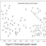 |
Figure 3: Estimated grade values
|
The horizontal axis (X) represents the calculated values of the Grade improved using AHL algorithm and the vertical axis (Y) represents the number of cases used for each grade category. The symbol ‘Δ’ represents the estimated “Grade values” for low grade tumor and ‘Δ’ for high grade respectively. For most of the cases, the values are distinct, which makes the classification easier.
To define the decision region for each case, the mean value m1 and m2 for each category is estimated.
The decision boundary was determined as the perpendicular bisector of the line joining m1 and m2. So the threshold is estimated as 0.92 for each grade category. The values lower the 0.92 are low grade (normal) and those values which are higher than 0.92 are high grade (abnormal). This procedure was repeated for many times. The average success rate has achieved.
The Table 2 shows the comparison of results for TNM grading and FCM grading:
Table 2: Comparison of FCM Grading Model with other Techniques
| Normal/ Abnormal cases | TNM + Biopsy | FCM Grading tool |
| Normal cases (85) | 85 | 77 |
| Abnormal cases (38) | 38 | 34 |
| Accuracy % for low grade tumor | 100 | 90.58% |
| Accuracy % for high grade tumor | 100 | 89.47% |
Conclusion
Researchers mainly focus on microscopic procedures to grade the tumor. And in many cases, the accuracy obtains only at the advanced stage where treatment becomes unsuccessful. The TNM system does not provide information on the biological characteristics. So a reliable method for categorization with excellent accuracy is essential. The proposed method used FCM grading model to categorize the tumor cases into low grade and high grade. Further, to improve the values, Active Hebbian Learning algorithm was used. The classification rate obtained was 90.48% and 89.47% for low grade and high grade respectively.
In future, features can be extracted using the feature extraction methods and can be given as input to the FCM.
Acknowledgements
The authors would like to thank Dr.T.P.Swamy.,MDS,Coimbatore and Dr.J.Sudha, MDS, Dental Surgeon, Surya Dental Clinic, Coimbatore for their suggestions in this work.
References
- K and Dr. K. Sankaranarayanan. Identification of Suspicious regions to detect Oral Cancers at an earlier stage.Int. J. of Advances in Engineering and Technology. 2012;3(1):84-91.
- Rastogi V, Puri N, Mishra S , Sharma R, Yadav L , Sabharwa R. Dilemmas in Grading Epidermoid Carcinoma. J. of Head and Neck Surgery. 2014;5(1):9–14.
CrossRef - Singh J. Histopathology of Oral Squamous Cell – carcinoma – A review. TMU J. Dent. 2014:1(4):141–144.
- Rama M, Krishnan M, Chakraborthy C, Kumar A.R. Wavelet based texture classification of oral histopathological sections.J. of Microscopy, Science, Technology, Applications and Education. 2010;2(1):897-906.
- K and Sankaranarayanan Dr.K. Comparison of Feature Extraction Techniques to classify Oral cancers using Image Processing. Int. J. of Application or Innovation in Engineering and Management. 2013; 2(6):456–462.
- Spyridonos E.I.P.P, Stylios C.D, Nikiforidis G.C and Groumpos P.P. Grading Urinary Bladder Tumors using Unsupervised Hebbian Algorithm for Fuzzy Cognitive Maps, Biomedical Soft Computing and Human Sciences. 2003;9(2).
- Chandrika R.R, Karthikeyan N, Karthik S. Texture Classification using Fuzzy Cognitive Maps for grading breast tumor. Asian J. of Information Technology. 2016;15(15):2709-2715.
- Sarabai T.D and Arthi Dr.K. Efficient Breast cancer classification using Improved Fuzzy Cognitive Maps with Csonn. Int. J. of Applied Engineering Research. 2016;11(4):2478-2485.
- B. Fuzzy Cognitive maps. Int. J. of Man – Machine studies. 1986;24:65 –75.
CrossRef - C.D and Groumpos .P.P. Fuzzy Cognitive Maps in Modelling Supervisory Control Systems. Journal of Intelligent and Fuzzy Systems. 2000;8.
- Bourgani E,Chrysostomos D, Stylios, Voula C, Georgopoulos, Manis G. A study on Fuzzy Cognitive Map structures for Medical Decision Support Systems, 8th. Conference of the European Society for Fuzzy Logic and Technology (EUSFLAT 2013), Atlantis press.744–751.
- Stylios E.C.D, Groumpos P.P. Active Hebbian learning algorithm to train Fuzzy Cognitive Map.International Journal of Approximate reasoning. 2004;37.

