
Manuscript accepted on :November 01, 2016
Published online on: --
Plagiarism Check: Yes
Sri Rahayu Santi and I Made Sukadana
Department of Chemistry-Faculty of Science and Maths. Corresponding Author E-mail. srahayusanti@gmail.com
DOI : https://dx.doi.org/10.13005/bpj/1070
Abstract
This study aimed to evidence the antiatherosclerosis of n-buthanol extract of gayam stem bark which were indicated through the decrease of 8-OHdG level and expression of ICAM-1 in Wistar rats that were treated with a high fat diet or hypercholesterolemia conditions within 4 months of observation. The study was initiated by preparing n-buthanol extract of gayam stem bark which was obtained from the partition of 95 g ethanol extract of gayam stem bark. A total of 30 g of n-buthanol concentrate extract was obtained and then was applied to Wistar rats for 4 months with the use of the posttest only control group design. Twenty-five Wistar rats were randomized into 5 groups, P0 (negative control), P1 (positive control, hypercholesterolemia), P2 (hypercholesterolemia + n-buthanol extract in the dose of 50 mg/kg bw), P3 (hypercholesterolemia + n-buthanol extract in the dose of 100 mg/kg bw), and P4 (hypercholesterolemia + n-buthanol extract in the dose of 150 mg/kg bw). The results showed that n-buthanol extract of gayam stem bark in the dose of 100 mg/kg bw was able to decrease the levels of 8-OHdG, while in the dose of 100-150 mg/kg bw the extract could decrease the expression ICAM-1 endhotel aorta in wistar rats hypercholesterolemia.
Keywords
Inocarpus fagiferus Fosb; Antiatherosclerosis; ICAM-1; 8-OHdG; Hypercholesterolemia
Download this article as:| Copy the following to cite this article: Santi S. R, Sukadana I. M. Antiatherosclerosis N-Buthanol Extract of Inocarpus Fagiferus Fosb Stem Bark Through The Decrease of 8-Ohdg Level and Expression Icam-1 in Rats Hypercholesterolemia. Biomed Pharmacol J 2016;9(3). |
| Copy the following to cite this URL: Santi S. R, Sukadana I. M. Antiatherosclerosis N-Buthanol Extract of Inocarpus Fagiferus Fosb Stem Bark Through The Decrease of 8-Ohdg Level and Expression Icam-1 in Rats Hypercholesterolemia. Biomed Pharmacol J 2016;9(3). Available from: http://biomedpharmajournal.org/?p=11098 |
Introduction
In Indonesia Inocarpus fagiferus Fosb as known as gayam or gatep (in Bali) is one of the herbs that empirically in the Fiji Islands is used as a drug to prevent heart disease and atherosclerosis (Sotheeswaran and Sharif, 1994). The stem bark of gayam were reported main contains compounds such as flavonoids and phenols with the total flavonoids and phenols respectively by 0.09% and 14.16%, that are able to capture the free radicals DPPH by 50% at a concentration of 20 ppm, and are able to inhibit the formation of lipid peroxide by 63.03%. The antioxidant activity of n-buthanol extract of gayam stem bark in vitro against DPPH is greater than the antioxidant activity of vitamin E which provides IC50 at a concentration of 25 ppm, as well as its ability to inhibit the formation of lipid peroxides by 50.39 % (Santi and Sukadana, 2015). Futhermore, the potential of antioxidant activity of n-buthanol extract of gayam stem bark were evidenced by decreasing of the plasm malondialdehide (MDA), total cholesterol, and triglyceride levels and increasing the SOD activity in dose of 50 mg/kg bw but in dose of 100 mg/kg bw decreasing of the plasm LDL cholesterol and increasing the plasm HDL cholesterol levels in the hypercholesterolemia wistar rats. (Santi et al., 2015).
The expression of ICAM-1 was a biomarker of early stage development of atherosclerosis (Price and Loscalzo, 1999; Naito et al., 2004; Lampros et al., 2012). The high intake of cholesterol causes increased reactive oxygen species (ROS) especially superoxide anion (×O2–) through activation of NADPH oxidase enzyme, so that the conditions of oxidative stress occurs (Madamanchi et al., 2004). This ROS molecules will oxidize LDL cholesterol to form mm-LDL cholesterol (lipid peroxide) are not stable in the intima layer and migration mm-LDL to endothelial cell causing dysfunction or cell damage. Endothelial cell dysfunction affects the expression of various inflammatory mediators (Kaliora et al., 2006), which is one of the increasingly expression of adhesion molecules ICAM-1 (Lampros et al., 2012). The high expression of cell surface adhesion molecules ICAM-1 causes high adhesion of leukocytes (lymphocytes, monocytes, and neutrophils) attached to the endothelial cells to transform into macrophages and it developed to lesion atherosclerosis (Davis, 2005). Dysfunction or cell damage can be occured on cell protein or nucleus that can be observed in the level of 8-OHdG plasm.
This study discusses the differences in the expression of ICAM-1 and the levels of 8-OHdG of each treatment group compared with the control group of hypercholesterolemia, as the marker of early stage atherosclerosis and cell protein damage due to lipid peroxidation (Stefan et al., 1996; Ahmed, 2001; Han et al., 2002)
Materials And Methods
The Research Materials
The plant material was obtained from Tabanan Bali and its classification has been determined.
Chemicals used in this study were ethanol (technical and pa), chloroform (technical and pa), n-buthanol, Kitt Rat-8-OHdG BT Lab. and blood plasm of wistar rat. Chemicals required for immunohistochemical analysis were aorta endhotel of Wistar rat, formaldehyde 10%, buffer citrate (PBS) 0,1 M (pH. 7,4), kit LSAB (Dako, Denmark), and Rabbit Anti-ICAM-1 Polyclonal Antibody (Bioss, Cat. Bs-0608R), and another chemical from Sigma-Aldrich (USA) ie.: methanol p.a, trypsine 0,25%, H2O2 3%, formaline buffer phosphate 10%, alcohol 70%, 90%, 96%, 100%, xylene, pure parafine, PBS, FBS 5%, Biotinylated Goat Anti-Polyvalent, Streptavidin peroxidase, aquabidest, hematoxyline Gill, and buffer tri sodium citrate. The determination of profile lipid was conducted at UPT. Balai Laboratorium Kesehatan Provinsi Bali. All of the rats were treated in accordance with the rules of the etical clerence of Ethics Committee of Animals Use in Research and Education of the Faculty of Veterinary Udayana University.
Research Instruments
The equipment used in this study was a set of glasses, extractor, blender, sieve, rotary vacuum evaporator, analytical balance, thermometer, micropipette, pipette, volumetric flask, stomach probes, syringes, desicators, test tubes, volumetric pipettes, rat cage, micro haematocrit tubes (Cat. 7493 21), EDTA Eppendorf tubes, sentrifuge 2-6E Sartorius, microtom rotary (Jung Histocut Leica 820), microscope binoculer Olympus CX41 with camera Olympus DP12, photographed with an Optilab Pro (Miconos, Indonesia) camera. Each preparation was photographed 4 times by in JPEG format using Optilab Raster Image Viewer 1.0 and 2.1 software (Miconos, Indonesia). microscope slide polylysine, coverslip, and elisa reader.
Researchers procedure
Preparation of n-buthanol extract of gayam stem bark and its appication on wistar rats
Procedure to prepared of n-buthanol extract of gayam stem as early reported (Santi et al., 2015). n-buthanol extract was applied at various doses i.e 50 mg/kg bw, 100 mg/kg bw, and 150 mg/kg bw. Twenty five wistar rats were randomized into 5 groups with posttest only control group design (Pocock, 2008) as follows:
P0= group of wistar rats fed with standard diet (negative control)
P1= group of wistar rats fed with a high-fat diet (positive control)
P2= group of wistar rats fed with a high-fat diet + n-buthanol extract of gayam stem bark in the dose of 50 mg/kg bw
P3= group of wistar rats fed a high-fat diet + n-buthanol extract of gayam stem bark in the dose of 100 mg/kg bw
P4= group of wistar rats fed with a high-fat diet + n-buthanol extract of gayam stem bark in the dose of 150 mg/kg bw
After 16 weeks, the blood plasm and aorta of all of the rats such as the control groups (P0 and P1) and the treatment groups (P2, P3, and P4) were drawn for expression ICAM-1 and 8-OHdG analyses. The difference of all variables were analyzed by one way Anova with a = 0.05.
Results and Discussion
Blood Plasm 8-OHdG Level
The average 8-OHdG level of rat blood plasm after 16 weeks for control and treatment groups based on Elisa analysis were presented in Figure 1.
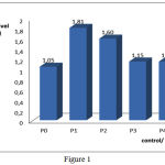 |
Figure 1 |
The average of 8-OHdG level Blood Plasm of treatment groups (P2, P3, and P4) toward P1. The difference mean of groups were: P0 vs P1, p<0.05; P2 vs P1, p>0.05; P3 vs P1, p<0.05; P4 vs P1, p<0.05; P2 vs P3, p>0.05; P2 vs P4, p>0.05; P3 vs P4, p>0.05
The analysis results of plasm 8-OHdG level showed that n-buthanol extract of gayam stem bark in doses of 100 until 150 mg/kg bw could decrease 8-OHdG level significantly (p<0.05) toward control group P1 but in dose of 50 mg/kg bw decrease level not significantly (p>0.05).
These condition was caused by the total flavonoid and phenol content in n-buthanol extract gayam stem bark able to captured ROS, an oxidator that caused macromolecule was oxidized as like guanine molecule in DNA, so that formed stable molecule (Chung and Xu, 1992). These ROS captured prevent to change guanine nucleotide in DNA into 8-hidroxy-2-dioxyguanosine (8-OHdG) compounds so it indirect to prevent DNA damage. In Figure 1 also shown that treatment group P3 and P4 in doses of 100 and 150 mg/kg bw respectively not difference significantly (p>0.05) toward control group P1 or hypercholesterolemia. This mean that only in doses of 100 mg/kg bw can decreases significanlty of 8-OHdG level (p<0.05).
Expression of ICAM-1 Aorta Endhotel
The average and description of expression ICAM-1 positive of rat aortic endothelial cells for control and treatment groups based on immunohistochemical analysis using Rabbit Anti-ICAM-1 polyclonal antibodies are presented in Figure 2 and Figure 3.
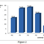 |
Figure 2 |
The average of ICAM-1 Expression of treatment groups (P2, P3, and P4) toward P1. The difference mean of groups were: P0 vs P1, p<0.05; P2 vs P1, p>0.05; P3 vs P1, p<0.05; P4 vs P1, p<0.05; P2 vs P3, p<0.05; P2 vs P4, p<0.05; P3 vs P4, p<0.05
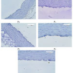 |
Figure 3 |
Aortic Endothelial Cells which Expression of the ICAM-1
The intake of high cholesterol diet (hypercholesterolemia) caused the increase of ICAM-1 expression significantly on P1 group (25.95± 0.69) compared with P0 group or standard diet (P0; 15.80±4.12). The decrease of expression significantly occured in treatment group 3 (P3; 20.30±.4.27) and group 4 (P4; 7.40±1.81) (p<0.05), as shown in Figure 1, while in dose of 50 mg/kg bw (P2) caused the increase expression not significantly. Hypercholesterolemia such as the P1 group will the release of platelet-activating factor (PAF), increase the synthesis and release of interleukin-1 (IL-1), and increase the release of tumor necrosis factor (TNF-a) which will stimulate polymorphonuclear leukocyte (PMNLs). This in turn produces reactive oxygen species (ROS), especially superoxide anion, an oxidative stress molecule. ROS can oxidize cholest-LDL molecules to their oxidized form of LDL (ox-LDL or mm-LDL) are not stable in the intima layer cholesterol to form mm-LDL cholesterol (lipid peroxide) are not stable in the intima layer and migration mm-LDL to endothelial cell causing dysfunction or endothelial cells damage in the walls of arteries and the aorta. Endothelial cell dysfunction affects the expression of various inflammatory mediators (Kaliora et al., 2006), which is one of the increasingly expression of adhesion molecules ICAM-1 (Lampros et al., 2012). The high expression of cell surface adhesion molecules ICAM-1 causes high adhesion of leukocytes (lymphocytes, monocytes, and neutrophils) attached to the endothelial cells to transform into macrophages then it developed to form lesions atherosclerotic (Davis, 2005). Really, the endothelial cells of blood vessels are very important as a barrier and transporting media of biochemical molecules such as from blood cells to tissues. They also play an important role in maintaining vascular homeostasis in response to the attacks of reactive free radical superoxide anion (Hosoya et al., 2005; Lin et al., 2005). Thus the expression of ICAM-1 can be used as a biomarker of early stage development of atherosclerosis (Price and Loscalzo, 1999; Naito et al., 2004; Lampros et al., 2012).
Antioxidant compounds in n-buthanol extract of gayam stem bark as like flavonoid and phenol in dose of 100-150 mg/kg bw can reduce the formation of lipid peroxides (mm-LDL). Decreasing lipid peroxidation products, the expression of adhesion molecules ICAM-1 also diminish, which means that the formation of atherosclerotic lesions is progressively reduced or inhibited. Tend to decrease of ICAM-1 expression if the groups of rat were given doses of n-buthanol extract more higher.
Conclusions
n-buthanol extract of gayam stem bark in the doses of 100mg/kg bw could prevent atherosclerosis by decreasing the 8-OHdG level in the hypercholesterolemia wistar rats.
n-buthanol extract of gayam stem bark in dose of 100-150 mg/kg bw could prevent atherosclerosis by decreasing the expression ICAM-1 aorta endhotel cell in the hypercholesterolemia wistar rats.
Acknowledgment
We would like to express our gratitude to DIKTI that provided us the fund throughout Hibah Fundamental (2016) and the Rector of Udayana University through the Head of LPPM that facilitated our research.
References
- Ahmed, E. 2001. Immune Mechanism in Atherosclerosis. Dissertation, ISSBN: 91-628-4612-4, Konferensrummet, Centrum for Molekular Medicin, Karolinska Sjukhuset
- Davis. 2005. Atherosclerosis an Inflamatory Process, Journal. Insur.Med, 37: 72-5
- Han, S.N., Leka, L.S., Lichtenstein, A.H., Ausman, L.M., Schaefer, E.J., and Meydani, S.N. 2002. Effect of Hydrogenated and Saturated, Relative to Polyunsaturated, Fat on Immune and Inflammatory Responses os adults with Moderate Hypercholesterolemia. Journal of Lipid Reasearh, 43, 3: 445-52
- Hosoya, T., Maruyama, A., Kang, M.L., Kawatani, Y., Shibata, T., Uchida, K., Itoh, K., Yamamoto, M. 2005. Differential Responses of the Nrf2-keap a System to Laminar and Oscilatory Shear Stresses in Endothelial Cells. The Journal of Biochemical Chemistry, 280(29): 27244-50
- Kaliora, A.C., Dedoussis, G.V.Z., Schmidt, H. 2006, Dietary Antioxidant in Preventing Atherogenesis. Atherosclerosis, 187:1-17
- Lampros, F., Georgios, A., Ioannis, S.V., Alkistis, P., Angeliki, M., Maria, K., Christos, V., Dimitrios, T., Dimitri, P.M., and Despina, P. 2012. Intercellular Adhesion Molecule (ICAM)-1 and Vascular Cell Adhesion Molecule (VCAM)-1 at the Early Stages of Atherosclerosis in a Rat Model. In vivo, 26: 243-50
- Lin, S.J., Shyue, S.K., Hung, Y.Y., Ku, H.H., Chen, J.W., Tam, K.B., Chen, Y.L. 2005. Superoxide Dismutase Inhibits the Expression of Vascular Cell Adhesion Molecule-1 and Intracellular Cell Adhesion Molecule-1 Induced by Tumor Necrosis Factor-Alpha in Human Endothelal Cells Through the JNK/p38 Pathways. Arterioscler Thromb Vasc Biol, 25(2): 334-40
- Madamanchi, N.R., Vendro, A., Runge M.S. 2004. Oxidative Stress and Vascular Disease. Arterioscler. Throm Vasc Biol, 25(1):29-38
- Naito, Y., Shimozawa, M., Manabe, H., Kuroda, M., Tomatsuri, N., Uchiyama, K., Takagi, T., Yoshida, N., and Yoshikawa, T. 2004. Inhibitory Effects of Red Wine Extracts on Endothelial-dependent Adhesive Interaction with Monocytes Induced by Oxysterol. Biol Res, 37:231-8
- Price, D.T., and Loscalzo, J. 1999. Cellular Adhesion Molecules and Atherogenesis. Am J Med, 107. 1: 85-97
- Santi, S.R dan Sukadana, I M. 2015. Aktivitas Antioksidan Total Flavonoid dan Fenol Kulit Batang Gayam (Inocarpus fagiferus Fosb), Jurnal Kimia, 9(2): 160-168
- Santi, S.R., Sukadana, I. M., Siaka, I M. 2015. The Potential of Total Flavonoids and Phenol Contents in Stem Bark of Gayam (Inocarpus Fagiferus Fosb) as an Antioxidant Through the Decrease of MDA Level, Increase of SOD Activities and Improvement of Lipid Profile in Rats Atherosclerosis Hypercholesterolemia, European Journal of Biomedical and Pharmaceutical Sciences, 2(6): 55-58
- Sotheeswaran, S., Sharif, M.R. 1994. Lipids from the Seeds of Seven Fijian Plant Species. Food Chemistry, 49: 11-3
- Stefan, J., Mikko, P.SA., Bengt, K., and Jan-Nilsson. 1996. Human Monocytes/ Macrophages Release TNF-a in Response to Ox-LDL. Arteriosclerosis, Thrombosis, and Vascular Biology, 16: 1573-9

