
Manuscript accepted on :August 11, 2016
Published online on: --
M. K. Masthan, N. Anitha, S. Hari Priya and N. Aravindha Babu
Department of Oral Pathology, Sree Balaji Dental College and Hospital, Bharath University, Pallikaranai, Chennai – 600100.
DOI : https://dx.doi.org/10.13005/bpj/1018
Abstract
In 1868, Paul Langerhans first described langerhans cell, they are dendritically shaped cells, which were located in epidermis of the squamous epithelia. Langerhans cells are seen in all suprabasal layer of stratified squamous epithelium. They play a vital role in local defense mechanisms in the epithelium. They are bone marrow derived dendritic cell and act as antigen presenting cells (APCs) of immune responses. APCs, the lymphocytes respond to specific microbes. In this article we have reviewed the physiology and mechanism of action of their role in different pathological conditions.
Keywords
Langerhans; immunological; stratum corneum
Download this article as:| Copy the following to cite this article: Masthan M. K, Anitha N, Priya S. H, Babu N. A. Langerhans Cells Pathophysiology- An Overview. Biomed Pharmacol J 2016;9(2). |
| Copy the following to cite this URL: Masthan M. K, Anitha N, Priya S. H, Babu N. A. Langerhans Cells Pathophysiology- An Overview. Biomed Pharmacol J 2016;9(2). Available from: http://biomedpharmajournal.org/?p=8191 |
Introduction
Langerhans cells (LC) lack desmosomal attachment and appear as clear cells in histological section, they are characterized by flask shape or rod like granules called birbeck granules. They have immunological function that identifies and process the antigenic material that enter into the epithelium from the external environment.These LC migrate from epithelium to regional lymph nodes.They are present in the suprabasal layer of stratified squamous epithelium.
Morphology And Distribution1
LCs are flattened disc shaped, which are aligned in a horizontal plane and parallel to the surface of the skin.They possess 5 to 9 dendrites.These dendrites liein the same horizontal plane.Dendrites from individual cells may overlap,and thereis no direct cell-to-cell contact.Dendritic processes can often be observed extending to the level of stratum corneum. In electron microscopy, LCs are 12 microns in diameter and have lobulated nuclei and cytoplasm. Specific cytoplasmic organelles are poor in vacuoles and organelles. Specific cytoplasmic organelles were first described by Birbeck hence called birbeck granules(100 nm to 1 mm in size). Birbeck granules seen in continuity with cytoplasmic membrane and are often clustered near the Golgi apparatus.There is always a conflict that these granules arise from Golgi apparatus as they have secretory function or it may arise from invagination of cytoplasmic membrane. LC are found in the epidermis of skin, in local lymph nodes, thymic epithelium and bronchial mucosa.In the skin they represent 1-2% of the entire epidermal population and seen only in the suprabasal area, with evendistribution. The cells have a mean c volume of 213µm3 and density about 1.6 x 10⁵ cells per mm2 of epidermis. With the dendrites, 25% of the surface area of skin covered by LC.Most of LCs contain one to several Birbeck granules in the epidermis some LCs does not possess the birbeck granules which are called “indeterminate cells” these are precursor cells to LCs. Loss of these organelles tend to increase their functional potential of LC.
Origin Of Langerhans Cells2
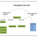
The Langerhans cells (LCs) origin from the bone marrow and then travel tothe epithelium to perform the function of antigen recognition and presentation. The myeloid and the lymphoid series cell type are CD34+ haematopoietic progenitor cell, which were differentiate into distinct Dendritic Cells (DC) subtypes and perform distinct functions. [2] Lymphoid progenitors give rise to precursors that differentiate intoplasmacytoid DCs, thymic DCs, and interdigitating DCs. Myeloid progenitors give rise to precursors of germinal centre DCs,interstitial DCs, monocyte/macrophage series, LCs, and peripheral blood DCs. This indicates that common precursors give rise to different subtypesDC, under specific environmental conditions.
 |
Figure
|
Function Of Langerhans Cells3
LCs can present antigen to helper and cytotoxic T-cells.
LCS present
⇓
Exogenous antigen
⇓
Via MHC class II
⇓
Present CD4+helper cells
LCs alsopossesses the MHC class I restricted cytotoxic (CD8+) T-cell responses, which depend on the expression of costimulatory molecules B7-1. In addition,by the production of cytokines, including IL-12 LCs stimulate NK cells. [5]
LC present in most tissue is unstable “immature” state thus unable to stimulate T-cells. Immature LCsthat express surface receptors of various antigens include toll-like receptors, C-type lectin receptors, Fc-g, and complement receptors6. LCs captured these antigen in peripheral tissues and processed to form MHC-peptide complexes7. Pathogens and inflammatory cytokines which alter LCs to be matured and mobilized. LCs migrate into the lamina propria and become CD83+ mature LCs are mobilized and matured in response to inflammatory cytokines and pathogen associated molecular patterns from oral mucosal pathogens. LCs migrate into the lamina propria and become CD83+ mature DCs. The maturation and migratory activity regulated by chemokine receptors, inflammatorychemokine/chemoattractants, and integrins.
Bacteria and their cell wall components (Lipopolysaccharides), and cytokines like IL-1b, GM-CSF, and TNF-a, everything stimulate DC maturation and mobilization but IL-10 blocks this process. LCs complete their maturation and attract T- and B-cells by secrete chemokine which maintain the viability of T lymphocytes7. Antigen may bind by B-cells, and then both B- and T-cells cluster with mature LCs, but activationin the T-cell area leading the immune responses. B-cell raises humoral immune response and form some antibody-secreting plasma cells but T-cells migrate to the site of antigen deposition, and mediate a cellular response.
Staining8
Hematoxylin and eosin staining
LCs appear as a clear cells- clear halo around their nuclei they present in suprabasal layers are also referred to as ‘high level clear cells
Special staining
Osmium iodide,Gold chloride, zinc osmium iodide and uranyl acetate-lead staining. Zinc osmium iodide, gives good staining, can also stains keratinocytes and melanocytes.
Enzyme histochemistry
ATPase, which is an excellent method for the identification of human LCs. Other enzymes5 nucleotidases adenosinediphosphatase (ADPase)and amino-peptidases.
Immuno histochemistry
LCs possess large number of cytoplasmic antigen used of antibodies in different techniques like, immunofluorescence or immunoperoxidase techniques. LCs is the high level of expression of MHC class II antigens also MHC class I antigens. Leukocyte common antigen (CD45) is also expressed by LCs and shows stresses their bone marrow origin. Many other antigens like CD11,CD25, CD29, CD1D and CD83,Fc-IgG, CD54, C3b/C4bare found in trace amount in subpopulations of LCs can detect by sensitive methods.CD1a immunolabeling is considered to be the most reliable method to identify the human LCs. Immunohistochemical markers CD1a, b, c and d express immature DCs.Advance studies shown that Langerin seems to be the more specific marker for LCs.
Electron microscopy
In electron microscope LCs appeared cleaved or folded nucleus and no tonofilaments or desmosomes The Birbeck granules present in their cytoplasm, resemble three-dimensional profile of a disc. The granule with vesicle at one end shows an appearance of ‘tennis racket’.
Langerhans Cells in Different Pathological Conditions
Gingivitis and periodontitis9-11
IncreasedLCs in gingival epithelium associated with gingivitis. LCs is five times more in gingivitis than normal gingiva in chronic adult periodontitis.Mature CD83+ DCs form immune conjugatesin the oral lymphoid foci with CD4+ T cells.
Human immunodeficiency virus infection
Acquired Immunodeficiency Syndrome and HIV related complex patients have decreased number of LCs.
Oral lichen planus
LCs was increased in lichen planus
Lichenoid drug eruptions
LCs was increased but lower the OLP
Recurrent aphthous stomatitis
LCsare increased in both recurrent aphthous stomatitis and Behcets syndrome.
Contact hypersensitivity
Contact hypersensitivity taking place in the epidermis Haptens-modified LCs can induce sensitization when particular immunizing conditions are applied.
Role of LCs in oral cancer
In oral cancer the increase in the number of LCs and also CD8 + lymphocytes and macrophages and in the epithelium
Radicular granuloma and cyst
Increase in the number of LCs, than other cysts
Conclusion
LCs as APCs play an important role in various oral pathologic conditions also initiates immune response. Many concepts about allow us to device specific immunotherapies and treatment modalities.
Reference
- The Normal Langerhans Cell and the LCH Cellin Macmillan Press Ltd., 1994 ‘Senior Lecturer/Honorary Consultant Dermatologist, Royal Postgraduate Medical School, Hammersmith Hospital, Ducane Road,London W12 ONN;. Department of Pathology, Childrens Hospital, Fifth Avenue, Pittsburgh PA 15213, USA.
- Pathophysiology of Langerhans cellsShweta Jaitley,TR SaraswathiDepartment of Oral Pathology and Microbiology, K D Dental College and Hospital, Mathura, Uttar Pradesh, India Department of Oral and Maxillofacial Pathology, Vishnu Dental College, Bhimavaram, Andhra Pradesh, India.
- Langerhans cells and their role in oral mucosal diseasesJuhi Upadhyay1,Ram B Upadhyay1, Panko Agrawal1, Shweta Jaitley1, Rita Shekhar Department of Oral and Maxillofacial Pathology, K.D. Dental College and Hospital, Mathura, India Department of Conservative Dentistry, K.D. Dental College and Hospital Matura, utter Pradesh , India.
- Inaba K.Immunologic properties of epidermal langerhans cells. Distinct requirement for stimulation of inprimed and sensitized T-lymphocytes. J Exp Med 1986; 164:605-13.
- Watts C. Capture and processing of exogenous antigens for presentation on MHC molecules. Annu Rev Immunol 1997; 15:821
- Schroeder H, Thelade J. Electron microscopy of normal human gingival epithelium. J Perodont Res 1986; 21:640-52.
- Schuler G, Steinman RM. Murine epidermal Langerhans cells mature into potent immunostimulatory dendritic cellsin vitro. J Exp Med 1985; 161:526-46.
- Datta SK, Redecke V, Prilliman KR, Takabayashi K, Corr M, Tallant T,et al. A subset of toll-like receptor ligands induces cross-presentation by bone marrow-derived dendritic cells. J Immunol 2003; 170:4102-10. [PUBMED]
- Van Kooyk Y, Engering A, Lekkerkerker AN, Ludwig IS, Geijtenbeek TB. Pathogens use carbohydrates to escape immunity induced by dendritic cells. Curr Opin Immunol 2004; 16:488-93.[PUBMED]
- Bos IR, Burkhardt A. Interepithelial cells of the oral mucosa. Light and electron microscopic observations in germfree, specific pathogen-free and conventionalized mice. J Oral Pathol 1980; 9:65-81.[PUBMED]
- DiFranco CF, Toto PD, Rowden G, Gargiulo AW, Keene JJ Jr, Connelly E. Identification of Langerhans cells in human gingival epithelium. J Periodontol 1985; 56:48-54. [PUBMED]

