Behnaz Esfandiari 1, Masoud Soliemani 2, Saeed Kaviani 2 and Kazem Parivar 1*
1Department of Biology, Science and Research Branch, Islamic Azad university, Tehran, Iran 2Department of hematology, Medical science faculty, Tarbiate Modares university, Tehran, Iran Corresponding Authors E-mail: kazem_parivar@yahoo.com
DOI : https://dx.doi.org/10.13005/bpj/906
Abstract
Neural tissue has limited capacity to regenerate. There is a critical need to explore strategies in regenerative medicine that use stem cell therapy to transplant differentiated cells into nervous system disease. Several long differentiation period techniques are now using for differentiate stem cells to neuron like cells.To evaluate a novel short time induction method for differentiate of human Adipose derived stem cells(hADSCs) to neuron like cells with bFGF, Forskolin and NGF in 8 days. Adipose stromal cells were cultured. The third passage cells were used for in vitro differentiation. Neuronal differentiation was induced by treatment of the hADSCs with basic fibroblastic growth factor(bFGF), Forskolin, Nerve Growth Factor(NGF) and 1% FBS for 8 days. Flocytometery analysis showed that ,99/4% of ADSCs will express CD105 marker,97/8% of stem cells will express CD90, 3/25% of ADSCs will express CD34 and 2/77% of cells will express CD45. hADSCs were induced to differentiate into neuron-like cells as evidenced by neural morphology and the presence of neuronal markers including Microtubule-associated protein(MAP2) and b-tubulinIII by Immunocytochemistry. RT-PCR and Real-time PCR analysis indicated the upregulation of MAP2, NSE and NFM genes. hADSCs can be readily obtained from a small amount of fat tissue with minimal injury. Our findings indicate that simultaneous presence of differentiation factors such as bFGF, Forskolin, and NGF led to rapid differentiation into neuron-like cells in 8 days.
Keywords
Adipose derived stem cells; Neuron Like Cell; Neuronal Markers
Download this article as:| Copy the following to cite this article: Esfandiari B, Soliemani M, Kaviani S, Parivar K. Rapid Neural Differentiation of Human Adipose Tissue-derived Stem Cells Using NGF, Forskolin and bFGF. Biomed Pharmacol J 2016;9(1) |
| Copy the following to cite this URL: Esfandiari B, Soliemani M, Kaviani S, Parivar K. Rapid Neural Differentiation of Human Adipose Tissue-derived Stem Cells Using NGF, Forskolin and bFGF. Biomed Pharmacol J 2016;9(1). Available from: http://biomedpharmajournal.org/?p=6666 |
Introduction
As of today, there is no effective treatment for injuries of the nervous system, but the differentiation of mesenchymal stem cells into nerve cells provides a potential remedy for the treatment of nervous system injuries (1). Mesenchymal stem cells are progenitor cells that can differentiate into different types of connective tissue lineages. These cells have the potential to maintain their differentiation capacity outside the body (2,3). The advantage of using mesenchymal stem cells over other stem cells is the useful application of their autologous properties (4,5). Mesenchymal stem cells can be derived from the patient’s own tissue, including bone marrow, adipose tissue, synovium and can differentiate into specific lineages (6,7). Although bone marrow is regarded as a source for mesenchymal stem cell extraction, characteristics such as damages to the patient during sample collection and the limited cell population after extraction, limits the use of bone marrow as a source for such cells. In contrast, adipose tissue is regarded as a rich source of mesenchymal stem cells with minimal invasive methods including lipo-aspiration, and produces a higher quantity of cells than bone marrow. The adipose tissue is also readily available with minimal injury to the person (8,9). Adipose stem cells are self renewal cells with high proliferation capacity and have the ability to differentiate into several mesenchymal lineages such as adipocytes, osteoblasts, myocytes, chondrocytes, endothelial cells, and cardiomyocytes (10,11). Recent studies suggest that these cells can also differentiate into non-mesodermal tissues including neuron-like cells. Because human adipose tissue is readily available in large quantities after local anesthesia with low pain, it can be regarded as a renewable source of mesenchymal stem cells (12,13). It has been proven that nerve tissue has a limited ability to regenerate and nerve regeneration after injury in adults is limited to specific areas of the brain, including the hippocampus, the ventricular zone, and the olfactory system (10). Thus, degeneration of neurons or glial cells or any other change in the extracellular matrix of nerve tissue, can lead to an extensive variety of clinical disorders. So, nervous tissue repair and regeneration has been an important challenge for researchers in the past decades (14). Regarding the accessibility of adipose tissue for patient with minimal injury and accessibility to higher volumes of sample than that of bone marrow, adipose tissue has been proposed as an alternative cell source for extracting mesenchymal stem cells (15). The differentiation ability of these cells to neuronal cells has been studied in the last few years using different induction methods. In a study by Tingting Huang and his
colleagues in 2006, the adipose mesenchymal stem cells were differentiated into neuron-like cells by inducing materials such as ethanol, KCl, valproic acid, hydrocortisone, and insulin (16). Also, the differentiation potential of these cells into neuronal cells by a mixture of valproic acid, butylated hydroxyanisole, insulin, and hydrocortisone has previously been demonstrated (17,18,19). in other researches by Chengcheng Ying and colleagues which was carried out in 2012 on the differentiation of rats’ adipose mesenchymal stem cells into nerve cells (12). The purpose of this study was to investigate the differentiation of hADSCs cells into neuron-like cells within a short period of time, because long differentiation period increases the possibility of cell contamination and damage owing to different reasons and may also induce cells’ aging. In the present study, hADSCs were differentiated into neuron-like cells using bFGF, Forskolin, and NGF for 8 days and neural markers were assessed by RT-PCR, Real-time PCR, and Immunocytochemistry.
Materials and Methods
Reagents
DMEM(Dulbecco’s Modified Eagles Medium,Gibco 12809116), FBS(Fetal Bovine Serum, Gibco 10360-108), penicillin-streptomycin (Gibco 15070-060,100U/ml), PBS(Phosphate Buffered Saline):79382 tables, SIGMA, Trypsin 0/2%(Gibco,25300), Para-formaldehyde(Sigma,16500), bFGF(Sigma,F0289), Forskolin(MP Biomedicals, LLC190669), NGF(Sigma,R4643), Collagenase, TypeІ(Gibco 10450-013), Triton x100(Sigma 9002-93-1), CD90(eBioscience 160187), CD105(eBioscience 180198), CD34(eBioscience1500190), CD45(eBioscience 1845160), Anti MAP2(Leinco Technologies,Inc), Anti ß tubullinІІІ(Santa Cruz Biotechnology), Goat anti-mouse IgG PE (Cedarlane 197276B0809), 4,6 diamidino-2phenylindole(DAPI 1:10,000 in PBS, Invitrogen), Trizol Reagent:T9424 Sigma_Aldrich, M_MuLV RT(Fermentas EP0441), Taq DNA polymerase: 200 Reaction, EP0402, with buffer, Maxima SYBER Green/ ROX qPCR Master Mix(2X) (Fermentas K0221)
Isolation and Expantion Of ADSCs
The adipose tissue was obtained through liposuction from volunteers who were 30-35 years old. The fatty tissues were washed comprehensively with PBS containing Pen/strep 1%, and then digested with collagenase, type I for 45 to 60 min at 37ºC with sporadic strong shaking. Enzyme activity was counteracted with DMEM containing 10% FBS and centrifuged at 1200 rpm for 10 min to obtain a cell pellet. The stromal cell pellet was plated in T75 flasks in DMEM supplemented with 20% FBS and 1% penicillin/streptomycin. Changing the medium after 72 h, allowed non-adhering and floating cells to be removed. Cells attained 80% confluence after 20 days. The hADSCs used for in vitro differentiation in the present investigation were from passage 3.
Flow Cytometry
ADSCs’ surface markers were analyzed by flow cytometry. Results of the analysis showed that the cells had high expression for markers CD90 and CD105 and low expression for CD45 and CD34 markers (13). The cells were initially trypsinized and added PBS / BSA 3% to cell pellet after centrifugation. Thereafter, proper concentration of antibodies was added to the cells in a dark environment and samples were incubated for 1 hour at 4°C and then cooled. Paraformaldehyde 1% was added to the samples and was analyzed by flow cytometry.
Neurogenic Differentiation
To differentiate hADSCs into neuron-like cells, cells of passage 3 were subjected to the differentiation inducers for 8 days. The differentiation protocol included factors such as Forskolin (10 μM), bFGF (10ng/ml), NGF (10 ng/ml) which were added to DMEM with 1% FBS. The cells culture medium were replaced every three days.
Immunocytochemistry
Immunocytochemistry determination for specific markers of nerve cells was carried out as follows: At first, differentiated cells were fixed with paraformaldehyde 4% for 20 min at 4°C. After washing with PBS buffer, they were made permeable for 5 min with Triton 100X, and then incubated for 45 min in goat serum5%. Initial antibodies, including MAP2 and b-tubulin ІІІ diluted in BSA/PBS0.2% were added and incubation was performed overnight. After washing and adding BSA/PBS1% for 30 min, secondary antibodies of anti-mouse IgG (PE) was added and incubated in the dark for 1 hr. Nuclear staining was performed with DAPI and samples were analyzed by fluorescence microscopy after washing.
Expression Of Neural Specific Genes
(RT-PCR) Analysis
Total RNA were extracted from cells using Trizol reagent according to the manufacturer’s protocol, RNA yields were appraised based on A260. RNA was reverse transcribted by utilizing random hexamer and M_MuLV RT. Then neural specific gene amplified by the polymerase chain reaction (PCR) using gene specific primers sets inscribed in Table 1. Thirty-two cycles were used for all genes and consisted of 2 min for first denaturation at 94°C, 30 s denaturation at 94°C, 45 s annealing at 60°C, 45 s extension at 72°C and 10 min final extension at 72°C. The house keeping gene ß-actin was used as an internal control. In subsequent analysis, PCR products were electrophoresed on agarose gel, afterwards gel bands were estimated by ACDSee photomanage software.
Table 1 : Sequences of primer pairs, annealing temperatures, sizes of PCR products, and cycle numbers used for PCR analysis.
| Name | host | Organ | Sequence
5` 3` |
Temperature
set up |
products’ Length
(base pair) |
| h- ENO2-F (NSE)
Neuron-specific Enolase |
Human | Neuron | GGA GAA CAG TGA AGC CTT GG | 60 | 238 |
| h- ENO2-R (NSE)
Neuron-specific Enolase |
Human | Neuron | GGT CAA ATG GGT CCT CAA TG | 60 | 238 |
| NFM-F
(Neuro filament Ш) |
Human | Neuron | TCA ACG TCA AGA TGG CTC TG | 60 | 189 |
| NFM-R
(Neuro filament Ш) |
Human | Neuron | TGT GTT GGA CCT TAA GCT TGG | 60 | 189 |
| MAP2-F
(Microtubule-Associated Protein2) |
Human | Neuron | AGT TCC AGC AGC GTG ATG | 60 | 97 |
| MAP2-R
(Microtubule-Associated Protein2) |
Human | Neuron | CAT TCT CTC TTC AGC CTT CTC | 60 | 97 |
| ß-actin – F | Human | Internal Control | CCT GGC GTC GTG ATT AGT G | 60 | 125 |
| ß-actin – R | Human | Internal Control | TCA GTC CTG TCC ATA ATT AGT CC | 60 | 125 |
Quantitative Real-Time PCR
One group of hADSCs were sustained in non-inductive control medium for 8 day, another group was induced toward the neurogenic lineages. The expression of MAP2, NSE and NFM was quantified for neurogenic samples. The same amount of total cellular RNA was used in reverse transcriptase reaction with random hexamer and M_MuLVRT. Quantitative Real-time PCR was carried out using Maxima SYBER Green/ ROX qPCR Master Mix (2X) and genes special primer. To normalize the gene expression levels, human b-actin expression was utilized.
Statistical Analysis
Data were obtained in quadruplicate, averaged and reported as mean ±standard deviation. Statistical analysis was carried out by the use of one way analysis of variance (ANOVA). A value of p ≤ 0.05 was found to be statistically significant.
Results
Human Adipose Mesenchymal Stem Cells
Adipose mesenchymal stem cells were seen as impure gatherings in the primary culture and they became pure in the next passages; mesenchymal cells were morphologically like fibroblast cells (spindle like) in pure colonies (Fig. 1). Because adipose tissue contains heterogeneous population of stromal cells, such as mature adipocytes, pre-adipose cells, fibroblasts, vascular smooth muscle cells, endothelial cells, monocytes, macrophages, and lymphocytes, they were seen as impure gatherings in primary cultures and became pure in the next passages.
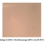 |
Figure 1: Morphology of ADSCs: The third passage ADSCs were 80–90 % confluent.(100X) |
Cell Surface Markers Of hADSCs
In this study, to ensure the purity of the derived mesenchymal stem cells, we assessed the expression of specific cell markers using flow cytometry. As mentioned, the adipose mesenchymal cells have high expression for CD90 and CD105 and minor expression for CD34 and CD45. The results showed that 99.4% of hADSCs expressed CD105 marker (Fig.2a.) and 97.8% of the stem cells expressed CD90 marker (Fig. 2b.), while CD34 marker was expressed in only 3.25% (Fig. 2c.) and CD45 marker in 2.77% (Fig.2d.) of these cells.
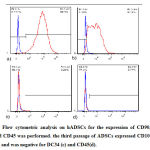 |
Figure 2: Flow cytometric analysis on hADSCs for the expression of CD90, CD105, DC34 and CD45 was performed. the third passage of ADSCs expressed CD105 (a) and CD90 (b), and was negative for DC34 (c) and CD45(d). |
Neuron-like Morphology of hADSCs
Neural differentiation happened in passage 3, when the cells had reached an acceptable purity, but had great growth and proliferation power. Induction of differentiation was performed in a eight day period in the presence of differentiation factors bFGF, Forskolin, and NGF. At the end of the eighth day, the cells had round and small cell body and processes like dendrites and axons (Fig. 3), while mesenchymal cells had spindle morphology like fibroblast cells (Fig. 1).
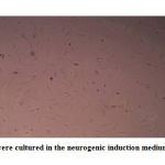 |
Figure 3: ADSCs were cultured in the neurogenic induction medium for 8days.(100X) |
Immunofluorescence Analysis
The expression of specific neuronal cells proteins were assessed using immunocytochemistry techniques. Observation of differentiated cells using fluorescent microscopy showed the presence of specific neuronal cells’ proteins, including ß-tubulin ІІІ (Fig. 4a.) and MAP2 (Fig. 4b.) in these cells.
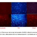 |
Figure 4: Fluorescence microscopic micrographs of hADSCs cultured on neuronal induction media for 8 days. differentiated cells were immunostained for a) b-tubulin and b) Map-2. |
Expression Of Neural-Specific Senes
PCR
Subsequent neural differentiation of hADSCs reverse transcription polymerase chain reaction analysis was used to discern the expression of neural differentiation in mRNA levels. The Induction medium was conducive for neurogenic differentiation and exposed the upward regulated expression of the neurogenic specific genes. RT-PCR analysis demonstration has increased mRNA expression for MAP2, NSE, and NFM genes. (Fig. 5)
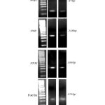 |
Figure 5: hADSCs were induced for 8days in the presence of neurogenic supplements.RT-PCR analysis of treated cells indicates that upregulation of specific gene expression as compared to untreated control cultures. ß-actin was assessed as internal controls. |
Real-time
Studies on neural gene expression with Quantitative Real-time PCR were performed to appraise the differentiation of hADSCs induced with specific culture medium. The results indicated that neural gene expression in hADSCs that were cultured in standard medium was low, but we observed high level of gene expression MAP2, NSE and NFM by using specific neural Induction medium after 8 days (Fig. 6a.b.c).
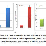 |
Figure 6: Real-time PCR gene expressions analysis of hADSCs proliferated in nerve differentiation and standard medium. Relative expression of a)Map2, b)NSE and c)NFM. *:indicate significant level of expression gene compared to hADSCs at p≤0.05. |
Discussion
Human adipose derived stem cells are cells with fibroblastic morphology that play an important role in stem cell therapy, because of its self-renewal, high proliferation capacity, ability to differentiate into several mesenchymal lineages, and devoid of embryonic stem cells’ problem such as ethical problems and risk of tumorigenesis. These cells have wider application in clinical research and treatment of nervous injuries than other cell sources. (11,14,20). The central nervous system is unable to recover itself, because mature neurons have a minor ability to reconstruct and also, neural stem cells do not have the needed ability to produce new efficient neurons (14,21,22).
Some reports show that mesenchymal stem cells can differentiate into neuronal cells in a culture medium under specific conditions (18,23). Chemical substances such as b-mercaptoethanol, BHA, Dimethylsulfoxide (DMSO) are used for neuronal differentiation of mesenchymal cells of the spleen, thymus, bone marrow, and adipose (14). In another study, a mixture of isobutylmethylxanthin, indomethacin, and insulin was used for neuronal induction of adipose mesenchymal cells (24,25,26). It has been shown that neuronal induction by chemical processes is reversible and after removal of differentiation medium, the cells morphology will return to their first form and express mesenchymal cells markers, which may have resulted from incomplete differentiation of cells (31). Also, the emerged neuron-like morphology and expression of neuronal protein can be due to cell shrinkage in response to stress factors, changes in the cytoskeleton and increase in the amount of antigen per unit area (27,28).
In 2012, Chengcheng Ying and colleagues differentiated rat ADSCs into nerve cells (12). Also, Sujeong Jang and colleagues put hADSCs under induction of bFGF for 8 days and then under induction of Forskolin to differentiate it into nerve cells (10). In present study The combination and selection Of Bfgf ,forskolin and NGF dosage depends on the research aim to investigate theexperimental method for neural differentiation in short period of time. the data approved that bFGF 10ng/ml, Forskolin 10 μM and NGF NGF 10 ng/ml were able to give rise to the maximum number of neuron like cells at day 8 of treatment, as the ADSCs were induced to differentiate. Our results revealed that combination of bFGF, Forskolin and NGF treatment increased the proportion of differentiated ADSCs to neuron like cells in short period of time. There is no question that the bFGF, Forskolinand NGF combination was a recuperated induction treatment for neuron like cell differentiation in comparison with either bFGF, Forskolin or NGF treatment alone as performed before. Long-term differentiation increases the risk of microbial and fungal contamination and increases cellular damage; meanwhile cells may not have the needed efficacy due to aging. The aged cells don’t have appropriate quality for induction and differentiation. Meantime frequent change of medium culture in long term differentiation process, cause separating cells from their bed .Therefor employ expedient short term differentiation method in present research ,reduces the risk of this problems.
Regarding differentiation of mesenchymal stem cells to neuronal cells, it can be concluded that there are two types of induction: fast and long-term. Fast induction occurs when cells are exposed to chemical factors, in which chemical induction causes tension in cells and result in contraction of cytoplasm and creates a morphology like neuron in cells (27,30). In the second type of induction, which needs more time, the created differentiation is more durable and cells are less exposed to tension, differentiation factors are used instead of chemical factors. As in previous studies using bFGF, growth factor was shown to initiate intracellular cascade routes of differentiation into neuron-like cells; in this study the mentioned factor was used. Moreover, bFGF is known as a factor for production of neural cells precursor with a high capacity to differentiate into nerve cells (10). NGF used in the induction protocol, plays a vital role in the life and survival of sympathetic system and sensory neurons and induce axonic growth and will also activate tropomyosin receptor kinase A (TrkA) that plays a vital role in cell induction and differentiation (32).
In this study, we tried to select an efficient method for the differentiation of adipose stem cells into neuron-like cells. So, we used another factor named Forskolin which increases intracellular levels of cyclic adenosine monophosphate (CAMP) by activating adenyl-cyclase enzyme and runs a set of signaling cascades for the process of differentiation (23).Nervous system repair is a complex biological phenomenon, as a result, the strategies for renewal and restoration of nerve tissue has attracted much attention, as it directly affects the patients’ quality of life. simultaneous presence of differentiation factors such as bFGF, Forskolin, and NGF induced phenotypic and morphological changes in cells towards neuron-like cells. The differentiated cells also show the morphology of nerve-like cells and expression of specific markers of nerve cells such as MAP2 and b-tubullin ІІІ demonstrated by immunocytochemical (ICC) staining . MAP2 and b-tubulin ІІІ are two proteins expressed in the early stages of neural differentiation. After induction of the cells with induced factors in this study, the differentiated cells expressed these proteins. furthermore, to ensure the nature of neuron-like cells, the expression of neuronal markers was evaluated using RT-PCR and Real-time PCR techniques. Administration of bFGF, Forskolin and NGF obviously increased the Expression MAP2,NSE and NFM in differentiated group for short time intervals in compared with the control group.
Previous studies showed that hADSCs naturally expresses nervous genes to some extent (10). Our results showed that inducing cells with special differentiation medium for one week, increased the expression of MAP2, NSE, and NFM.
It should be noted that in the present study, after the process of differentiation, cells morphology changed to neuron-like cell after a week but their survival was limited to only one week after differentiation, this may cause cell cycle arrest and the loss of self-renewal ability of the cells after the differentiation process.
Conclusions
In conclusion, combined bFGF, Forskolin, and NGF significantly enhanced the rate differentiation of ADSCs to neuron like cells in short period of time , which may supply support for the efficacious application of ADSCs in neurodegenerative disease and cell transplantation. However, more assessments, like electrophysiology, patch clamp studies of cell transplantation are required to identify the capabilities and true identity of the induced cells.
References
- Tomioka Sh, Nagahama T, Tokuyama R, Tatehara S, Satomura K ( 2012) Nerve growth factor increases electrical activity of neural cells derived from murine bone marrow stromal cells. Neuroendocrinology Letters 33,177-182
- Short B, Brouard N, Occhiodoro-Scott T, Ramakrishnan A, Simmons P ( 2003) Mesenchymal Stem Cells. Archives of Medical Research 34,565–571.
- Yamanaka Sh. (2012) Induced Pluripotent Stem Cells: Past, Present, and Future. Cell stem cell 10,678-684
- Ben-Ami E, Berrih-Aknin S, Miller A ( 2011) Mesenchymal stem cells as an immunomodulatory therapeutic strategy for autoimmune diseases. Autoimmunity Reviews 10,410–415
- Patel SA, Sherman L, Munoz J, Rameshwar P ( 2008)Immunological properties of mesenchymal stem cells and clinical implications. Immunol. Ther. Exp 56,1-8.
- Pountos I, Corscadden D, Emery P, Giannoudis PV (2007) Mesenchymal stem cell tissue engineering:Techniques for isolation,expansion and application. Injury, Int. J. Care Injured 38,S23-33
- Barry FP, Murphy JM ( 2004) Mesenchymal stem cells: clinical applications and biological characterization. The International Journal of Biochemistry & Cell Biology 36,568–584.
- Furth ME, Atala A.( 2009) Stem cells sources to treat diabetes. J Cell Bioche 106,507-511.
- Pittenger M, Mackay A, Beck S, Jaiswal R, Douglas R, Mosca J, Moorman M, Simonetti D, et al( 1999) Multilineage potential of adult human mesenchymal stem cells. Science 284,143-147.
- Jang S, Cho HH, Cho YB, Park JS, Jeong S (2010) Functional neural differentiation of human adipose tissue-derived stem cells using bFGF and forskolin. BMC Cell Biology 11,1-13.
- Bassi G, Pacelli L, Carusone R, Zanoncello J, Krampera M ( 2012) Adipose-derived stromal cells (ASCs). Transfusion and Apheresis Science 47,193-8.
- Ying C, Hu W, Cheng B, Zheng X, Li S (2012) Neural Differentiation of Rat Adipose-Derived Stem Cells in Vitro. Cell Mol Neurobiol 32,1255–63.
- Strem B, Hicok K, Zhu M, Wulur I, Alfonso Z, Schreiber R, Fraser J, Hedrick M (2005). Multipotential differentiation of adipose tissue-derived stem cells. Keio J med 54,132-41.
- Prabhakaran M, Venugopal JR, Ramakrishna S (2009)Mesenchymal stem cell differentiation to neuronal cells on electrospun nanofibrous substrates for nerve tissue Biomaterials 30,4996–5003.
- De Ugarte DA, Morizono K, Elbarbarya A, Alfonso Z, A.Zuk P, Zub M, Dragoo J, Ashjian P, et al ( 2003) Comparison of Multi-Lineage Cells from Human Adipose Tissue and Bone Marrow. Cells Tissues Organs 174,101–109.
- Huang T, He D, Kleiner G, Kuluz J(2007) Neuron-like Differentiation of Adipose-Derived Stem Cells From Infant Piglets in Vitro. The Journal of Spinal Cord Medicine 30,S35-40.
- Safford KM, Hicok KC, Safford SD, Halvorsen YC, Wilkison WO, Gimble JM, Rice HE (2002) Neurogenic differentiation of murine and human adipose derived stromal cells.Biochem Biophys Res Commun 294,371-379.
- Safford KM, Safford SD, Gimble JM, Shetty AK, Rice HE(2004) Characterization of neuronal/glial differentiation of murine adipose-derived adult stromal cells. Exp Neurol 187,319-328.
- Guilak F, Lott KE, Awad HA, Cao Q, Hicok KO, Fermor B, Gimble J (2006) Clonal analysis of the differentiation potential of human adipose-derived adult stem cells. J Cell Physiol 206,229-237.
- Zuk P, Zhu M, Ashjian P, De Ugrate D, Huang J, Mizuno H, Alfonso Z, Fraser J, et al(2002) Human Adipose Tissue Is a Source of Multipotent Stem Cells. Molecular Biology of the Cell 13,4279–4295.
- Xu Y, Liu Zh, Liu L, Zhao C, Xiong F, Zhou C, Li Y, Peng F, et al. (2008) Neurospheres from rat adipose-derived stem cells could be induced into functional Schwann cell-like cells in vitro. BMC Neurosci 9,1-10.
- Kingham PJ, Kalbermatten DF, Mahay D, Armstrong SJ, Wiberg M, Terenghi G (2007)Adipose-derived stem cells differentiate into a Schwann cell phenotype and promote neurite outgrowth in vitro. Experimental Neurology 207,267-274.
- Anghileri E, Marconi S, Pignatelli A, Cifelli P, Galie M, Sbarbati A, Krampera M, Belluzzi O, et al (2008) Neuronal differentiation potential of human adipose-derived mesenchymal stem cells. Stem Cells Dev 17,909-916.
- Ning H, Lin G, Lue T, Lin CS (2006) Neuron-like differentiation of adipose tissue-derived stromal cells and vascular smooth muscle cells. Differentiation 74,510-518.
- Ashjian PH, Elbarbary AS, Edmonds B, DeUgarte D, Zhu M, Zuk PA, Lorenz P, Benhaim P, et al( 2003) In vitro differentiation of human processed lipoaspirate cells into early neural progenitors. Plast Reconstr Surg 111,1922-1931.
- Fujimura J, Ogawa R, Mizuno H, Fukunaga Y, Suzuki H(2005) Neural differentiation of adipose-derived stem cells isolated from GFP transgenic mice.Biochem Biophys Res Commun 333,116-121.
- Yang F, Murugan R, Wang S, Ramakrishna S(2005) Electrospining of nano/micro scale Poly (L- lactic acid) aligned fibers and their Potential in neural tissue engineering. Biomaterials 26,2603-2610.
- Lu P, Blesch A, Tuszynski M (2004) Induction of Bone Marrow Stromal Cells to Neurons: Differentiation,Trans Differentiation,or Artifact. Journal of Neuroscience Research 77,174-191.
- Muñoz-Elías G, Woodbury D, Black I (2003) Marrow stromal cells,mitosis,and neuronal differentiation: stem cell and precursor functions. Stem Cells 21,437-448.
- Croft AP, Przyborski SA(2006) Formation of neuron by non-neural adult stem cell: Potential mechanism implicates and artifact of growth in culture. Stem Cells 24,1841-1851.
- Zurita M, Bonilla C, Otero L, Aguayo C, Vaquero J( 2008) Neural trans differentiation of bone marrow Stromal cells obtained by chemical agents is a short-time reversible phenomenon. Neuroscience research 60,275-280.
- Liu F, Xuan A, Chen Y, Zhang J, Xu L, Yan Q, Long D (2014) Combined effect of nerve growth factor and brain‑derived neurotrophic factor on neuronal differentiation of neural stem cells and the potential molecular mechanisms. Molecular medicine reports 10,1739-45.


