Sri Durga1, L. Rajasekar2, Okram Robin3
1Senior Lecturer, Dept Of Orthodontic, Tagore Dental College and Hospital 2Reader, Department of Orthodontics, Sree Balaji Dental College and Hospital, Bharath University, Pallikaranai, Chennai-600100 3Okram Robin, Post Graduate, Department of Orthodontics, Sree Balaji Dental College and Hospital, Bharath University, Pallikaranai, Chennai-600100
DOI : https://dx.doi.org/10.13005/bpj/692
Keywords
Second; Premolar; Extraction; Case; Report
Download this article as:| Copy the following to cite this article: Durga S, Rajasekar L, Robin O. Usefullness of Laser in Oral and Maxillofacial Surgery. Biomed Pharmacol J 2015;8(October Spl Edition) |
| Copy the following to cite this URL: Durga S, Rajasekar L, Robin O. Usefullness of Laser in Oral and Maxillofacial Surgery. Biomed Pharmacol J 2015;8(October Spl Edition). Available from: http://biomedpharmajournal.org/?p=3454> |
Introduction
To correct the malocclusion is the chief aim of an orthodontist. For each individual, ideal facial esthetics would result when the teeth were placed in ideal occlusion. Malocclusion is caused by two ways either spacing or crowding in which crowding is the most common cause.Treatment of a crowded arch requires space gaining. This has been achieved through two ways of treatment – extraction or non extraction modality. In extraction , premolars are the most common tooth being spared in orthodontic treatment1.Conveniently located between the anterior and posterior segments, premolars would appear to be the obvious choice for correcting crowding and anterior-posterior discrepancies.Different premolar extraction patterns provide space in different locations of the arches. For example, this extra space can be used to reduce protrusion or to camouflage skeletal Class II or Class III problems. An extraction pattern can also be selected that removes abnormally small or large premolars that contribute to a Bolton discrepancy.Second-premolar extractions often are utilized in situations that present with mild anterior crowding and no protrusion2, posterior crowding anterior open bites3, or when the molar anchorage needs to be intentionally lost4.TSALD is the most important factor necessitating the decision to extract premolars.The basic indication for second premolar extraction is when there is moderate anterior crowding with no protrusion and the patient has good facial balance.Removing the second premolars will give enough space to resolve minor crowding while not changing the profile. It also leaves the incisors in their original positions over basal bone without inclining them labially, which is undesirable.
Diagnosis and Etiology
The patient was a 16 year old man with a chief complaint of forwardly placed upper front teeth. The pretreatment photographs and clinical examination showed average lower facial height, deficient chin and decreased clinical FMA with a straight profile and straight divergence. On intra oral examination there was a mesiobuccal rotation of 15 and spacing between 22 and 23.Mesiobuccaly Rotated 15 has created a midline shift in upper arch of 1.5 mm towards left in the transeverse plane. With overbite of 3mm and overjet of 4mm. The lateral cephalometric analysis showed a Class I skeletal base with an average mandibular plane angle and proclined maxillary and mandibular teeth. IMPA 105 degree reflected a compensatory proclination of the mandibular incisors. The panoramic radiograph showed unerupted third molars and the periodontal state was good. Molar relationship was Class I on both the sides with a canine Class I and incisor Class I relationship on both sides. The Bolton tooth ratio analysis indicated tooth material excess in the mandible by 1.1 mm in the anteriors and overall tooth material excess in the mandible was 0.3 mm.
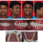 |
Figure 1: Pre-treatment extra and intra oral photographs |
Pre-treatment extra and intra oral photographs
Based on the findings the patient was diagnosed as Angle’s skeletal Class I base with an Angle’s dentoalveolarClass I malocclusion, with decreased mandibular plane angle and bi-dental protusion , mesiobuccal rotation in relation to 15 and spacing in relation to 21 and 22 with upper midline shift.
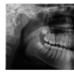 |
Figure 2 |
Treatment Objectives
The treatment objectives of this patient were (1) to correct proclination of incisors (2) to achieve ideal overjet and overbite (3) to maintain Class I canine and molar (4) to maintain soft tissue profile.
Treatment Progress
Before the orthodontic treatment, the maxillary and mandibular second premolars were extracted. Anchorage concern in this case was moderate to establish Class I molar relation. 0.022 x 0.028-in MBT PEA brackets were bonded to both arches. Five months after bracket bonding, leveling and aligning in the maxilla and mandible were complete.
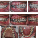 |
Figure 3: Space closure with 0.019 x 0.025” SS Posted archwires |
Space closure with 0.019 x 0.025” SS Posted archwires
Space closure was initiated with 0.019 x 0.025 in stainless steel wire to maintain torque control. Brass wire bent into the form of “C” shaped hooks were soldered into the 0.019 x 0.025 in wire just distal to the lateral incisor brackets. These posted archwires were incorporated with reverse curve of spee and posterior segment of the wire was detorqued (to avoid unnecessary palatal cusp plunging down in the maxillary molar) and they were inserted for a month without activation, to allow the incorporated torque to establish itself upon the maxillary anteriors(as some amount of anchor loss is expected during retraction).
Active tiebacks were given across the maxillary molar hooks and the soldered posts simultaneously class-II blue elastics were given to achieve Class I Canine relation. After 18 months of active treatment, the molar and canine relationships on both sides were Class I and the midline deviation was also corrected. At the end of space closure, settling was achieved with 0.018 in Stainless steel sectional wires and kobayashi hooks in the form of triangular red elastics for 2months. The appliances were removed after 20 months of treatment. Begg’s wraparound retainer was placed in the maxillary arch and a fixed lingual retainer was placed in the mandibular arch.
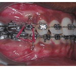 |
Figure 4: Settling with Triangular elastics |
Settling with Triangular elastics
Treatment Results
Proclination, which was the patient’s chief complaint was eliminated.the dental midlines were aligned with the facial midlines. The posterior occlusal relationships were improved to achieve Class I canine and molar relationships on both sides. More ideal overjet and overbite relationships were established.
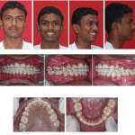 |
Figure 5: Post treatment Extra and Intra oral photos |
Post treatment Extra and Intra oral photos
Discussion
In the treatment of a Class I patient extraction only in the maxilla or both the arches is a common method to correct crowding , protrusion and occlusal relationships. The basic indication for second premolar extraction is when there is moderate anterior crowding. There are certain effects of premolar extraction which includes incisor angulation, incisor retraction, molar protraction, occlusal plane, vertical dimension, tooth size and arch length discrepancy and arch changes. On evaluation of Bolton’s analysis it was found that extraction of all second premolars and upper second and lower first premolars did not create tooth-size discrepancies with the overall ratios, whereas extraction of all first premolars and upper first and lower second premolars created discrepancies with the overall ratios that were outside the range of 87.0% to 89.0%, described by Bolton. According to De Angelis concept extraction of second premolar was preffered because it was better esthetic , created an harmonious posterior Bolton relationship. Second premolar extraction procedure required increased time for root paralleling which could be counteracted by incorporating a mesial tip in the first premolar bracket by altering the position while bonding the brackets
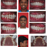 |
Figure 6 |
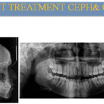 |
Figure 7: Post Treatment CEPH and OPG |
Refrences
- Proffit W, Forty year review of extraction frequency at a university orthodontic clinic, Angle Orthod 1994; 407– 14.
- SchoppeRj , An analysis of second premolar extraction procedures, Angle Orthod 1964; 34: 292 – 302.
- Brandt S, Safirstein G R, Different extractions for different malocclusions , Am J Orthod 1975; 68: 15 – 41.
- De Castro N, Second premolar extraction in clinical practice, Am J Orthod 1974; 65:115 – 37.


