Sunil Chandy Varghese1, C. Deepak2
1Dept. Of Orthodontics, Tagore Dental College & Hospital, Chennai, India 2Department of Orthodontics, Sree Balaji Dental College and Hospital, Bharath University, Pallikaranai, Chennai-600100
DOI : https://dx.doi.org/10.13005/bpj/697
Abstract
Nasal airway patency and malocclusion have long been Interrelated. It is obvious that severe malocclusion must make it difficult for the individual to breathe, chew, swallow, and speak. The Reverse Of This Could Also Be True! Alterations or adaptations in function can be an etiologic factor for malocclusion, by influencing the pattern of growth and development and thereby resulting in malocclusion. This article attempts to compile the views supporting and opposing nasal obstruction as a cause for malocclusion.
Keywords
Nasal airway; Problems; Alterations or adaptations
Download this article as:| Copy the following to cite this article: Varghese S. C, Deepak C. Airway Problems and its Related Disorders- An Orthodontist Perspective. Biomed Pharmacol J 2015;8(October Spl Edition) |
| Copy the following to cite this URL: Varghese S. C, Deepak C. Airway Problems and its Related Disorders- An Orthodontist Perspective. Biomed Pharmacol J 2015;8(October Spl Edition). Available from: http://biomedpharmajournal.org/?p=3491> |
Introduction
Mouth Breathing
Etiology
anatomical-dns, congenital
microgenia
pathological-adenoids, tonsils,
Habitual
“Adenoid Facies”—coined at GUY’S hospital,London,
constitutes the following- long face, constricted upper dental arch, exposed upper incisors, receded lower jaw, short upper lip, associated habits
Obstructive Sleep Apnea
Definition
Condition caused either by complete occlusion or partial collapse of the upper airway despite the presence of simultaneous respiratory effort. Cessation occurs at the level of nostrils and mouth. Condition is considered pathologic when the episodes last for at least ten seconds and at a frequency of 30 times or more during 7 hrs. of nocturnal sleep in REM and especially in non REM stages of sleep.
Types
- Central Apnea: cessation of diaphragmatic excursions
- Upper Airway Apnea: obstruction to air flow pass the oro-pharynx but with persistent diaphragm movements.
- Mixed Apnea: cessation of air flow and absent respiratory effort early in the episode, followed by unsuccessful attempts at respiration later in the episode.
Role of Genioglossus Muscle in Osa
OSA is characterized by recurrent upper air way occlusion during inspiration. The genioglossus muscle is believed to contribute to this. genioglossus muscle activity has been demonstrated in phase with inspiration during sleep.
Preferential activation of this muscle is correlated with pharyngeal opening and resolution of apnea. A dynamic relationship between supraglottic pressure and genioglossus muscle amplitude has been postulated to explain upper airway occlusion in subjects with OSA.
Effects of Osa
- SYMPTOMS during sleep
- Snoring
- Abnormal motor activity
- Disturbed nocturnal sleep
- Sensation of choking
- Heart burn
- Nocturia
- Heavy sweating
Signs
- large tongue
- elongated soft palate
- reduced pharyngeal length
- decreased posterior air space
- increased gonial angle
- increased upper and lower facial height
- steep occlusal plane
- elongated upper and lower incisors
Diagnosis
Is by Polysomnography
Measurements are made to assess sleep stages of breathing and gas exchange to detect sleep stages.
PSG ensures the no. of apnic episodes per hour of sleep expressed by respiratory (Disturbance Index)measurements of chest and abdominal efforts and oxygen saturation.
Airway measurement by cephalometric 3D imaging – lateral pharyngeal dimension.
Treatment
Medical
- Weight loss is beneficial
- Nasal vaso constriction sprays
- Withdrawal of respiratory depressing alcohol (antihistamines and tranquilizers)
Surgical
- Uvulo palato pharnygoplasty
- Tracheostomy
- Expansion hyoid plasty
- Mandibular advancement
- Sectioning of hyoid
Diagnosis of Mouth Breathing
- Clinical Examination:
- ask patient to hold water in the mouth
- use double sided mouth mirror orcotton wisps
- facial pattern – long face with incompetent lips not necessary indicate mouth breathing pattern.
Cephalometric Analysis
Mc NAMARA airway analysis upper,lower
- Upper pharyngeal width – the point on posterior outline on soft palate to closest point on pharyngeal wall – 15 to 20 mm in width.values 2mm or less indicate airway impairment
- Lower pharyngeal width from point of intersection of posterior border of tongue and inferior border of mandible to the closet point on posterior pharyngeal wall – 11 to 14mm usually values are high due to anteriorly positioned tongue as the adenoids are enlarged.
Other Cephalometric Findings
- vertical growth pattern
- increased ANB
- increased gonial angle
- decreased mandibular length
- steep MP angle
- over erupted upper posterior Segments
 |
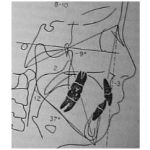
Figure 1 |
Other Diagnostic Tests
- Spirometry
- Oximeter– to evaluate oxy-Hb level
- Rhinomanometry-instrument used to measure nasal patency
STEDMAN’S medical dictionary defines it as “study of nasal obstruction and nasal airflow characteristics
- Pneumotachograph– device consisting of flow meter, pressure measuring manifold, and a recording instrument.
- Respirometry-study of both nasal and oral respiratory function
- SNORT – simultaneous nasal and oral respiratory technique
Effects of Airway Obstructions
Head Posture Changes
Beni Solow and Antje Tallgren
Extension of the head in relation to the cervical column was found in connection to large anterior facial ht. and small post. Facial height, small anterio-posterior dimension, large mandibular inclination to anterior cranial base & to nasal plane, facial retrognathism, large cranial base angle and small nasopharyngeal space.
- Ricketts(1968)-
reported subjects with enlarged adenoid with extension of head &forward and downwardly positioned tongue.
- Ninima & Cole
noted 5 degree increase in cranio facial angle associated with nasal obstruction.
- Mandubular Rotation
In response to enlarged adenoids which occupy the posterior pharyngeal space the tongue gets anteriorly positioned leading to downward and backward rotation of the mandible. The ANB angle increases, MP angle increases, LAFH increases-Long Face Syndrome.
Treatment Options
- Treating the etiologic factors
Tonsillectomy
Adenoidectomy
Correction of DNS
Nasal polyps
- Orthodontic
Oral Screen
Rapid Maxillary Expansion
Mandibular Advancement
- Surgical
hyoid bone repositioning
Bi-jaw advancement
mandibular advancement
References
- Wong ML, Sandham A, Ang PK, Wong DC, Tan WC, Huggare J. Craniofacial morphology, head posture, and nasal respiratory resistance in obstructive sleep apnoea: An inter-ethnic comparison. Eur J Orthod. 2005;27:91–97.
- Schlenker WL, Jennings BD, Jeiroudi MT, Caruso JM. The effects of chronic absence of active nasal respiration on the growth of the skull: A pilot study. Am J Orthod Dentofacial Orthop. 2000;117:706–713.
- Lopatiene K, Babarskas A. [malocclusion and upper airway obstruction] Medicina (Kaunas) 2002;38:277–283.
- Lyberg T, Krogstad O, Djupesland G. Cephalometric analysis in patients with obstructive sleep apnoea syndrome: Ii. Soft tissue morphology. J Laryngol Otol. 1989;103:293–297.
- Tangugsorn V, Skatvedt O, Krogstad O, Lyberg T. Obstructive sleep apnoea: A cephalometric study. Part ii. Uvulo-glossopharyngeal morphology. Eur J Orthod. 1995;17:57–67.
- Maltais F, Carrier G, Cormier Y, Series F. Cephalometric measurements in snorers, non-snorers, and patients with sleep apnoea. Thorax. 1991;46:419–423.
- Zucconi M, Ferini-Strambi L, Palazzi S, Orena C, Zonta S, Smirne S. Habitual snoring with and without obstructive sleep apnoea: The importance of cephalometric variables. Thorax.1992;47:157–161.
- Pae EK, Lowe AA, Sasaki K, Price C, Tsuchiya M, Fleetham JA. A cephalometric and electromyographic study of upper airway structures in the upright and supine positions. Am J Orthod Dentofacial Orthop. 1994;106:52–59.
- Guilleminault C, Stoohs R. Obstructive sleep apnea syndrome in children. Pediatrician. 1990;17:46–51.
- Bacon WH, Turlot JC, Krieger J, Stierle JL. Cephalometric evaluation of pharyngeal obstructive factors in patients with sleep apneas syndrome. Angle Orthod. 1990;60:115–122.


